
Vascular territory segmentation using mutual clustering information from image space and label space

a technology of applied in the field of vascular territory segmentation using mutual clustering information from image space and label space, can solve the problems of increasing the risk of ischemic stroke, reducing the blood supply to the subserved parts of the brain, and quantitative mapping of blood flow from individual source arteries is still not practical in the clinical setting

Active Publication Date: 2014-09-18
WAKE FOREST UNIV HEALTH SCI INC
View PDF3 Cites 4 Cited by
  • Summary
  • Abstract
  • Description
  • Claims
  • Application Information

AI Technical Summary

Benefits of technology

The invention provides methods, systems, circuits, and workstations for automatically segmenting an image volume into separate vascular territories using mutual clustering in image and label space. This technology can be useful for MRI brain scans and evaluating large artery diseases, cerebral vascular disease, and carotid stenosis. It can also be used for routine brain scans for neurological evaluations and stroke treatment. The invention provides a color map that indicates the associated feeding artery per voxel in unique colors. The segmentation analysis can be carried out using a processor associated with an MR Scanner and / or at least one clinician workstation. The invention also includes image and data processing circuits and a computer program code for implementing this technology.

Problems solved by technology

Carotid or vertebrobasilar stenosis restricts distal blood flow, which decreases blood supply to the parts of the brain subserved by these vessels, and increases the risk of ischemic stroke.
Although invasive CT-based methods have been used for qualitative assessment of vascular territory perfusion, quantitative mapping of blood flow from individual source arteries is still not practical in the clinical setting.
Vascular territory mapping using arterial spin labeling (ASL) has been proposed, but currently typically requires complicated planning prior to scanning and extensive post-processing, which hinders the practical clinical use of these methods.
The source location of a territory is often wide-spread due to noise and large vessel diameters, becoming more complicated when the labeling plane includes a large number of source arteries.

Method used


Image

  • Vascular territory segmentation using mutual clustering information from image space and label space
  • Vascular territory segmentation using mutual clustering information from image space and label space
  • Vascular territory segmentation using mutual clustering information from image space and label space

Examples


Embodiment Construction

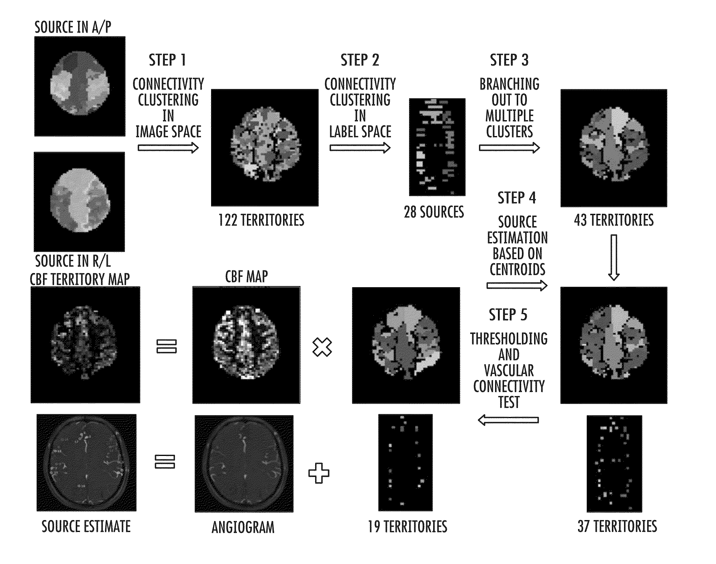
[0005]Embodiments of the invention are directed to methods, systems and circuits that can automatically segment an image volume into separate vascular territories using mutual clustering in image and label space.

[0006]Embodiments of the invention provide systems, methods, circuits, workstations and methods suitable for automated vascular territory mapping for resolving source locations and to determine if multiple sources in different perfusion territories are from a single artery.

[0007]Embodiments of the invention may be particularly useful for MRI brain scans for evaluation of large artery diseases, cerebral vascular disease, and carotid stenosis and may also be useful for stroke, especially thromboembolic stroke, and / or for evaluation of treatments or clinical trials.

[0008]Embodiments of the invention may be implemented as a routine brain scan for neurological evaluations due to the automated processing and short MRI signal acquisition time required for vascular mapping that can ...


PUM


Abstract

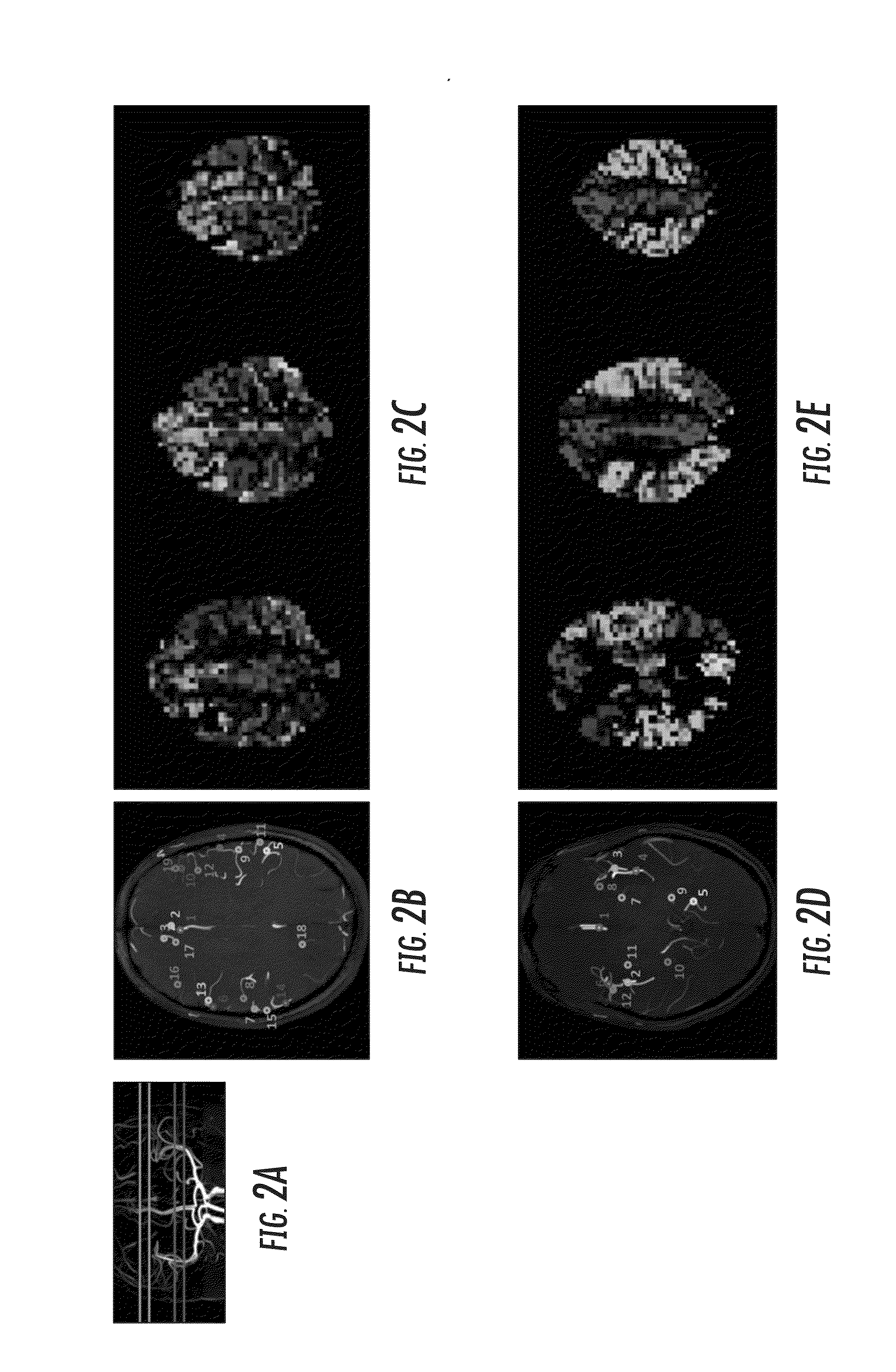
Methods, systems, computer programs, circuits and workstations are configured to generate at least one two-dimensional weighted CBF territory map of color-coded source artery locations using an automated vascular segmentation process to identify source locations using mutual connectivity in both image and label space.

Description

RELATED APPLICATIONS[0001]This application claims the benefit of and priority to U.S. Provisional Application Ser. No. 61 / 788,826, filed Mar. 15, 2013, the contents of which are hereby incorporated by reference as if recited in full herein.BACKGROUND[0002]Carotid or vertebrobasilar stenosis restricts distal blood flow, which decreases blood supply to the parts of the brain subserved by these vessels, and increases the risk of ischemic stroke. Surgical intervention with carotid endarterectomy or endovascular angioplasty / stenting is generally pursued if the diameter of the lumen of the internal carotid artery (ICA) is reduced more than 70%, which is typically documented by noninvasive imaging. Collateral circulation increases in the brain as a normal physiologic mechanism to by-pass and compensate for the blockage in the main artery. In some cases, this increased collateral flow can supply enough oxygenated blood to maintain adequate cerebral perfusion for supporting brain function in...

Claims


Application Information

Patent Timeline
IPC IPC(8): G06T7/00
CPCG06T7/0012G06T7/155G06T2207/10081G06T2207/10088G06T2207/30101G06T7/90A61B5/0042A61B5/026A61B5/0263A61B5/743A61B5/7445G06T11/008G06T2207/30016
Inventor JUNG, YOUNGKYOO
Owner WAKE FOREST UNIV HEALTH SCI INC
Learn More