
Detection device, detection strip, and detection system

a detection device and detection strip technology, applied in the field of detection devices, detection strips, detection systems, can solve the problems of inability to analyze eye fluid samples, inability to apply general blood testing to assess the activity of eye diseases, and inability to sample eye fluid for analysis, etc., to achieve easy and fast operation, improve permeability and water retention ability, and simplify the process

Inactive Publication Date: 2014-10-23
NATIONAL TSING HUA UNIVERSITY
View PDF1 Cites 0 Cited by
  • Summary
  • Abstract
  • Description
  • Claims
  • Application Information

AI Technical Summary

Benefits of technology

The present invention provides a detection device that is easy and fast to operate. The device uses a reaction portion with fiber-based material and anti-growth factor antibodies to detect antigens in body fluids like angiogenic factors. The detection process is direct and intuitive for users as there is no need for additional instruments. The fiber-based material has good permeability and water retention ability, which is useful for small samples. Overall, the detection device of the present invention provides a convenient and efficient solution for clinical applications.

Problems solved by technology

When the concentration of VEGF increases, lesions of diabetic retinopathy or macular degeneration become worse and may even lead to blindness.
However, due to complexity and specialization of the nervous structure of the eyes, general blood testing is not applicable to assess lesion activity of eye diseases.
Besides, there is no suitable method to sample eyes fluid for analysis of the growth factors in lesions.
Although these two drugs can be applied for the disease, the molecular detection devices applied for eye disease are still poor.
Hence, the concentration of angiogenesis in eye cannot be effectively detected or tracked, not to mention adequately assess the amount of drug and mode of administration facilities.
However, the detection of traditional method needs to obtain detection sample by drawing blood of patients.
Otherwise, relatively large number of reagents is required, and longer time is required for detection, which virtually extending the time for diagnosing and increasing the burden on the patient.
The detection equipments of traditional ELISA require high cost.
Furthermore, traditional ELISA needs to be processed in the laboratory environment with specific apparatus, which is unfavorable for clinical detection.

Method used


Image

  • Detection device, detection strip, and detection system
  • Detection device, detection strip, and detection system
  • Detection device, detection strip, and detection system

Examples


experiment 1

Detection of the VEGF by Paper-Based Detection Device Applying ELISA System

[0062]According to the present invention, it first provides a chromatography filter paper plate. After moistening the paper plate, VEGF antigens are added thereon and stand for 5 to 7 minutes. Then, bovine serum albumin (BSA) is added as blocking agents to prevent non-specific binding. After standing for 5 to 7 minutes, anti-VEGF antibodies which conjugate HRP are added to react with the antigens for 7 to 10 minutes. Next, the second blocking agent Streptavidin is added to react with reagents for 7 to 10 minutes. Finally, the chromatography filter paper plate is rinsed. Meanwhile, the solution comprising 3, 3′, 5, 5′-tetramethylbenzidine (TMB) and H2O2 are also added thereon until dry. After capturing the images of the paper plate, the images can be analyzed for gaining information.

[0063]As shown in FIG. 6A and FIG. 6B, they are calibration curves illustrating logarithmic value of the VEGF antigen concentrati...

experiment 2

The Detection of VEGF Concentration Produced by Diabetes Patients with Retinopathy Symptom by Paper-Based Detection Device Applying ELISA System

[0064]Determined by the above-mentioned method, the average VEGF concentration of the diabetes patients with retinopathy symptom is 740.1±267.7 pg / mL (total samples=14).

experiment 3

The Detection of VEGF Concentration Produced by Age-Related Macular Degeneration Patients by Paper-Based Detection Device Applying ELISA System

[0065]Determined by the above-mentioned method, the average VEGF concentration of the age-related macular degeneration patients is 383±155.5 pg / mL (total samples=17).


PUM

PropertyMeasurementUnit
pore sizeaaaaaaaaaa
pore sizeaaaaaaaaaa
concentrationaaaaaaaaaa

Abstract

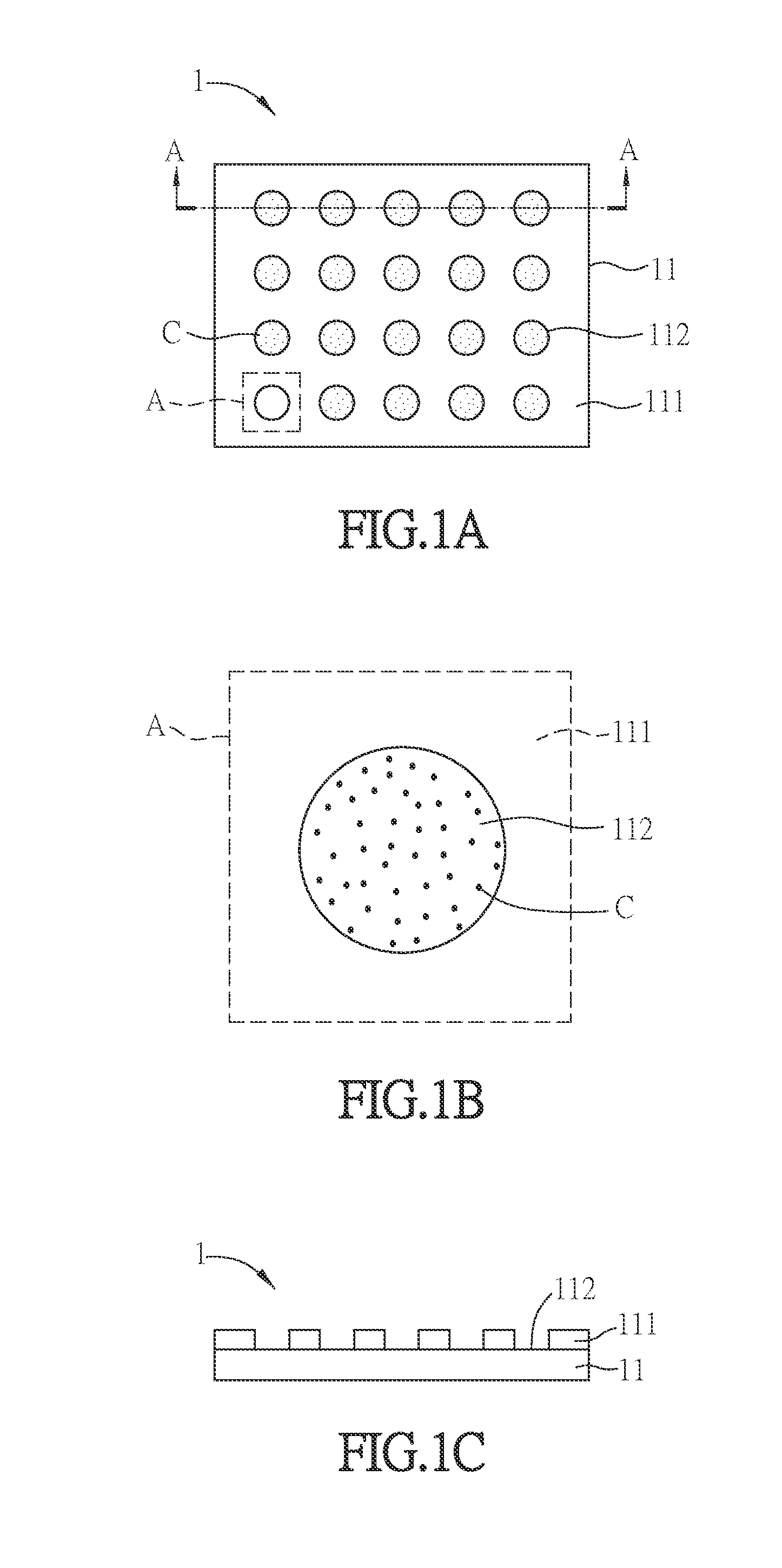
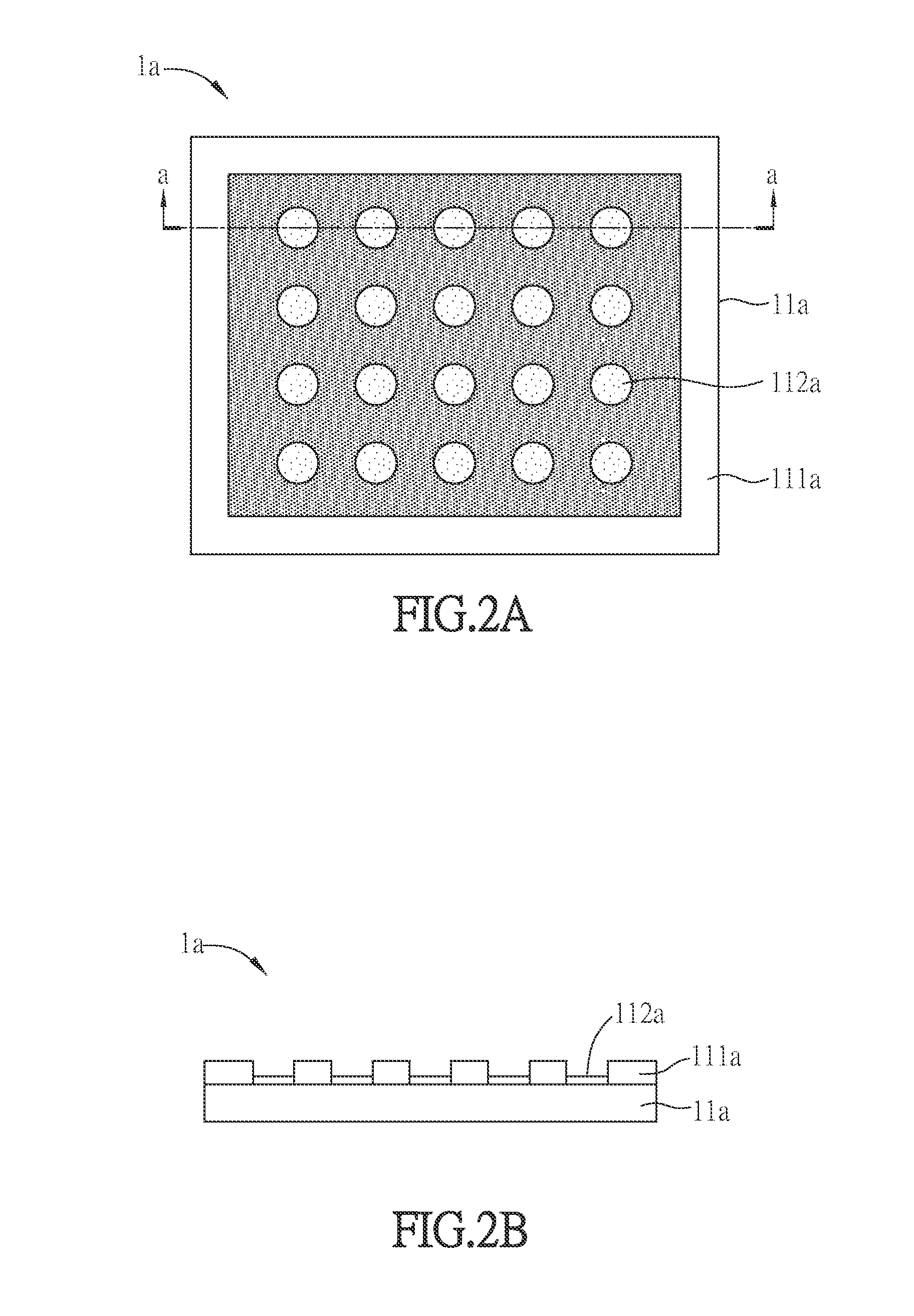
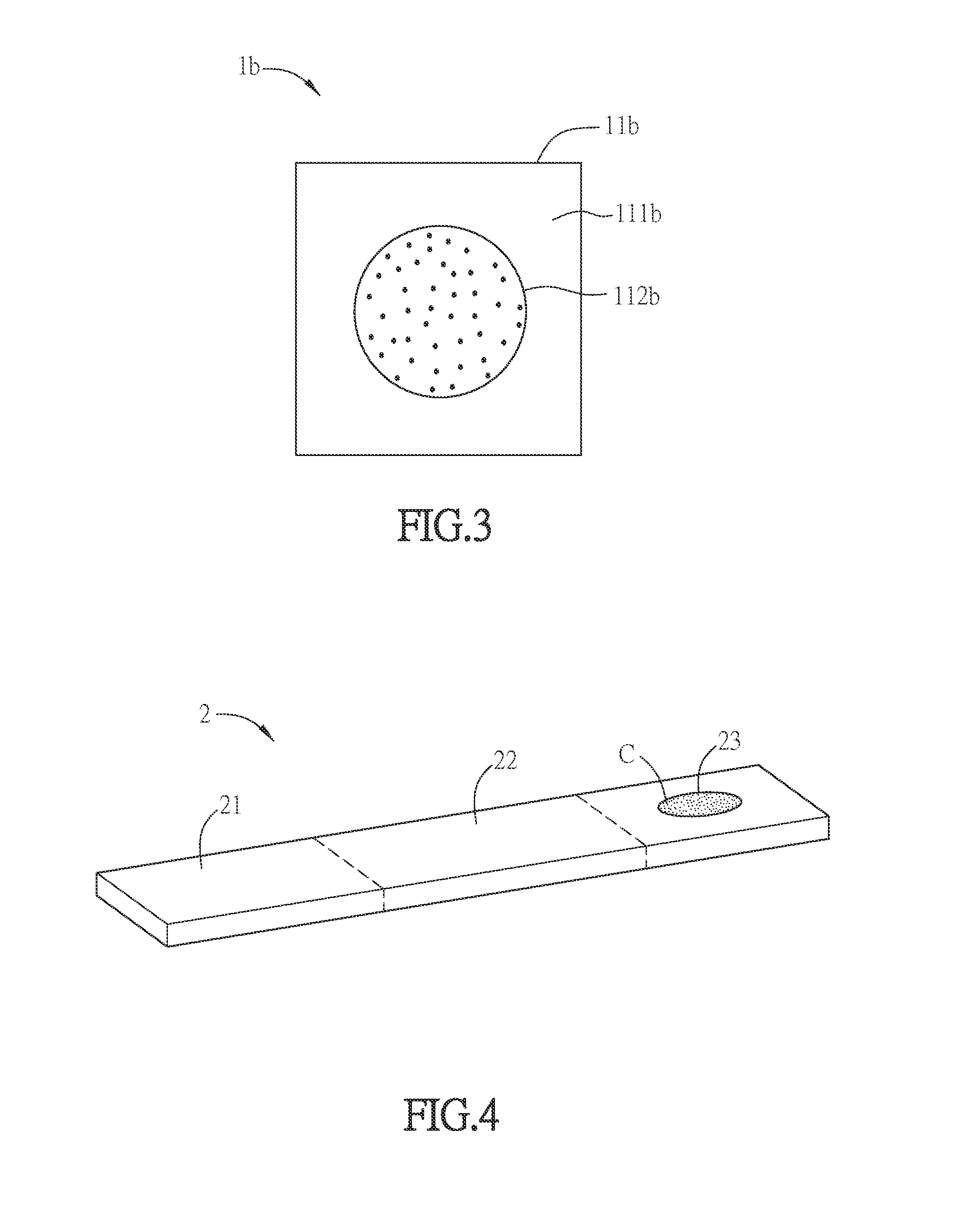
A detection device applied for detecting body fluids includes a substrate and a plurality of anti-growth factor antibodies. The substrate includes at least one reaction portion. The reaction portion includes a fiber-based material. Anti-growth factor antibodies are disposed on the fiber-based material. The present invention further provides a detection strip and a detection system for detecting body fluids. The present invention is advantageous for easy operation, lower amount of reagents and rapid analysis.

Description

CROSS REFERENCE TO RELATED APPLICATIONS[0001]This application is a Continuation-In-Part of U.S. application Ser. No. 13 / 867,409 filed on Apr. 22, 2013, which claims priority under 35 U.S.C. §119(a) on Patent Application No(s). 102100580 filed in Taiwan, Republic of China on Jan. 8, 2013, the entire contents of which are hereby incorporated by reference.BACKGROUND OF THE INVENTION[0002]1. Field of Invention[0003]The present invention relates to a detection device, a detection strip, and a detection system, and more particularly, to a detection device, a detection strip, and a detection system applied for detecting eye vascular lesion.[0004]2. Related Art[0005]With the development of technology and biotechnology medical care, the average human lifespan has significantly extended. With the changing of lifestyle, the number of patients suffering from diabetes and macular degeneration also increases. These two diseases are proved to be highly relevant to the concentration of vascular end...

Claims


Application Information

Patent Timeline
Patent Type & Authority Applications(United States)
IPC IPC(8): G01N33/543
CPCG01N33/54306G01N33/74G01N33/558G01N2333/475G01N2333/49G01N33/54388
Inventor HSU, MIN-YENYANG, CHUNG-YAOCHENG, CHAO-MIN
Owner NATIONAL TSING HUA UNIVERSITY
Learn More