Estimating Pharmacokinetic Parameters in Imaging

a pharmacokinetic and imaging technology, applied in the field of molecular imaging and mathematical modeling, can solve the problems of complex, interdisciplinary effort, and difficulty in designing, performing, and interpreting imaging studies

Active Publication Date: 2014-10-30
INVICRO
View PDF5 Cites 4 Cited by
  • Summary
  • Abstract
  • Description
  • Claims
  • Application Information

AI Technical Summary

Benefits of technology

[0006]The present invention encompasses methods for estimating physiological parameters relevant to drug development and patient treatment from imaging data. The present methods can be used to estimate tumor and contrast agent properties from molecular imaging data, parameters which may be used in accelerating the development of cancer therapeutics, improving patient selection and stratification for clinical trials and treatment regimes, and monitoring patient responses to treatment. Thus, the featured methods can be used to improve not only the design and preclinical development of therapeutics, but also the clinical use of tumor targeting therapeutics and complementary biomarkers. More specifically, the methods allow in vivo molecular imaging to be used to quantitatively estimate physiological parameters such as antigen expression levels, contrast agent internalization rate, tumor vascularity, and contrast agent binding affinity. Furthermore, sub-organ quantitative analysis of tumors allows characterization of the variability of these tumor properties in vivo.

Problems solved by technology

Designing, performing, and interpreting these imaging studies is a complex, interdisciplinary effort.

Method used


Image

  • Estimating Pharmacokinetic Parameters in Imaging
  • Estimating Pharmacokinetic Parameters in Imaging
  • Estimating Pharmacokinetic Parameters in Imaging

Examples


example 1

[0053]The Krogh cylinder geometry, on which our model is based, has been validated in the literature to predict both average tumor uptake of antibodies (Jackson et al., Br. J. Cancer 80:1747-1753, 1999; Baxter and Jain, Br. J. Cancer 73:447-456, 1996) as well as antibody microdistribution in tumors (Thurber et al., J. Nucl. Med. 48:995-999, 2007). We have compared model simulations to experimental data and have found that the model correlates very well with published experimental data for radiolabeled peptides and proteins. The model simulations were performed in MATLAB using the method of lines and the stiff ODE solver ode15s. Input parameters (e.g. binding affinity, blood clearance, internalization rate) were estimated from the literature. The sum of the radioactivity associated with free, bound, and internalized ligand was compared to the published total radioactivity in the tumor as assessed by ex vivo gamma counting. In addition to preclinical data, we have found that the model...

example 2

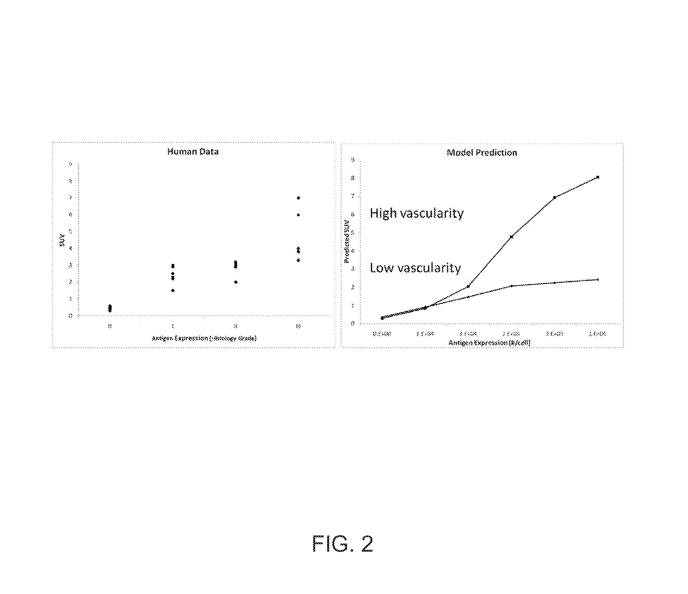
[0056]Guanylyl cyclase C (GCC) is expressed on normal and malignant intestinal epithelial cells and is an attractive target for antibody-drug conjugate therapy. 5F9 has been identified as a picomolar-affinity antibody to GCC. We evaluate the dynamic in vivo distribution of 111In-labeled 5F9 antibody in xenograft mice bearing tumors with different levels of antigen expression using microSPECT / CT. A distributed model of molecular transport in tumors was used to estimate antigen density and tumor vascularity of GCC-expressing tumors in vivo. To investigate the accuracy of model estimations, vascular density was experimentally measured by vascular casting. Subcutaneous tumors were established in mice with a GCC-expressing cell line (GCC-293), a primary tumor line (PHTX-09C), and an antigen-negative cell line (HEK-293). ˜500 μCi 111In-labeled 5F9 was injected into tumor-bearing mice (n=5). Mice were imaged by microSPECT / CT at 3, 24, 48, 96, and 144 hr post-injection. Vascular casting exp...


PUM


Abstract

A method is provided for estimating a parameter of physiological significance. One or more images are provided of a tissue in a subject to whom a dose of a contrast agent (CA) has been administered, using a computer equipped with image processing software, the concentration or relative concentration of the agent in a region or regions of interest in the tissue is determined, thus generating concentration data. The time-based behavior of concentrations of CA within the tissue is determined using a pharmacokinetic model that is based on a set of pharmacokinetic model parameters. Using computer code, the pharmacokinetic model is fit to the concentration data, varying one or more parameters, such that a best fit estimate of a parameter of physiological significance is provided.

Description

CROSS-REFERENCE TO RELATED APPLICATIONS[0001]This application claims the benefit of the filing date of U.S. provisional application No. 61 / 421,079, which was filed Dec. 8, 2010, which is expressly incorporated by reference herein in its entirety.FIELD OF THE INVENTION[0002]The present invention relates to molecular imaging and mathematical modeling and more particularly to methods that use these techniques in concert to assess various physiological parameters relevant to drug development and patient treatment. For example, the present methods can be used to estimate tumor vascularity, contrast agent internalization rate, contrast agent binding affinity, and binding site concentration from molecular imaging data.BACKGROUND[0003]It is well known that studies are conducted in animals during preclinical drug development to determine in vivo pharmacokinetics, pharmacodynamics, efficacy, and toxicity, and that these studies are used in an effort to predict effective drug concentrations in...

Claims


Application Information

Patent Timeline
Patent Type & Authority Applications(United States)
IPC IPC(8): G06T7/00
CPCG06T7/0012G06T7/0016G06T2207/10104G06T2207/10108G06T2207/30096G06T2207/30104G16C20/30
Inventor ORCUTT, KELLY DAVISHOPPIN, JOHNHESTERMAN, JACOBLACKAS, CHRISTIAN
Owner INVICRO
Why Eureka
Try Eureka