
Device and process to confirm occlusion of the fallopian tube

a fallopian tube and fallopian tube technology, applied in the field of medical devices and processes, can solve the problems of using hysterosalpingograms, requiring radiology equipment for hsg, sending patients out, etc., and achieve the effect of confirmating the intratubal occlusion

Inactive Publication Date: 2014-10-30
BOSTON SCI SCIMED INC
View PDF14 Cites 4 Cited by
  • Summary
  • Abstract
  • Description
  • Claims
  • Application Information

AI Technical Summary

Benefits of technology

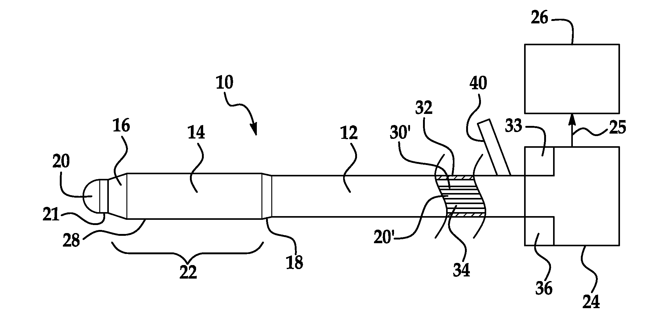
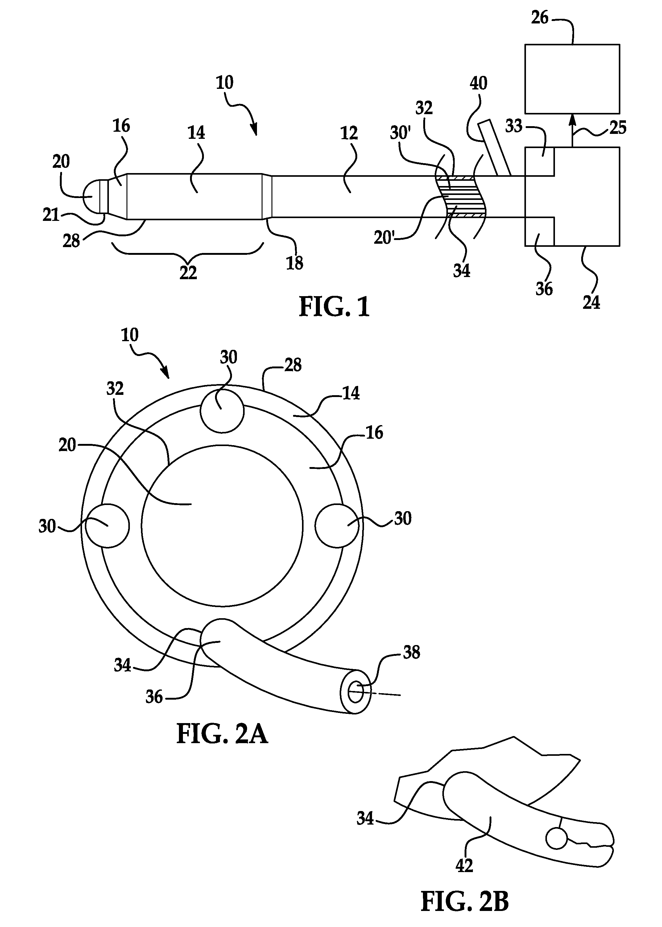
The patent describes a device that can be used to confirm the placement of an implant in a fallopian tube. The device includes a shaft with a visualization modality, such as fiber optics or sound waves, to provide visual or acoustic imaging of the tube. The device can also be used to guide the placement of the implant and evaluate its integrity. The process of visualization can be completed using a hysteroscope or other instruments. The images generated through the visualization techniques are shown to a user or physician through an ocular, video headgear, or video display.

Problems solved by technology

However, there are several disadvantages to using the hysterosalpingogram.
These limitations include the HSG requires radiology equipment and therefore most often is not done in the office of the gynecologist, and instead a patient is routinely sent to a separate radiology department to have the HSG procedure performed.
Sending a patient out of the office is counterproductive in that a procedure conducted in office allows for better patient management, and increases the likelihood that the patient will return for a necessary follow-up procedure.
Also, in while an in-office procedure increases patient convenience and reduces cost to both the healthcare system and to the patient; HSG is not amenable to these benefits.
Furthermore, partly due to the fact that many HSG interpretative radiologists are not as familiar with the tortuosity and topography of the fallopian tube as reproductive specialists, HSG procedures have a high false negative rate of 30% (World Health Organization (WHO) (1986) “Comparative Trial Of Tubal Insufflation, Hysterosalpingography And Laparoscopy With Dye Hydrotubation For Assessment Of Tubal Patency”Fertil.
In addition, patients often complain of pain and can be allergic to the dye used in the HSG procedure.
HSG is also prone to spread infection through the dye infusion into the fallopian tube; as between 1-3% of all women who undergo HSG develop some type of infection soon after the procedure (Eric Daiter, MD Pelvic Factor Infertility: Uterine Cavity Infertility (2009)).
Furthermore, transvaginal ultrasound can only be used on implants which are echogenic and therefore would not work on all materials used, such as implanted silicone matrices or to visualize tissue condition proximal to an implant.

Method used


Image

  • Device and process to confirm occlusion of the fallopian tube
  • Device and process to confirm occlusion of the fallopian tube
  • Device and process to confirm occlusion of the fallopian tube

Examples


Embodiment Construction

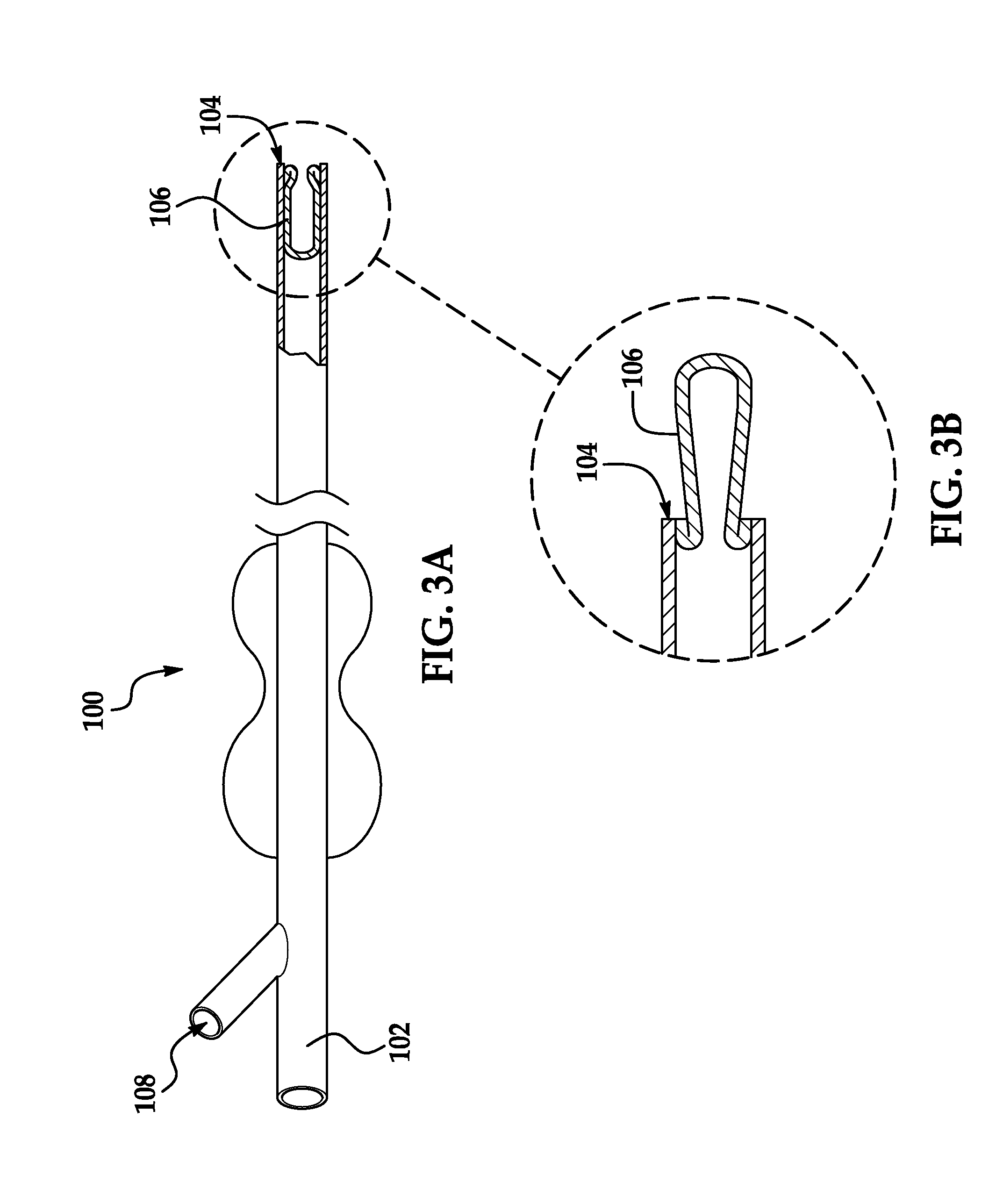
[0017]An inventive device and process is provided that has utility to confirm successful occlusion of the fallopian tube intratubal, including but not limited to purposeful occlusions caused by the implantation of polymer matrices such as silicones; or by the implantation of metal coils, such as coils made from nickel titanium alloy or stainless steel; or other implant materials, such as polyethylene terephthalate (PET), poly(ethylene oxide) (PEO) and poly(butylene terephthalate) copolymers (PBT), polyamides, or combinations thereof. Embodiments of the present invention allow a physician to determine the degree of tissue reaction and whether or not the implantation has been properly captured by the tissue, as well as the integrity of the implant itself. In specific embodiments of the present invention, an inventive device is used to guide the placement of an implantation.

[0018]The term “physician” is used herein to include all appropriate medical practitioners and explicitly inclusi...


PUM


Abstract

A device is provided to confirm intratubal occlusion in a subject of a fallopian tube having an inner diameter that includes a tubular shaft having a distal end and an interior lumen. An examination head is joined to the distal end of the shaft. A visualization modality in the examination head provides visual or acoustic imaging of the fallopian tube. A power source for the visualization modality is provided. A handle is provided for control of the device. An ex vivo imager of an ocular, video headgear, or a video display is provided in communication with the visualization modality. A process for evaluating an intratubal implant in a fallopian tube through optical or sonic wave visualization is also provided.

Description

CROSS-REFERENCE TO RELATED APPLICATIONS[0001]This application is a non-provisional application that claims priority benefit of U.S. Provisional Application Ser. No. 61 / 559,120, filed Nov. 13, 2011 the contents of which are hereby incorporated by reference.FIELD OF THE INVENTION[0002]The present invention in general relates to a medical device and process and system inclusive thereof, and in particular to a device and intratubal process for confirming the occlusion of a subject fallopian tube.BACKGROUND OF THE INVENTION[0003]The ostium (plural ostia) of the fallopian tube may refer to the proximal or distal opening of the tube. The proximal tubal opening (ostium) is located within the uterus at the uterotubal junction and accessible via hysteroscopy. Occlusion at this opening is referred to as proximal tubal occlusion. The distal tubal opening (or abdominal ostium) is the opening in the infundibulum of uterine tube into the abdominal cavity. In ovulation, the oocyte enters the fallop...

Claims


Application Information

Patent Timeline
IPC IPC(8): A61B5/00A61B8/12
CPCA61B5/4325A61B5/0084A61B5/0073A61B8/12A61L27/14A61B1/303A61B2017/4233A61B2090/3735A61B2090/3782A61B2090/3614
Inventor SARNA, SURBI
Owner BOSTON SCI SCIMED INC
Learn More