
Disposable endoscope

a disposable endoscope and endoscope technology, applied in the field of endoscopes, can solve the problems of fatal disease in patients when it is reused, high cost, and high cost, and achieve the effects of preventing cross-contamination caused by repeated use, simple structure of disposable endoscopes, and reducing costs

Inactive Publication Date: 2014-11-27
OPTIMEDE
View PDF9 Cites 10 Cited by
  • Summary
  • Abstract
  • Description
  • Claims
  • Application Information

AI Technical Summary

Benefits of technology

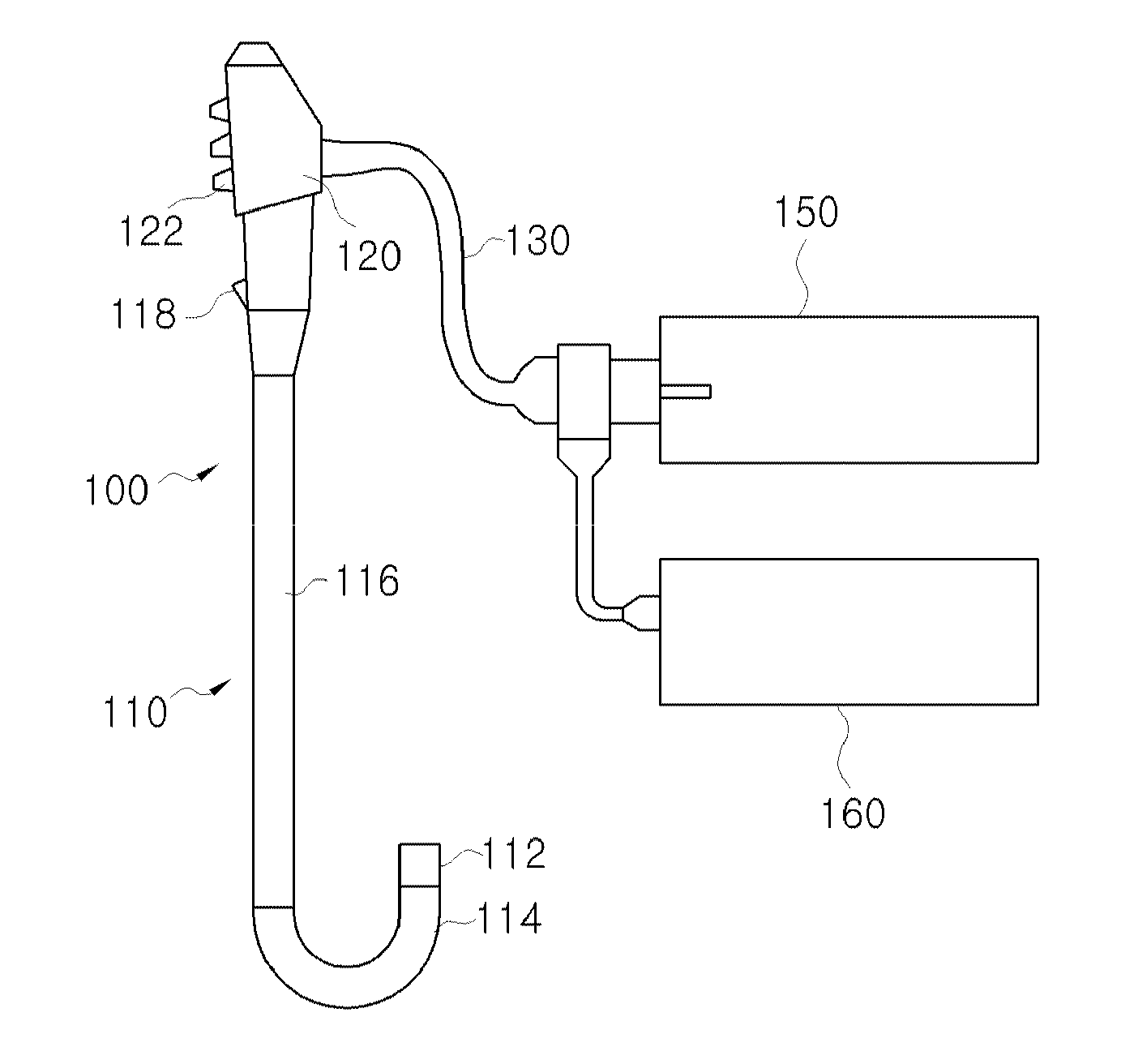
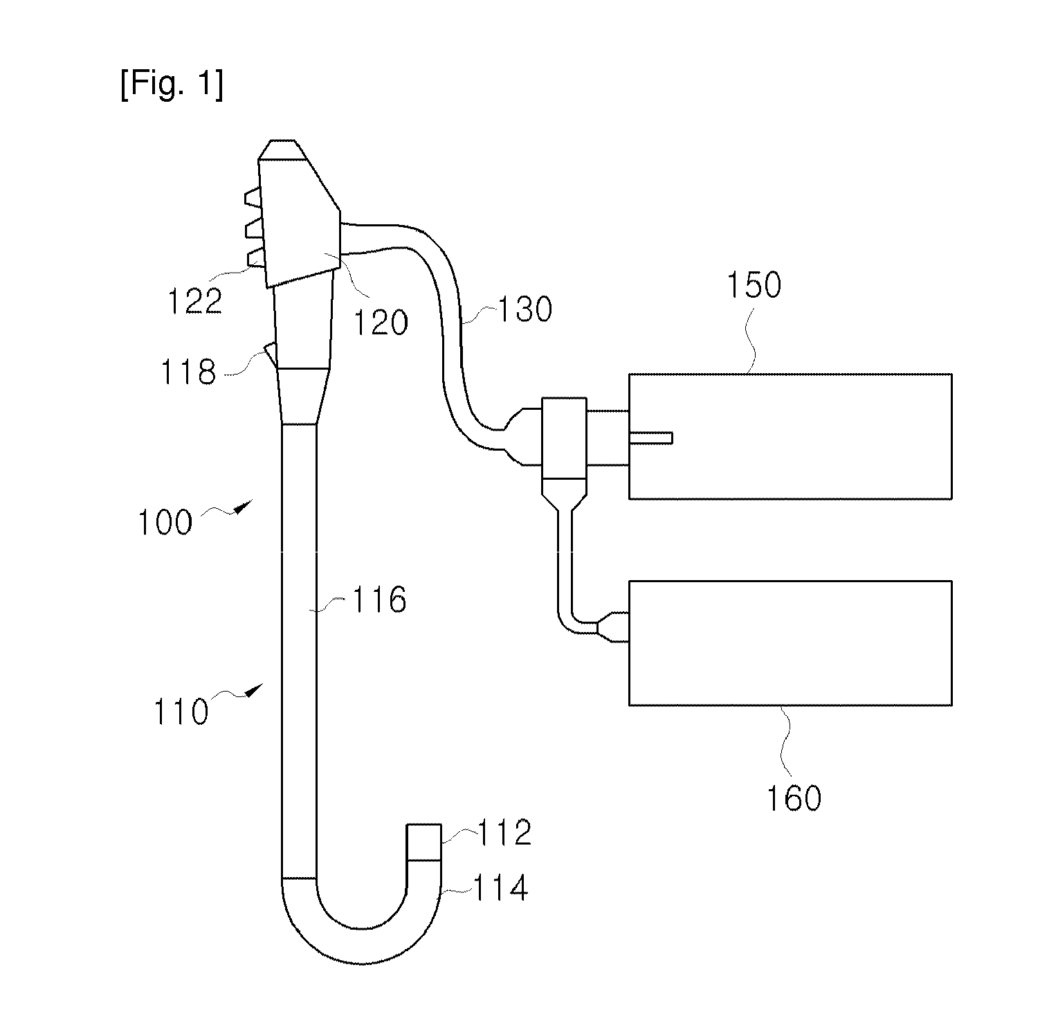
The present invention relates to a disposable endoscope that can be used once to prevent cross-contamination, reducing the risk of infection for patients. The endoscope has a simple structure to lower production costs. It also allows for precise control of photography for accurate diagnosis and high-resolution photographing using multiple mirrors and lenses to improve patient treatment and therapy quality.

Problems solved by technology

However, the biggest problem in endoscopes according to the related art is a risk of cross-contamination that occurs when an endoscope that has been used once in a patient's treatment is reused.
Thus, when the probe portion of the endoscope is not properly sterilized, the probe portion may cause a fatal disease in the patient when it is reused.
Thus, there are technical limitations, such as the need of a post-process including sterilization and incomplete prevention of the risk of causing a disease.

Method used


Image

  • Disposable endoscope
  • Disposable endoscope
  • Disposable endoscope

Examples


Embodiment Construction

[0032]Hereinafter, exemplary embodiments of the present invention will be described in detail.

[0033]FIG. 3 illustrates a configuration of a whole endoscope system to which a disposable endoscope is applied, according to an exemplary embodiment of the present invention, and FIG. 4 illustrates a configuration of main parts of the disposable endoscope illustrated in FIG. 3, and FIG. 5 illustrates a combined state of adjustment mirrors.

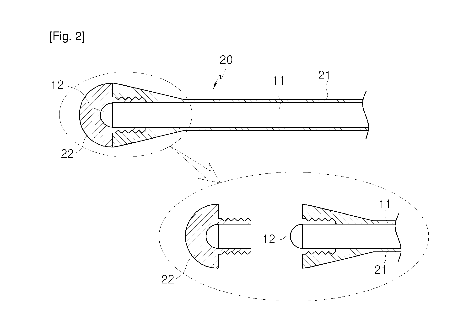
[0034]First, as illustrated, a disposable endoscope according to an exemplary embodiment of the present invention is largely configured of a main body 200, a light emitting fiber 220, a light receiving fiber 230, a protective tub 240, and a light adjustment unit 250.

[0035]First, the main body 200 is connected directly to a monitor M or is connected to the monitor M via a computer or an image processing device (not shown), and a light emitting portion 211 and a light receiving portion 212 that are portions for transmitting an image are arranged in front of...


PUM


Abstract

Provided is a disposable endoscope that enables simple treatment by capturing an image inside a body as a moving picture or photo, and in particular, that enables replacement of a probe portion inserted into the body and that preemptively prevents infection in patients due to repeated usage. The disposable endoscope includes: a main body that is connected to a monitor, and in front of which a light emitting portion and a light receiving portion are arranged in parallel; a light emitting fiber that guides light from the light emitting portion toward an object to be examined; a light receiving fiber that guides light reflected from the object to be examined toward the light receiving portion; and a light adjustment unit that is placed within the protective tube that is adjacent to ends of the light emitting fiber and the light receiving fiber and that adjusts a position of light toward the object to be examined or light reflected from the object to be examined, wherein the light receiving fiber guides light that is reflected from the object to be examined through the light adjustment unit toward the light receiving portion. A probe portion inserted into a body of a patient or a user is used only once so that cross-contamination caused by repeated usage can be prevented in advance. A structure of the disposable endoscope is simple so that costs can be reduced due to reduction in a unit cost of production. A portion in which photographing of a moving picture or photo is substantially performed can be precisely controlled so that a precise diagnosis can be carried out. Photographing of a moving picture or photo with high resolution can be performed using a plurality of mirrors and lenses so that the quality of a patient's treatment or therapy can be improved.

Description

TECHNICAL FIELD[0001]The present invention relates to an endoscope, and more particularly, to a disposable endoscope that enables simple treatment by capturing an image inside a body as a moving picture or photo, and in particular, that enables replacement of a probe portion inserted into the body and that preemptively prevents infection in patients due to repeated usage.BACKGROUND ART[0002]In general, medical endoscopes (hereinafter referred to as endoscopes) are medical instruments that are inserted into a hollow organ or cavity of a body and enable examination of the interior of the hollow organ or cavity of the body without performing direct surgery. Endoscopes have made great contributions toward medical development since being devised by Kussmaul in 1863.[0003]In terms of a development procedure of endoscopes, first generation endoscopes that showed a rough image by combining images formed on each optical fiber through a bundle of optical fibers developed into second generatio...

Claims


Application Information

Patent Timeline
Patent Type & Authority Applications(United States)
IPC IPC(8): A61B1/00A61B1/07G02B6/26
CPCA61B1/00103G02B6/262G02B2006/12104A61B1/07A61B1/00165A61B1/00096A61B1/00101A61B1/04A61B1/06
Inventor KIM, HUN TAE
Owner OPTIMEDE
Learn More