
Fully automated vascular imaging and access system

Inactive Publication Date: 2015-03-05
VASCULOGIC
View PDF1 Cites 119 Cited by
  • Summary
  • Abstract
  • Description
  • Claims
  • Application Information

AI Technical Summary

Benefits of technology

The present invention provides a small, portable device that can withdraw and introduce fluid into blood vessels. This device improves patient comfort, increases practitioner safety, and helps to streamline procedures in hospitals. Overall, it offers a more efficient and effective solution for common medical procedures.

Problems solved by technology

Currently, venipunctures are executed manually by trained personnel, but there are problems inherent with these processes.
Many times locating a vein or artery is a challenge, especially in young, elderly, dark-skinned, and obese patients whose veins are small, fragile, rolling, or obscured by melanin or subcutaneous fat.
To complicate matters, multiple attempts at needle insertion may be required, due either to the inexperience of the person obtaining the sample, and / or from difficulty in locating the target vessel.
Success depends heavily on the procedure, patient type, and clinician's experience, and mistakes result in multiple punctures, therapy delays, adverse effects, pain, and stress for the patient, the family, and the medical staff.
Venipuncture is furthermore the leading cause of bloodborne pathogen transmission among US healthcare workers.
Annual reports estimate 600,000-800,000 accidental needlestick injuries in the U.S., a percentage of which can lead to serious or fatal infections.
In total, failed vessel punctures are estimated to cost the U.S. health care system $4.7 billion / year.
Unfortunately, the extents of training, experience, and skill vary widely between healthcare personnel.
Although this technology may provide methods of viewing veins externally, it does not provide any depth representation of the veins under the skin, is frequently ineffective for difficult venous access patients, and does not address the frequency of needle-induced bloodborne pathogen transmission among practitioners.
These technologies are furthermore limited to peripheral cannula insertions as they lack the imaging depth needed to visualize deeper central vessels.
Although ultrasound based technologies have the advantage of providing greater imaging depth to allow peripheral and central vessel access, the effective use of these technologies requires a substantial amount of clinical training and is frequently subject to human error.
However, these systems are not conducive to providing automated venous or arterial access.
Despite significant advances in the fields of medical robotics and computerized surgery, currently there is no technology that is able to directly address the fundamental problems of patient and practitioner safety for vessel punctures.
Furthermore there is no technology for complete automated cannula insertion for the purpose of access to peripherally or centrally located blood vessels.
Finally there is no technology that combines an imaging system with a robotically driven for the purpose of blood vessel puncture.

Method used


Image

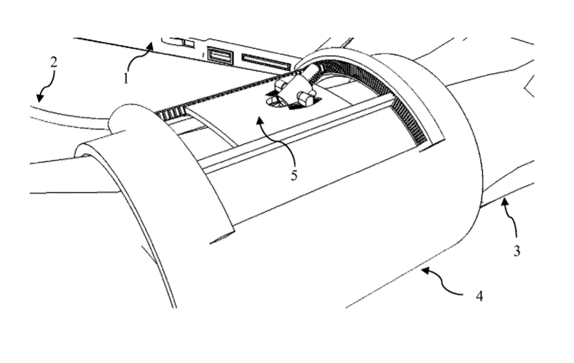
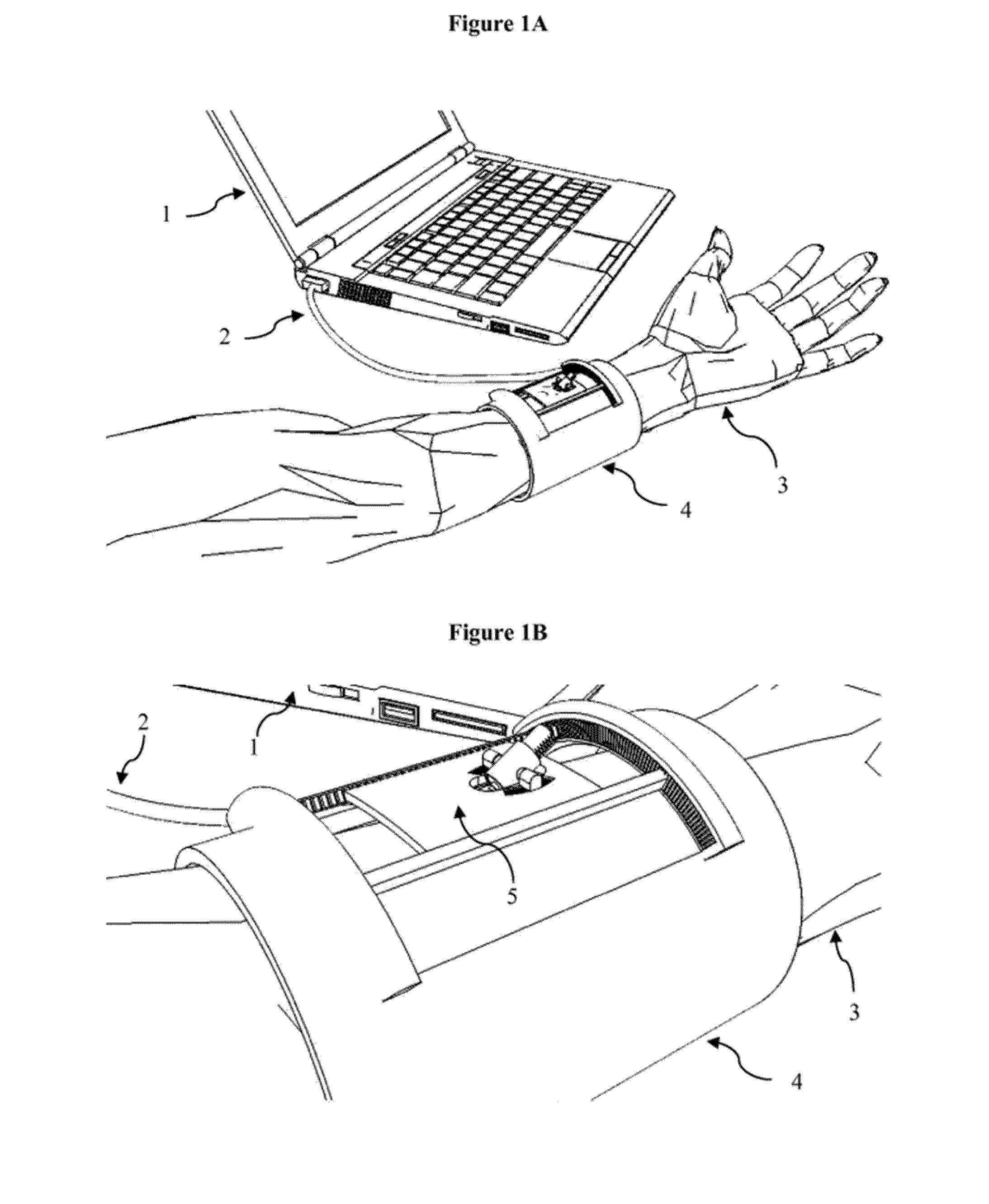
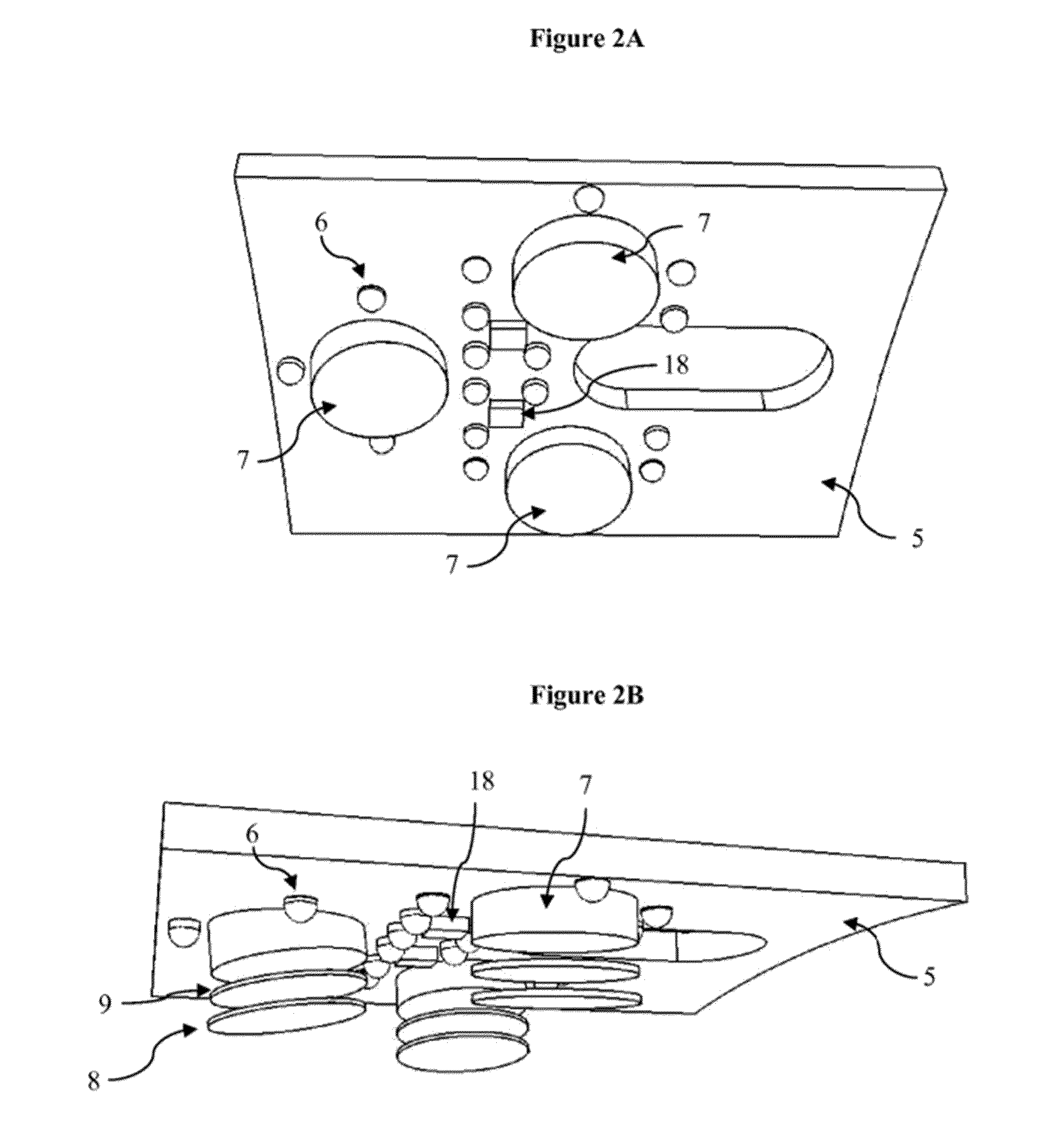
  • Fully automated vascular imaging and access system
  • Fully automated vascular imaging and access system
  • Fully automated vascular imaging and access system

Examples


Embodiment Construction

[0059]The invention is directed in part to a fully automated system for peripheral vein imaging and access. The system preferably includes an imaging system for providing continuous and real-time imaging of blood vessels, said imaging system comprising at least one of optical, acoustic, photoacoustic, or tactile imaging. The automated system further preferably comprises image processing software for generating a continuous and real-time three-dimensional (3D) computer model of the blood vessels based on the imaging system, and selecting an optimal vessel target based on visual and anatomical information for inserting a needle into the selected vessel target. The system further comprises a robotic effector comprising a needle, a needle attachment unit, and a needle actuation system that positions the needle at the selected vessel target located by the image processing software, and a computer connected to the imaging system and robotic effector, said computer directing, continuously ...


PUM


Abstract

The present invention is directed to a self-contained, fully automated vascular imaging and access device, methods of imaging, mapping, and analyzing three-dimensional views of blood vessels, and methods for providing continuous and real-time communication with a robotically actuated needle and a computer interface.

Description

BACKGROUND OF THE INVENTION[0001]The first step in many clinical interventions is to establish venous or arterial access to the bloodstream, a procedures that is critical for a wide range of clinical applications, including blood draws, diagnostic tests, and the delivery of medication, nutrients, and other fluids through the bloodstream.[0002]Currently, venipunctures are executed manually by trained personnel, but there are problems inherent with these processes. Successful placement of the cannula requires the coordination of complex deductive and visuomotor tasks in real time. Many times locating a vein or artery is a challenge, especially in young, elderly, dark-skinned, and obese patients whose veins are small, fragile, rolling, or obscured by melanin or subcutaneous fat. To complicate matters, multiple attempts at needle insertion may be required, due either to the inexperience of the person obtaining the sample, and / or from difficulty in locating the target vessel. Success dep...

Claims


Application Information

Patent Timeline
IPC IPC(8): A61B19/00A61B5/15
CPCA61B19/2203A61B19/5244A61B2019/5246A61B2019/2207A61B5/150206A61B17/3403A61B2017/3409A61B2017/3413A61B5/15003A61B5/150748A61B34/32A61B34/30A61B90/11A61B2090/3616A61B90/37A61B2034/2065
Inventor MAGUIRE, TIMCHEN, ALVIN
Owner VASCULOGIC
Learn More