Anti-PD1 Antibodies and their Use as Therapeutics and Diagnostics

a technology of anti-pd1 antibodies and diagnostics, applied in the field of anti-pd1 antibodies and their use as therapeutics and diagnostics, can solve the problems of poor prognosis of patient survival outcom

Active Publication Date: 2015-03-19
BEIGENE SWITZERLAND GMBH
View PDF0 Cites 313 Cited by
  • Summary
  • Abstract
  • Description
  • Claims
  • Application Information

AI Technical Summary

Benefits of technology

[0004]Expression of PD-1 was frequently found in immune cells such as T-cells, B-cells, monocytes and natural killer (NK) cells. It was rarely expressed in other human tissues, such as muscle, epithelium, neuronal tissues, etc. Furthermore, high level of PD-1 expression is often associated with activation of immune cells. For example, when human T-cell line, Jurkat, was activated by phytohaemagglutinin (PHA) or phorbol ester (12-O-tetradecanoylphorbol-13-acetate, or TPA), the expression of PD-1 was up-regulated visible in Western Blot (Vibharka et al., 1997 Exp Cell Res 232:25-28). The same phenomenon was observed in stimulated murine T- and B-lymphocytes and in primary human CD4+ T-cells upon stimulation by anti-CD3 antibody (Agata et al., 1996 Int Immunol 8:765-772; Bennett et al., 2003 J Immunol 170:711-118). The increase of PD-1 expression following stimulation of T effector cells redirects the activated T-effector cells towards exhaustion and reduced immune activities. Therefore, PD-1 mediated inhibitory signal plays an important role in immune tolerance (Bour-Jordan et al., 2011 Immunol Rev 241:180-205).

Problems solved by technology

More frequently, the increased expression of PD-1 and PD-L1 in those cancers is associated with poor prognosis of patient survival outcome.

Method used


Image

  • Anti-PD1 Antibodies and their Use as Therapeutics and Diagnostics
  • Anti-PD1 Antibodies and their Use as Therapeutics and Diagnostics
  • Anti-PD1 Antibodies and their Use as Therapeutics and Diagnostics

Examples


example 1

Generation of Anti-PD-1 Monoclonal Antibody

[0125]Anti-PD-1 monoclonal antibodies (mAbs) were generated based on conventional hybridoma fusion technology (Kohler and Milstein 1976 Eur J Immunol 6:511-519; de St Groth and Sheidegger 1980, J Immunol Methods 35:1-21; Mechetner 2007 Methods Mol Biol 378:1-13) with minor modifications. MAbs with high binding activities in enzyme-linked immunosorbent assay (ELISA) and fluorescence-activated cell sorting (FACS) assay were selected for further characterization

[0126]PD-1 Recombinant Protein for Immunization and Binding Assays

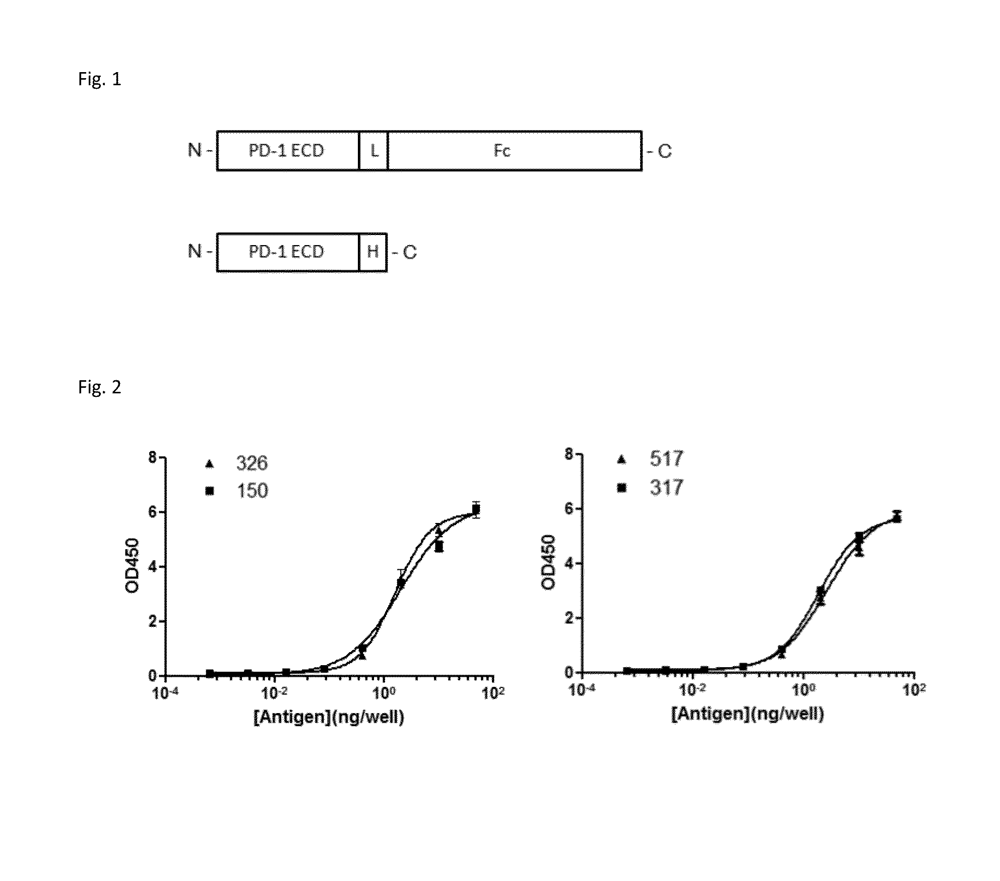
[0127]Expression plasmid containing full-length human PD-1 cDNA was obtained from Origene (Cat. No. SC117011, NCBI Accession No: NM—005018.1, Beijing, China). The extracellular domain consisting of amino acid (AA) 1-168 of PD-1 (SEQ NO. 1, SEQ NO. 2) was PCR-amplified, and subcloned in pcDNA3.1-based expression vector (Invitrogen, Carlsbad, Calif., USA) with C-terminus fused either to a His6 tag or to the γFc domain of hu...

example 2

Comparison of Binding Activities Among Anti-PD-1 Antibodies

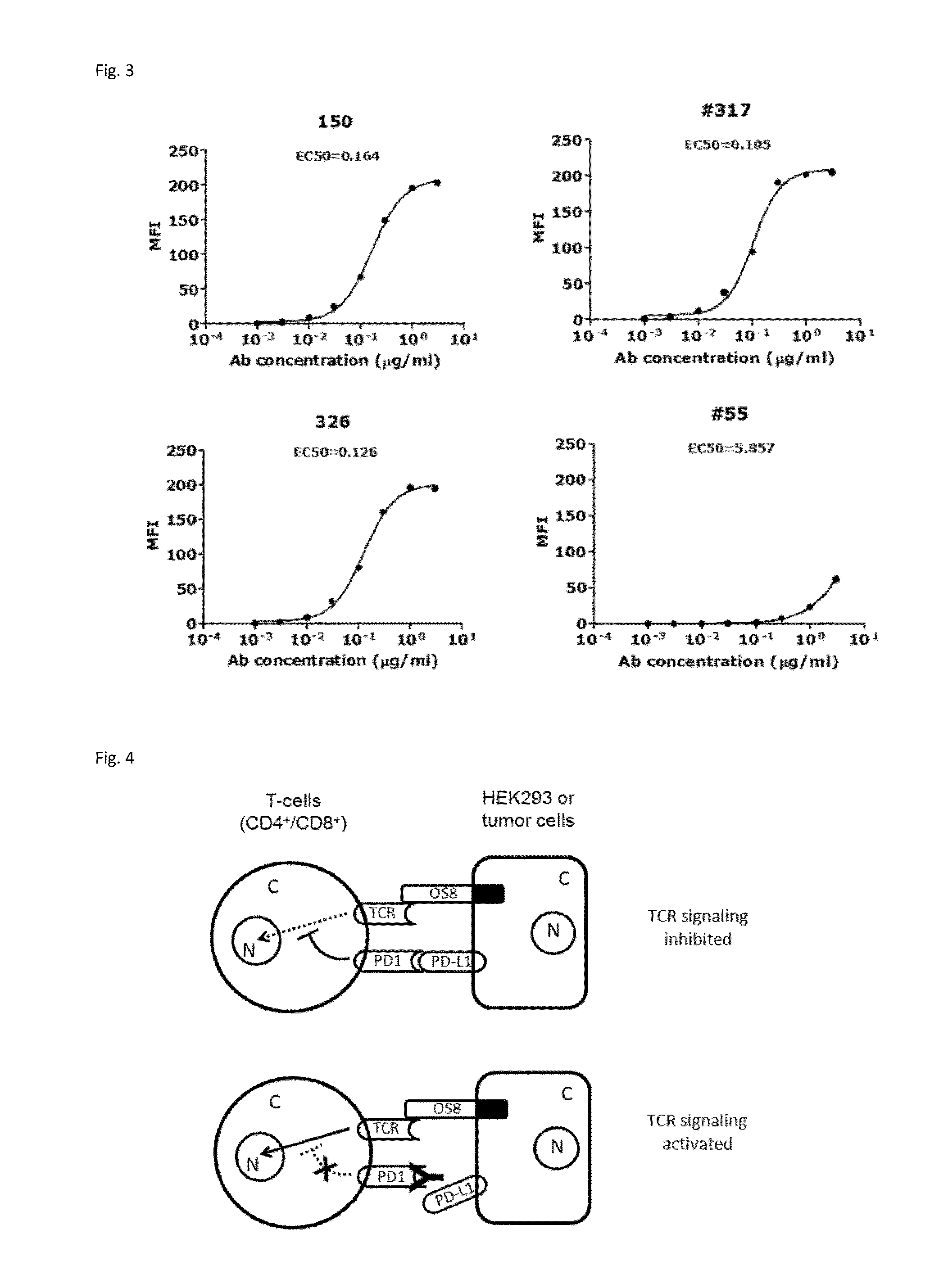
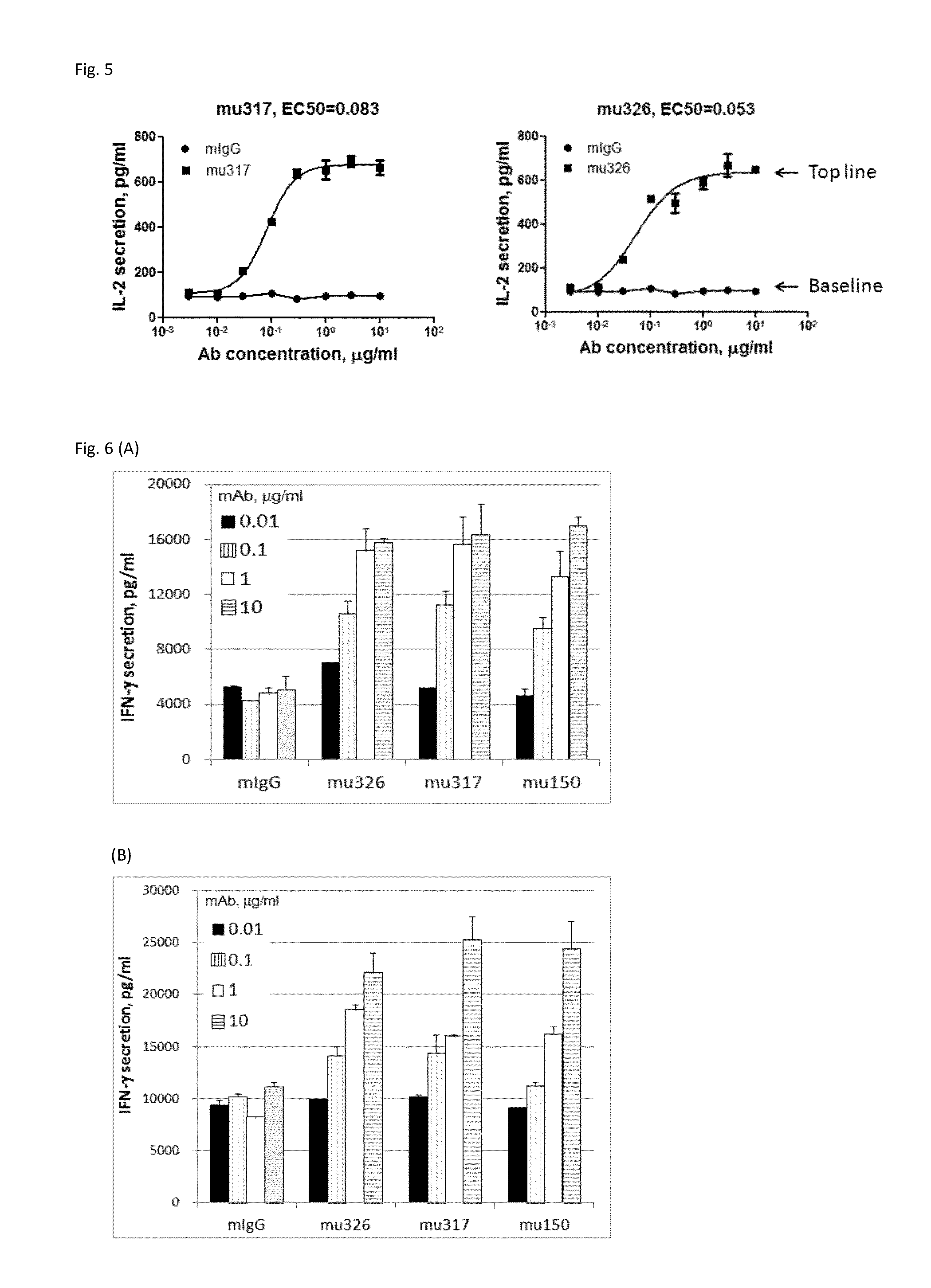
[0140]Through screening thousands of hybridomal clones we identified some top monoclonal antibodies (mAb), which bind to human PD-1 with high specificity and strength. As shown in ELISA assay (FIG. 2), three of the top antibodies elicited such binding strength and specificity. FACS analysis results demonstrated the selected monoclonal antibodies bind to the native PD-1 proteins expressed on cell surface. Murine mAb317 (mu317), mu326 and mu150 showed concentration-dependent binding activity, and their binding EC50 (Effective concentration at 50% activity) was significantly lower than that of the control mu55 (FIG. 3).

[0141]Assess mAb Binding Affinity by Surface Plasmon Resonance (SPR)

[0142]The mAbs with high binding activities in ELISA and FACS, as well as with potent functional activities in the cell-based assays (herein) were examined for their binding kinetic constant in real time binding reactions. Murine anti-PD-1 mAbs w...

example 3

Functional Activity of Anti-PD-1 Antibodies in Human T Cells

[0149]Generation of Stable Cell Lines

[0150]Retroviral packaging cell line PT67, human T cell lines HuT78 and HEK293 were obtained from the American Type Culture Collection (ATCC, Rockville, Md.). A HuT78 subline HuT78 / PD-1 that expresses PD-1 was generated by retroviral transduction using pFB-neo vector (Strategene / Agilent Tech, Santa Clara, Calif.) containing the PD-1 gene, according to the protocol described previously (Zhang et al. 2005 Blood 106: 1544-1551). The T cell engager, a membrane-anchored chimeric Ab (OS8), was constructed by fusing the single chain variable fragment (scFv) of an anti-human CD3 mAb OKT3 (Kipriyanov et al. 1997, PEDS 10:445-453) to the C-terminal domain (113-220) of mouse CD8α (NCBI Accession No: NP—001074579.1) which includes hinge, transmembrane and cytoplasmic domains. By doing so, anti-CD3 scFv is anchored to cell surface as a T cell activator. Human PD-L1, PD-L2 and OS8 cDNAs were sub-clone...


PUM

PropertyMeasurementUnit
volumeaaaaaaaaaa
concentrationaaaaaaaaaa

Abstract

Provided are antibodies that specifically bind to Programmed Death-1 (PD1, Pdcd-1, or CD279) and inhibit PD1-mediated cellular signaling and activities in immune cells, antibodies binding to a set of amino acid residues required for its ligand binding, and uses of these antibodies to treat or diagnose cancer, infectious diseases or other pathological disorders modulated by PD1-mediated functions.

Description

CROSS REFERENCE TO RELATED APPLICATION[0001]This application is a division of U.S. Ser No. 14 / 076,214, filed Nov. 10, 2013 (now US), and a CON of PCT / CN2013 / 083467, filed Sep. 13, 2013.INTRODUCTION[0002]Programmed Death-1 (PD-1, also termed as CD279) is a 55 KD receptor protein related to CD28 / CTLA4 co-stimulatory / inhibitory receptor family (Blank et al., 2005 Cancer Immunol Immunother 54:307-314). The genes and cDNAs coding for PD-1 were cloned and characterized in mouse and human (Ishida et al., 1992 EMBO J 11:3887-3395; Shinohara et al., 1994 Genomics 23:704-706). The full length PD-1 contains 288 amino acid residues (NCBI accession number: NP—005009). Its extracellular domain consists of amino acid residues 1-167, and the cytoplasmic C-terminal tail comprises residues 191-288, which has two hypothetical immune-regulatory motifs, an immunoreceptor tyrosine-based inhibitory motif (ITIM; Vivier et al., 1997 Immunol Today 18:286-291) and an immunoreceptor tyrosine switch motif (ITSM...

Claims


Application Information

Patent Timeline
Patent Type & Authority Applications(United States)
IPC IPC(8): C07K16/28
CPCC07K16/2803C07K2317/569C07K2317/70C07K2317/565A61K39/39591C07K16/2818A61K2039/505C07K2317/567C07K2317/55C07K2317/52C07K2317/34C07K2317/76C07K2317/94C07K2317/92C07K2317/71C07K2317/24A61K2300/00C07K2317/33C07K2317/53C07K2317/73A61P25/00A61P31/12A61P35/00
Inventor LI, KANGZHANG, TONGSONG, JINGXU, LANLANLIU, QIPENG, HAO
Owner BEIGENE SWITZERLAND GMBH
Why Eureka
Try Eureka