
Medical image processing apparatus, method of operating the medical image processing apparatus, and medical image processing program

a medical image processing and image processing technology, applied in the field of method of operating the medical image processing apparatus, and medical image processing program, can solve the problems of increasing cost, low reliability of posture data appended inability to append the above posture data to three dimensional images, etc., to achieve the effect of increasing cos

Inactive Publication Date: 2015-03-26
FUJIFILM CORP
View PDF6 Cites 4 Cited by
  • Summary
  • Abstract
  • Description
  • Claims
  • Application Information

AI Technical Summary

Benefits of technology

The present invention aims to provide a medical image processing apparatus, method, and program that automatically obtains posture data of a subject when a three dimensional image of the subject is imaged, without user input or an increase in cost. This is achieved by extracting at least one organ region from the three dimensional image of the subject and obtaining directional data based on its position or shape. The posture data of the subject on the stage is then obtained based on the region of the stage and the directional data of the subject. Overall, this technology achieves automated and reliable posture data appended to three dimensional images.

Problems solved by technology

However, there is a possibility that a user will input erroneous posture data, and the reliability of the posture data appended to three dimensional images will be low.
In addition, there are cases in which the aforementioned posture data cannot be appended to three dimensional images, depending on the imaging apparatus.
However, it is necessary for a plurality of cameras to be provided in this method, which will result in an increase in cost.

Method used


Image

  • Medical image processing apparatus, method of operating the medical image processing apparatus, and medical image processing program
  • Medical image processing apparatus, method of operating the medical image processing apparatus, and medical image processing program
  • Medical image processing apparatus, method of operating the medical image processing apparatus, and medical image processing program

Examples


Embodiment Construction

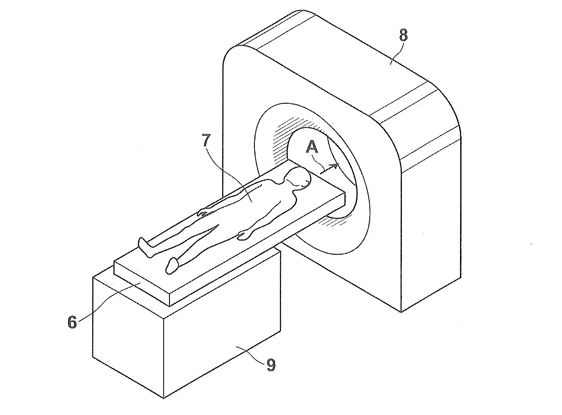
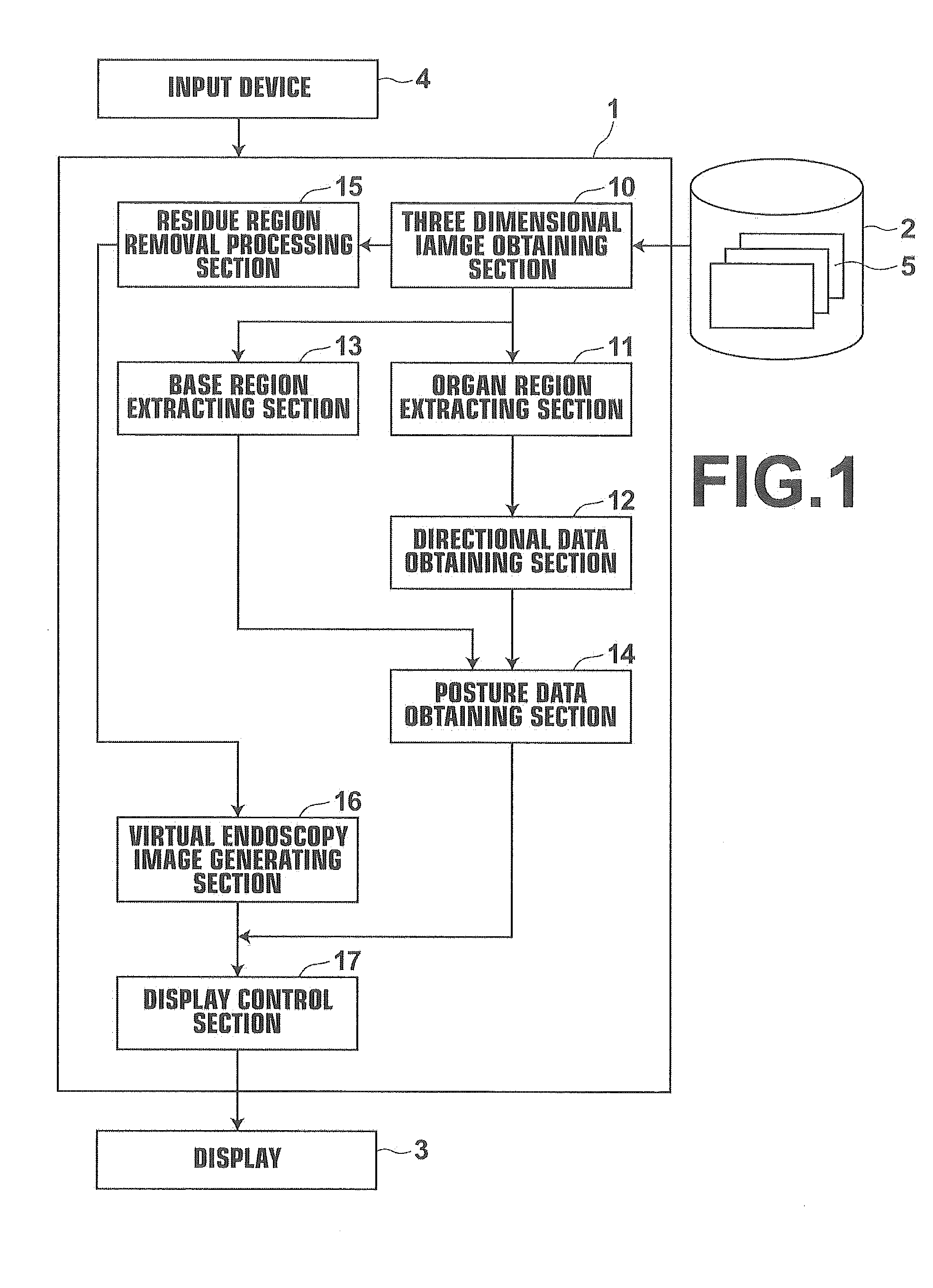
[0055]Hereinafter, a medical image diagnosis assisting system that employs an embodiment of a medical image processing apparatus, the method of operating the medical image processing apparatus, and a medical image processing program of the present invention will be described in detail with reference to the attached drawings. FIG. 1 is a block diagram that illustrates the schematic configuration of the medical image diagnosis assisting system of the present embodiment.

[0056]As illustrated in FIG. 1, the medical image diagnosis assisting system 1 of the present embodiment is equipped with a virtual endoscopy image display control apparatus 1, a three dimensional image storage server 2, a display 3, and an input device 4.

[0057]The virtual endoscopy image display control apparatus 1 is a computer, in which a virtual endoscopy image display program that includes the embodiment of the medical image processing program of the present invention is installed.

[0058]The virtual endoscopy image ...


PUM


Abstract

An organ region extracting section that extracts at least one organ region from a three dimensional image of a subject; a directional data obtaining section that obtains directional data of the subject based on one of the position of the organ region within the body of the subject and the shape of the organ region; a stage region extracting section that extracts a region of a stage on which the subject is placed from the three dimensional image; and a posture data obtaining section that obtains posture data of the subject on the stage, based on the region of the stage and the directional data of the subject; are provided.

Description

CROSS REFERENCE TO RELATED APPLICATIONS[0001]The present application claims priority under 35 USC §119 to Japanese Patent Application No. 2013-197896 filed on Sep. 25, 2013. The above application is hereby expressly incorporated by reference in its entirety, into the present application.BACKGROUND OF THE INVENTION[0002]1. Technical Field[0003]The present invention is related to a medical image processing apparatus, a method of operating the medical image processing apparatus, and a medical image processing program that obtain posture data of a subject based on a three dimensional image obtained by imaging the subject.[0004]2. Background Art[0005]Recently, lumen organs such as the large intestine, the small intestine, and the stomach of patients are extracted from three dimensional images imaged by modalities such as a CT (Computed Tomography) apparatus. The extracted three dimensional images of the lumen organs are employed to perform image diagnosis.[0006]For example, it is possibl...

Claims


Application Information

Patent Timeline
IPC IPC(8): A61B5/11A61B5/00
CPCA61B5/1121A61B5/72A61B5/0073A61B5/0082A61B5/1071A61B5/7425A61B5/7435A61B6/032A61B6/466A61B6/5217A61B6/5223G16H50/30
Inventor HAMADA, AKIHIRO
Owner FUJIFILM CORP
Learn More