In addition, there is continued effort to develop computer code that can automatically analyze an
ultrasound image scan to compute these
metrics, though the use of such algorithms is not yet widespread.
However, two factors in particular militate against this expansion in use and benefit:
training needs, and (at least in India) heavy regulation against the possibilities for misuse.
(While it is possible for an expert to acquire a scan directly, the expert's time is a limited and costly resource, best not used in acquisition, where travel and other capital and personnel costs are required.)
Particularly in developing countries, there is a lack of well trained personnel in the field.
(This mismatch, also typical with the more advanced volume scans, greatly increases the cognitive difficulty of their tasks.)
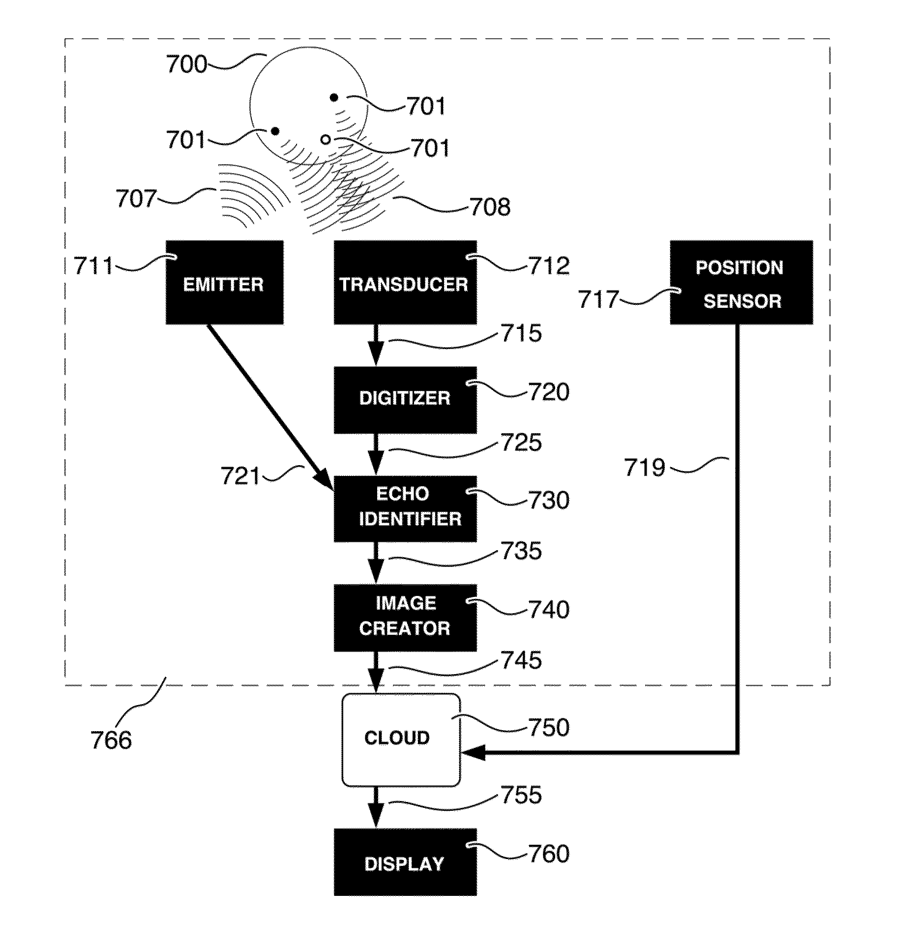
Software may be used to provide automatic corrections based on automated
image analysis for orientation and operation of the distal sensing devices, but as long as the goal remains optimality for one or more discrete slices, it is hard to operate without
human judgment, and individual skill transfer of such judgment from expert to trainee, because anatomical
clarity plays a large role in defining such optimality.
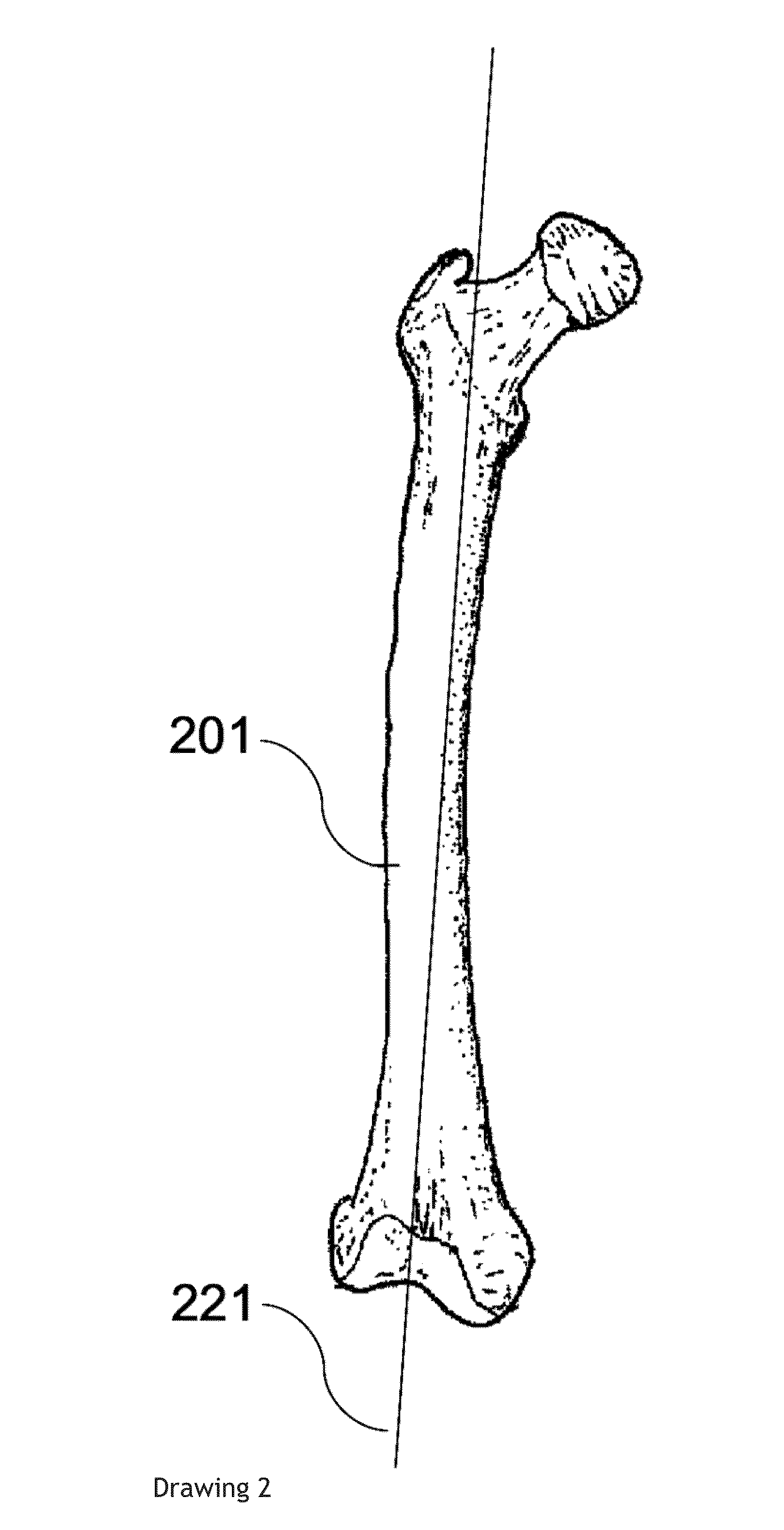
It is equally problematic to estimate the length of the
humerus (a smaller and more delicate bone though geometrically slightly simpler), and parameters such as
head circumference, occipitofrontal
diameter and abdominal circumference are even more difficult.
Since that feedback involves anatomical knowledge, it creates a
bottleneck of available ultrasound operators and a difficulty in training more, but there is also a problem with the fact that such feedback requires that the device be operated by somebody who from moment to moment, sees its output as an anatomical image.
This causes a significant obstacle to
dissemination of the new generation of highly portable cheap scanners.
We refer particularly to India, but the problem is widespread.
However, if a
local operator or clinical worker can see the images of a
fetus, that person can be bribed to reveal the sex.
To
attack that problem, the Indian government has also severely restricted the deployment of
ultrasonic imaging equipment for any purpose whatever, particularly in remote areas, outside the larger and more controllable clinics.
In several states, the sale of
portable ultrasound is a criminal offence, because it can abet in the crime of sex determination.
These restrictions render the technology unavailable for ethical diagnostic purposes, even those unrelated to
pregnancy, and prevent both businesses and non-profit organizations from supplying ultrasound services in many areas of India and other countries.
If the rural operator could acquire clinically useful data which is then transferred to a controlled clinic, using equipment that does not enable the operator to access images that reveal
fetal sex, such restrictions would be unnecessary.
However, since the present
system requires that the operator sees the current image (in order to use anatomical awareness to improve it), it cannot simultaneously blind the operator to
fetal sex.
A similar problem arises with the privacy of such images: while most countries have privacy laws restricting the availability of
patient information (the Health Insurance Portability and Accountability Act of 1996 in the US being a notable example), the more people see a medical datum, the greater the risk that it is shared improperly.
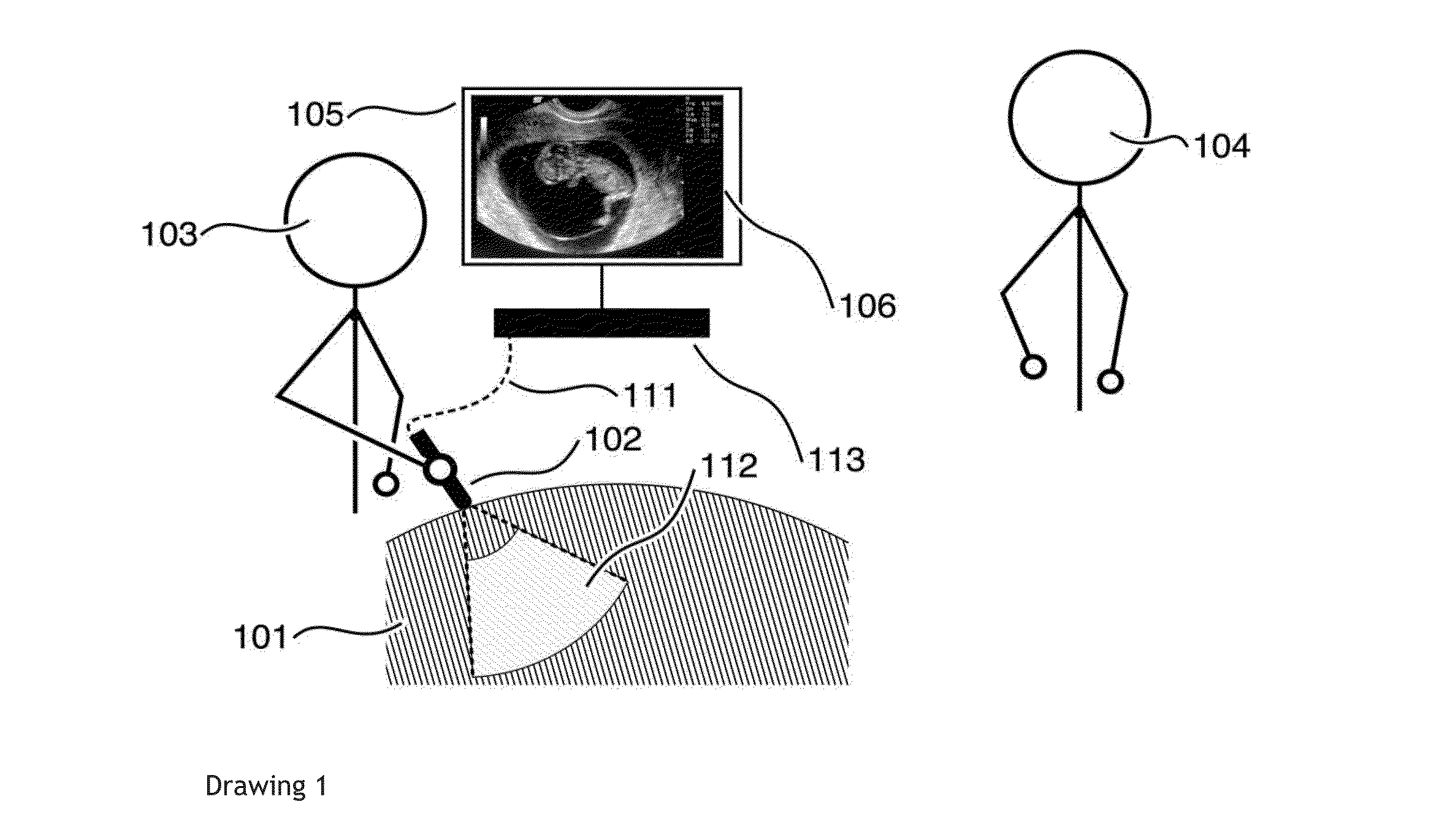
The paper “Interactive Training
System for
Medical Ultrasound” by C J Banker and P C Pedersen illustrates well the current method of guidance, by visual anatomical feedback imaging the interior of a real or
virtual patient: the twin disadvantages of this are the considerable training required, and the (less demanding) utility for
fetal sex determination, which in some situations is illegal.

