Coupled segmentation in 3D conventional ultrasound and contrast-ehhanced ultrasound images

a 3d ultrasound and contrast-ehanced technology, applied in the field of three-dimensional ultrasound imaging, can solve the problems of manual segmentation of organs in a 3d image, inability to realize clinical practice, and inability to provide satisfying methods,

Active Publication Date: 2015-07-30
KONINKLJIJKE PHILIPS NV
View PDF5 Cites 15 Cited by
  • Summary
  • Abstract
  • Description
  • Claims
  • Application Information

AI Technical Summary

Benefits of technology

[0024]The goal of the method is then to find the best and , using the image I information. This is done by minimizing the following energy:
[0025]In the first term, also called data fidelity, H is the Heaviside function (H(x)=1 if x>0 and 0 if x<0) which means that the integral is actually only inside the deformed object. r(x) is an image-based function that returns at each point a negative (respectively positive) value if the voxel is likely to be outside (respectively inside) the object of interest. For ambiguous regio

Problems solved by technology

However, it is often tedious to manually segment an organ in a 3D image.
While quantification and visualization tools are relatively available for 2D images, 3D volumes analysis is often done by hand through tedious procedures impossible to rea

Method used


Image

  • Coupled segmentation in 3D conventional ultrasound and contrast-ehhanced ultrasound images
  • Coupled segmentation in 3D conventional ultrasound and contrast-ehhanced ultrasound images
  • Coupled segmentation in 3D conventional ultrasound and contrast-ehhanced ultrasound images

Examples


Embodiment Construction

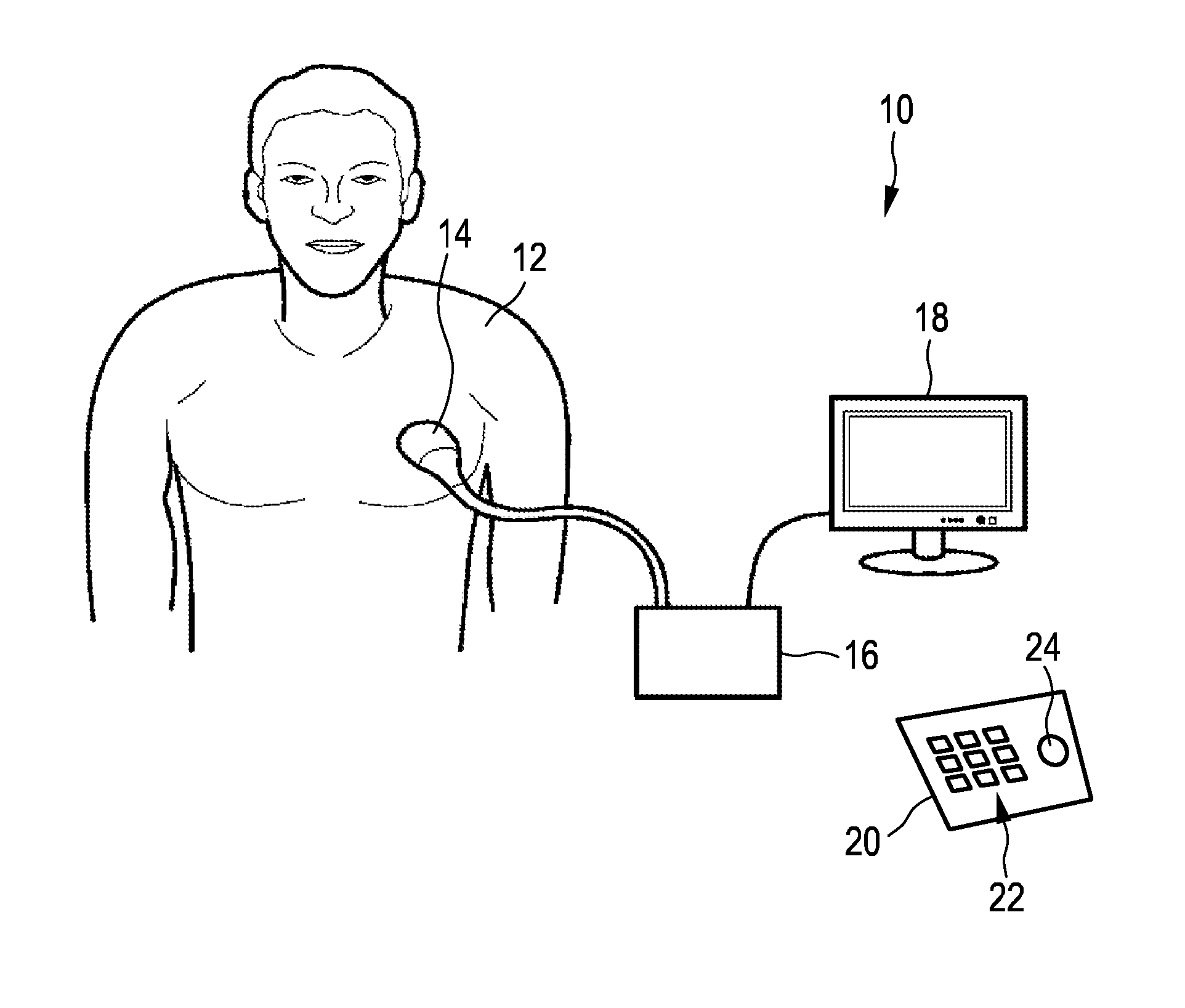
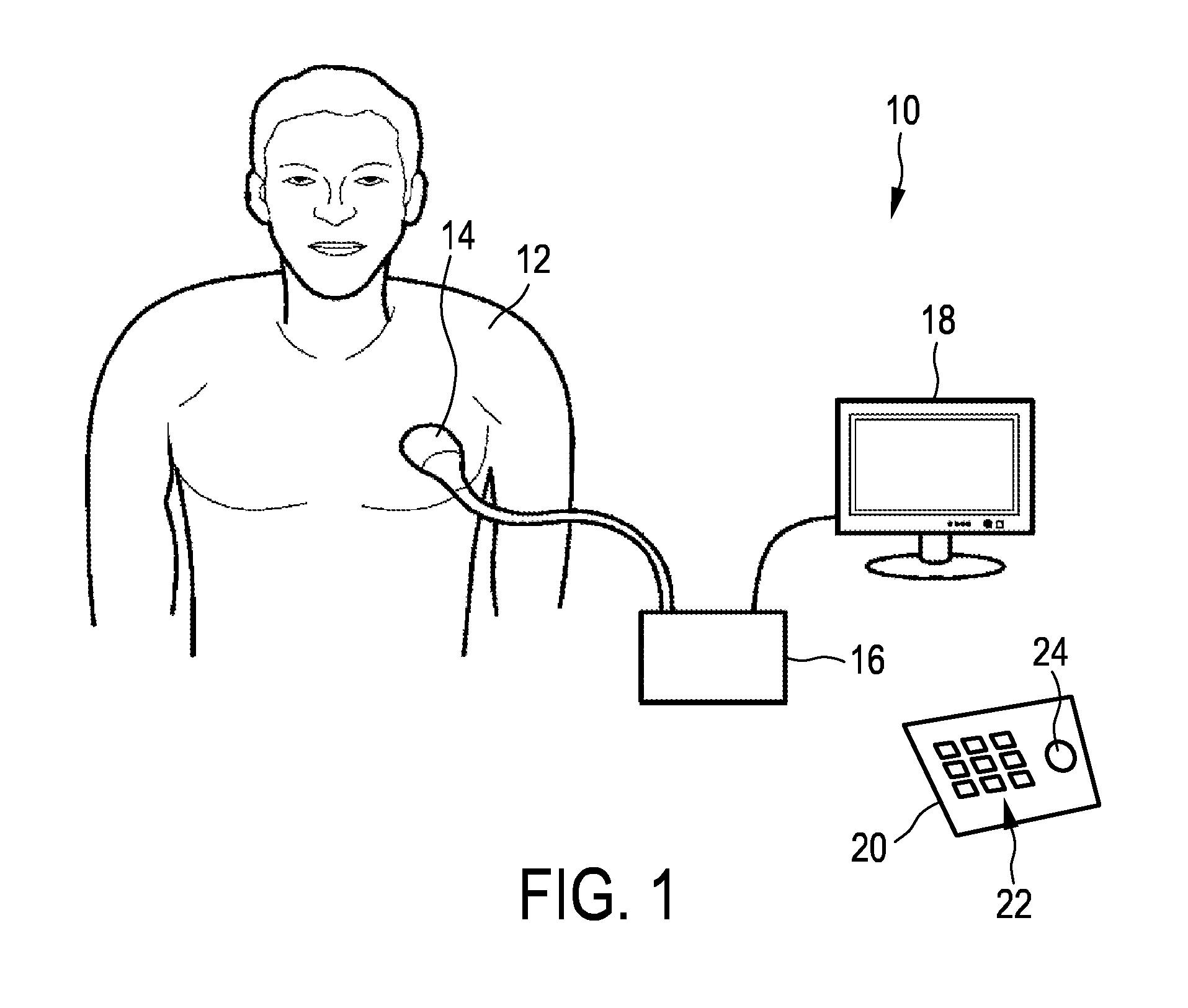
[0065]FIG. 1 shows a schematic illustration of an ultrasound system 10 according to an embodiment, in particular a medical ultrasound three-dimensional imaging system. The ultrasound imaging system 10 is applied to inspect a volume of an anatomical site, in particular an anatomical site of a patient 12. The ultrasound system 10 comprises an ultrasound probe 14 having at least one transducer array having a multitude of transducer elements for transmitting and / or receiving ultrasound waves. In one example, the transducer elements each can transmit ultrasound waves in form of at least one transmit impulse of a specific pulse duration, in particular a plurality of subsequent transmit pulses. The transducer elements can for example be arranged in a one-dimensional row, for example for providing a two-dimensional image that can be moved or swiveled around an axis mechanically. Further, the transducer elements may be arranged in a two-dimensional array, in particular for providing a multi-...


PUM


Abstract

The present invention relates to an ultrasound imaging system (10) for inspecting an object (97) in a volume (40). The ultrasound imaging system comprises an image processor (36) configured to conduct a segmentation (80) of the object (97) simultaneously out of three-dimensional ultrasound mage data (62) and contrast-enhanced three-dimensional ultrasound image data (60). In particular, this may be done by minimizing an energy tem taking into account both the normal three-dimensional ultrasound image data and the contrast-enhanced three-dimensional image data. By this, the normal three-dimensional ultrasound image data and the contrast-enhanced three-dimensional image data may even be registered during segmentation. Hence, this invention allows a more precise quantification of one organ in two different modalities as well as the registration of two images for simultaneous visualization.

Description

FIELD OF THE INVENTION[0001]The present invention relates to three-dimensional ultrasound imaging. In particular, the current invention relates to image processing and the segmentation of objects in the scanned volume.BACKGROUND OF THE INVENTION[0002]In three-dimensional ultrasound imaging, or volume imaging, the acquisition of a three-dimensional image is accomplished by conducting many two-dimensional scans that slice through the volume of interest. Hence, a multitude of two-dimensional images is acquired that lie next to another. By proper image processing, a three-dimensional image of the volume of interest can be built out of the multitude of two-dimensional images. The three-dimensional information acquired from the multitude of two-dimensional images is displayed in proper form on a display for the user of the ultrasound system.[0003]Further, so-called live three-dimensional imaging, or 4D imaging, is often used in clinical applications. In live three-dimensional imaging, a r...

Claims


Application Information

Patent Timeline
IPC IPC(8): G06T7/00G06T3/00
CPCG06T7/0097G06T7/0024G06T3/0006G06T2207/30056G06T2207/30084G06T2207/10136G06T2207/20112G06T7/0085G06T7/12G06T7/174G06T2207/20116G06T2200/04G06T2207/20161G06T7/149G06T7/11
Inventor PREVOST, RAPHAEL MANUA MICHELARDON, ROBERTO JOSEMORY, BENOIT JEAN-DOMINIQUE BERTRAND MAURICE
Owner KONINKLJIJKE PHILIPS NV
Why Eureka
Try Eureka