Regenerating functional neurons for treatment of disease and injury in the nervous system

a functional neuron and neuron technology, applied in the direction of drug compositions, peptide/protein ingredients, genetic material ingredients, etc., can solve the problem of no method available to reverse glial scars for brain repair

Active Publication Date: 2015-09-10
PENN STATE RES FOUND
View PDF3 Cites 14 Cited by
  • Summary
  • Abstract
  • Description
  • Claims
  • Application Information

AI Technical Summary

Benefits of technology

[0299]The effects of reactive glia-neuron conversion on the injury areas were examined. Axon fibers increased significantly in the injured region with infected with retrovirus construct pCAG-NeuroD1-IRES-GFP, compared to control areas infected with retrovirus construct pCAG-GFP. Consistent with the generation of new neurons in the injury site, axon fibers labeled by SMI312 increased significantly in the region infected with retroviral vector pCAG-NeuroD1-IRES-GFP compared to the region infected with the GFP control vector, n=9 animals. FIG. 2A shows representative images illustrating GFP fluorescence in cells infected with a retrovirus construct encoding GFP alone, pCAG-GFP, top left or in cells infected with retroviral vector pCAG-NeuroD1-IRES-GFP, bottom left, at 12-16 DPI. SMI 312 immunofluorescence labels axons in the same fields, shown in the corresponding middle panels top and bottom. The overlap of GFP fluorescence and SMI 312 immunofluorescence in the same field is shown in the corresponding right panels, scale bar, 20 μm. FIG. 2B is a graph showing results of quantitation of axon fibers in the injured region infected with retrovirus vector pCAG-NeuroD1-IRES-GFP showing NeuroD1-induced conversion, compared to the control injured region infected with pCAG-GFP, showing a significant increase in axon fibers in the injured region infected with retrovirus construct encoding NeuroD1-GFP (n=9).
[0300]Glial cell secreted CSPG significantly decreased after NeuroD1-induced conversion, n=9 animals. FIG. 2C shows representative images illustrating GFP fluorescence in cells infected with a retrovirus construct encoding GFP alone, top left or in cells infected with retrovirus vector pCAG-NeuroD1-IRES-GFP, bottom left, at 12-16 DPI. CSPG immunofluorescence in the same fields is shown in the corresponding middle panels top and bottom. The overlap of GFP fluorescence and CSPG immunofluorescence in the same field is shown in the corresponding right panels, scale bar, 20 μm. FIG. 2D is a graph showing results of quantitation of CSPG in the injured region infected with retrovirus vector pCAG-NeuroD1-IRES-GFP showing NeuroD1-induced conversion, compared to the control injured region infected with retrovirus construct encoding GFP alone, showing a significant decrease in CSPG in the injured region infected with retrovirus vector pCAG-NeuroD1-IRES-GFP (n=9).
[0301]In contrast, the blood vessels, labeled by dextran-Texas Red, significantly increased in the region infected with retrovirus vector pCAG-NeuroD1-IRES-GFP compared to the GFP control, n=6 animals. FIG. 2E shows representative images illustrating GFP fluorescence in cells infected with a retrovirus construct encoding GFP alone, top left or in cells infected with retrovirus construct pCAG-NeuroD1-IRES-GFP, bottom left, at 12-16 DPI. Dextran-Texas Red (Dextran-TR) fluorescence labels blood vessels in the same fields, shown in the corresponding middle panels top and bottom. The overlap of GFP fluorescence and Dextran-TR fluorescence in the same field is shown in the corresponding right panels, scale bar, 20 μm. FIG. 2F is a graph showing results of quantitation of area of blood vessels in the injured region infected with retrovirus construct pCAG-NeuroD1-IRES-GFP showing NeuroD1-induced conversion, compared to the control injured region infected with retrovirus construct encoding GFP alone, showing a significant increase in area of blood vessels in the injured region infected with retrovirus construct pCAG-NeuroD1-IRES-GFP (n=6).
[0302]At early stage of conversion, at 2 DPI, some cells infected with retrovirus construct pCAG-NeuroD1-IRES-GFP showed weak but clear GFAP expression (arrow). Gap junctions were detected at 2 DPI between astrocytes and newly converted neurons. FIG. 2G shows representative images illustrating NeuroD1-GFP fluorescence in the left panel, GFAP immunofluorescence in the same field, shown in the middle left panel, arrow, Cx43 immunofluorescence in the same field shown in the middle right panel, arrowheads, and the overlap of NeuroD1-GFP fluorescence, GFAP immunofluorescence and Cx43 immunofluorescence in the same field, shown in the right panel, scale bar, 10 μm.

Problems solved by technology

Currently there is no method available to reverse glial scar for brain repair.

Method used


Image

  • Regenerating functional neurons for treatment of disease and injury in the nervous system
  • Regenerating functional neurons for treatment of disease and injury in the nervous system
  • Regenerating functional neurons for treatment of disease and injury in the nervous system

Examples


example 1

Mouse Cortical Astrocyte and NG2 Culture

[0271]For astrocyte culture, postnatal (P3-P5) mouse cortical tissue was dissociated and plated onto 25 cm2 flasks. Cells were cultured for 5-6 days, and flasks were rigorously shaken daily to remove neurons and non-astrocytic cells. After reaching confluence, astrocytes were centrifuged for 5 min at 1,000 rpm, re-suspended, and plated on poly-D-lysine (Sigma) coated coverslips (12 mm) Astrocyte culture medium contained DMEM / F12 (Gibco), 10% fetal bovine serum (Gibco), penicillin / streptomycin (Gibco), 3.5 mM glucose (Sigma), and supplemented with B27 (Gibco), 10 ng / mL epidermal growth factor (EGF, Invitrogen), and 10 ng / mL fibroblast growth factor 2 (FGF2, Invitrogen).

[0272]For mouse NG2 culture, the cortical tissue of postnatal mice (P3-P5) was dissociated and plated in 25 cm2 flasks coated with poly-D-lysine (Sigma). The cells were maintained in DMEM / F12 (Gibco) with 10% fetal bovine serum (Gibco) for 9 days, with a medium change every 3 day...

example 2

Human Cortical Astrocyte and Microglia Culture

[0273]Human cortical astrocytes (HA1800) were purchased from ScienCell (California). Cells were subcultured when they were over 90% confluent. For subculture, cells were trypsinized by TrypLE™ Select (Invitrogen), centrifuged for 5 min at 1,000 rpm, re-suspended, and plated in a medium consisting of DMEM / F12 (Gibco), 10% fetal bovine serum (Gibco), penicillin / streptomycin (Gibco), 3.5 mM glucose (Sigma), and supplemented with B27 (Gibco), 10 ng / mL epidermal growth factor (EGF, Invitrogen), and 10 ng / mL fibroblast growth factor 2 (FGF2, Invitrogen). The astrocytes were cultured on poly-D-lysine (Sigma) coated coverslips (12 mm) at a density of 50,000 cells per coverslip in 24-well plates (BD Biosciences). Human primary microglial cells were obtained from Clonexpress, Inc. (Maryland). The cells were cultured in DMEM / F-12 (Gibco) supplemented with 5% FBS, 10 ng / ml of macrophage colony-stimulating factor (M-CSF, Invitrogen), 10 ng / mL epiderm...

example 3

Retrovirus Production

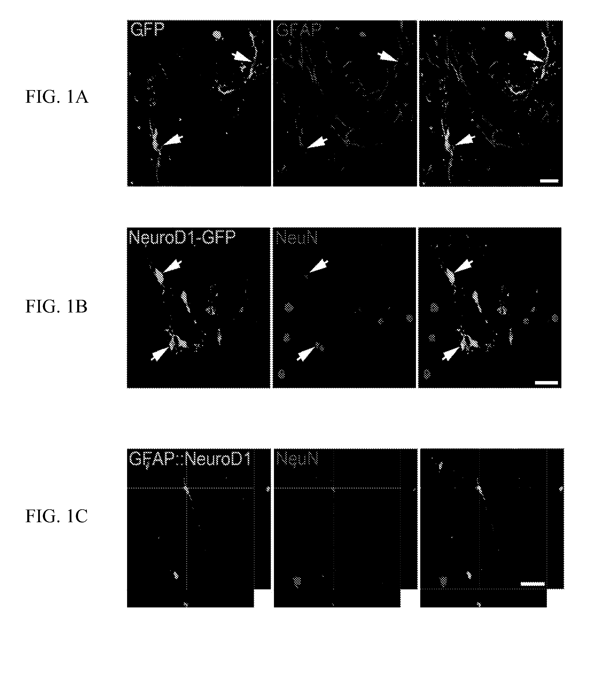
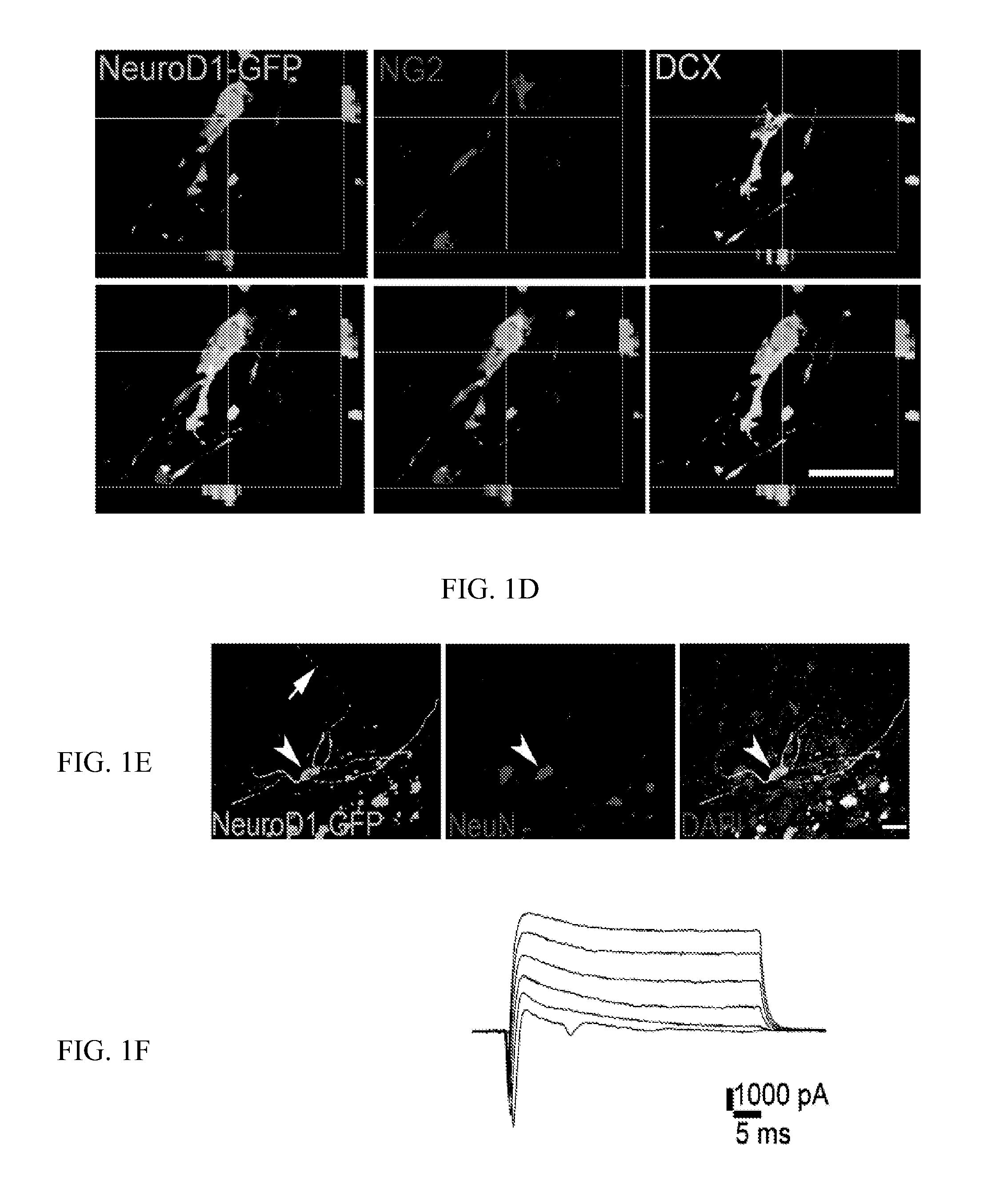
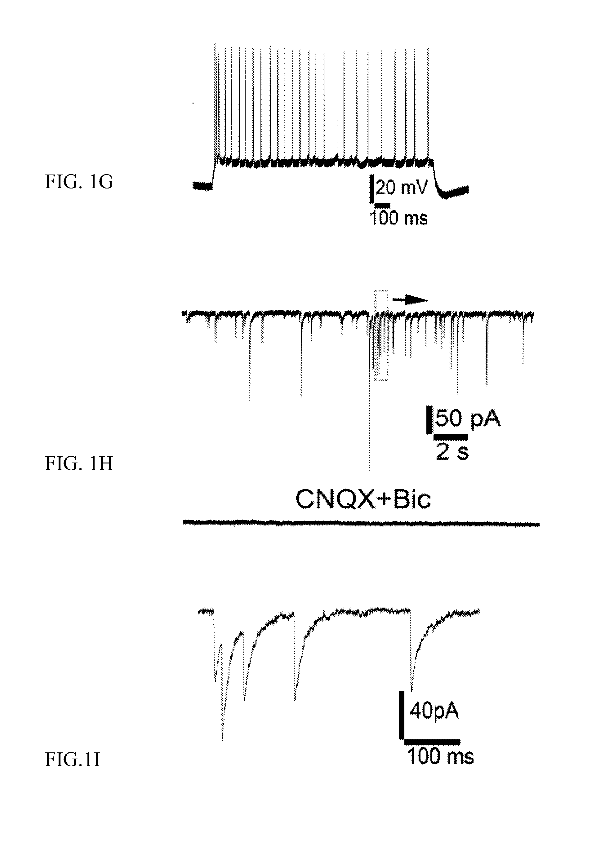
[0274]The mouse NeuroD1 gene was subcloned from the pAd NeuroD-I-nGFP construct, described in Zhou, Q. et al., Nature 455(7213):627-632, 2008 (Addgene) and inserted into a pCAG-GFP-IRES-GFP retroviral vector described in Zhao, C. et al., J. Neurosci., 26(1):3-11,2006 to generate pCAG-NeuroD1-IRES-GFP retroviral vector. The sequence of CAG-NeuroD1-IRES-GFP is shown herein as SEQ ID NO:9. A control retrovirus construct, pCAG-GFP, encodes GFP and not NeuroD1.

[0275]The human GFAP promoter gene was subcloned from hGFAP Promoter-Cre-MP-1 (Addgene) and replaced the CAG promoter to generate pGFAP-NeuroD1-IRES-GFP and pGFAP-GFP-IRES-GFP retroviral vectors.

[0276]The human NG2 promoter was subcloned and replaced the CAG promoter to generate hNG2-NeuroD1-IRES-GFP and hNG2-GFP-IRES-GFP retroviral vectors.

[0277]The mouse Lcn2 promoter was subcloned and replaced the CAG promoter to generate mLcn2-NeuroD1-IRES-GFP and mLcn2-GFP-IRES-GFP retroviral vectors. Reactive glial cells ...


PUM

PropertyMeasurementUnit
speedaaaaaaaaaa
flow rateaaaaaaaaaa
pHaaaaaaaaaa

Abstract

Methods for producing new neurons in the brain in vivo are provided according to aspects of the present invention which include introducing NeuroD1 into a glial cell, particularly into a reactive astrocyte or NG2 cell, thereby “converting” the reactive glial cell to a neuron. Methods of producing a neuronal phenotype in a glial cell are provided according to aspects of the present invention which include expressing exogenous NeuroD1 in the glial cell, wherein expressing exogenous NeuroD1 includes delivering an expression vector, such as a viral expression vector, including a nucleic acid encoding the exogenous NeuroD1 to the glial cell.

Description

REFERENCE TO RELATED APPLICATIONS[0001]This application claims priority from U.S. Provisional Patent Application Ser. No. 61 / 673,471, filed Jul. 19, 2012 and 61 / 762,506, filed Feb. 8, 2013, the entire content of which is incorporated herein by reference.GOVERNMENT SUPPORT[0002]This invention was made with government support under Grant No. MH083911, awarded by the National Institutes of Health. The Government has certain rights in the invention.FIELD OF THE INVENTION[0003]General aspects of the invention relate to the in situ conversion of glial cells to functional neuronal cells in the central nervous system (CNS), and the methods to transforming glial cells to neuronal cells both in vitro and in vivo.BACKGROUND OF THE INVENTION[0004]The central nervous system in mammals is largely unable to regenerate itself following injury. Neurons are often killed or injured as a result of an injury or neurological condition, such as a disease or other pathology. It is well known that glial cel...

Claims


Application Information

Patent Timeline
Patent Type & Authority Applications(United States)
IPC IPC(8): A61K48/00A61K38/17
CPCA61K38/1709A61K48/005A01K2227/105A01K2267/0356C12N2740/13043A61P25/00C12N15/86
Inventor CHEN, GONGZHANG, LEIWU, ZHENGCHEN, YUCHENWANG, FANGUO, ZIYUAN
Owner PENN STATE RES FOUND
Why Eureka
Try Eureka