Quantitative measurement of human blastocyst and morula morphology developmental kinetics

a blastocyst and morula technology, applied in the field of quantitative measurement of human blastocyst and morula morphology development kinetics, can solve the problems of low birth rate, miscarriage, and well-documented adverse outcomes for both mother and fetus, and achieve the effect of minimizing the time to pregnancy

Active Publication Date: 2015-09-24
ARES TRADING SA
View PDF0 Cites 17 Cited by
  • Summary
  • Abstract
  • Description
  • Claims
  • Application Information

AI Technical Summary

Benefits of technology

[0014]In one embodiment, the measuring step is automated. In another embodiment, the imaging acquires images that are digitally stored. In one embodiment, the imaging employs darkfield illumination, brightfield illumination, or a combination of the two imaging modalities. Other modalities can also be used such as phase contrast, Hoffman modulation contrast, differential interference contrast, polarized light, fluorescence, single or multiplane or combinations thereof. In some embodiments, darkfield illumination may be used to provide enhanced image contrast for subsequent feature extraction and image analysis.
[0019]One aspect of the invention provides methods for ranking the likelihood of an embryo or blastocyst being euploid within a patient's available embryos or blastocysts, so that the probability of selecting a euploid embryo for transfer is maximized, and time to pregnancy is minimized. In some aspects of the invention, methods are provided for ranking the likelihood that an embryo is euploid, or will become a euploid blastocyst. In such aspects, compaction and / or cavitation and / or expansion and / or collapse parameters of an embryo are measured to arrive at a parameter measurement which can be employed to provide ranking of the likelihood of the embryo to be euploid or become a euploid blastocyst, thereby providing a means for selecting the embryo with highest likelihood being euploid for transfer. In some embodiments, the compaction and / or cavitation and / or expansion and / or collapse parameter is a morphological event that is measurable by time-lapse microscopy.

Problems solved by technology

Infertility is a common health problem that affects 10-15% of couples of reproductive-age.
Multiple gestations have well-documented adverse outcomes for both the mother and fetuses, such as miscarriage, pre-term birth, and low birth rate.
Potential causes for failure of IVF are diverse; however, since the introduction of IVF in 1978, one of the major challenges has been to identify the embryos that are most suitable for transfer and most likely to result in term pregnancy.
The understanding in the art of basic embryo development is limited as studies on human embryo biology remain challenging and often exempt from research funding.
These differences and many others make it inappropriate to directly extrapolate from one species to another.
In spite of such differences, the majority of studies of preimplantation embryo development derive from model organisms and are difficult to relate to human embryo development (Zernicka-Goetz, M.
However, potential risks of these methods also exist in that they prolong the culture period and disrupt embryo integrity (Manipalviratn S, et al.
However, manual review of time-lapse videos is labor-intensive and time-consuming.
Furthermore, manual assessment is subject to high inter- and intra-observer variations.
This is especially problematic for time-lapse parameters that involve morula and / or blastocyst stage events (e.g., compaction, cavitation, expansion and collapse), as these events occur in a gradual fashion, and it is difficult for human observers to mark each event beginning and end points in a consistent and reproducible manner.
Not withstanding the recent developments in time lapse imaging that allow clinicians to select embryos with greater developmental potential based on timing parameters of the first few cell cycles, current embryo selection relies primarily on morphological evaluations which are very subjective and offer limited predictive value of embryo viability.
Failure to correctly identify the most viable embryos can lead to unsuccessful IVF treatment or multiple gestation pregnancy.

Method used


Image

  • Quantitative measurement of human blastocyst and morula morphology developmental kinetics
  • Quantitative measurement of human blastocyst and morula morphology developmental kinetics
  • Quantitative measurement of human blastocyst and morula morphology developmental kinetics

Examples


example 1

Quantitative Measurement of Blastocyst Expansion and Collapse Dynamics

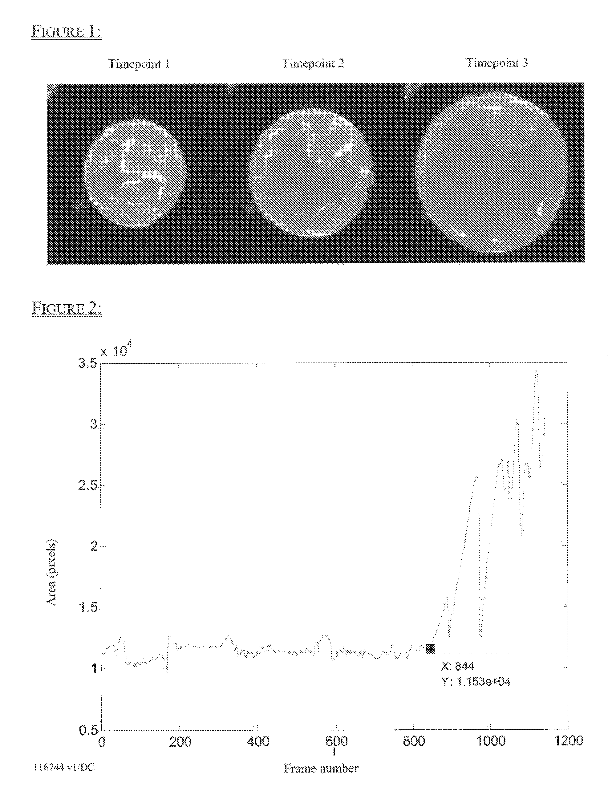
[0148]In order to quantitatively measure blastocyst expansion and collapse dynamics, embryos were imaged using a time-lapse imaging system through day 5 at an image frequency of 5 minutes per frame. First, the embryo outer boundary segmentation was generated for every frame in a time lapse image sequence as illustrated in FIG. 1. The area of the embryo outer boundary segmentation was then calculated and plotted against time (FIG. 2). Timings, durations, and frequencies of blastocyst expansion and collapse were then analyzed. Increases in area over time indicate blastocyst expansion, and decreases in area over time indicate collapse. This data demonstrates that a single image feature, the area of the embryo outer boundary segmentation, can be used to identify the onset and resolution of expansion and collapse.

example 2

Quantitative Measurement of Compaction and Cavitation Dynamics

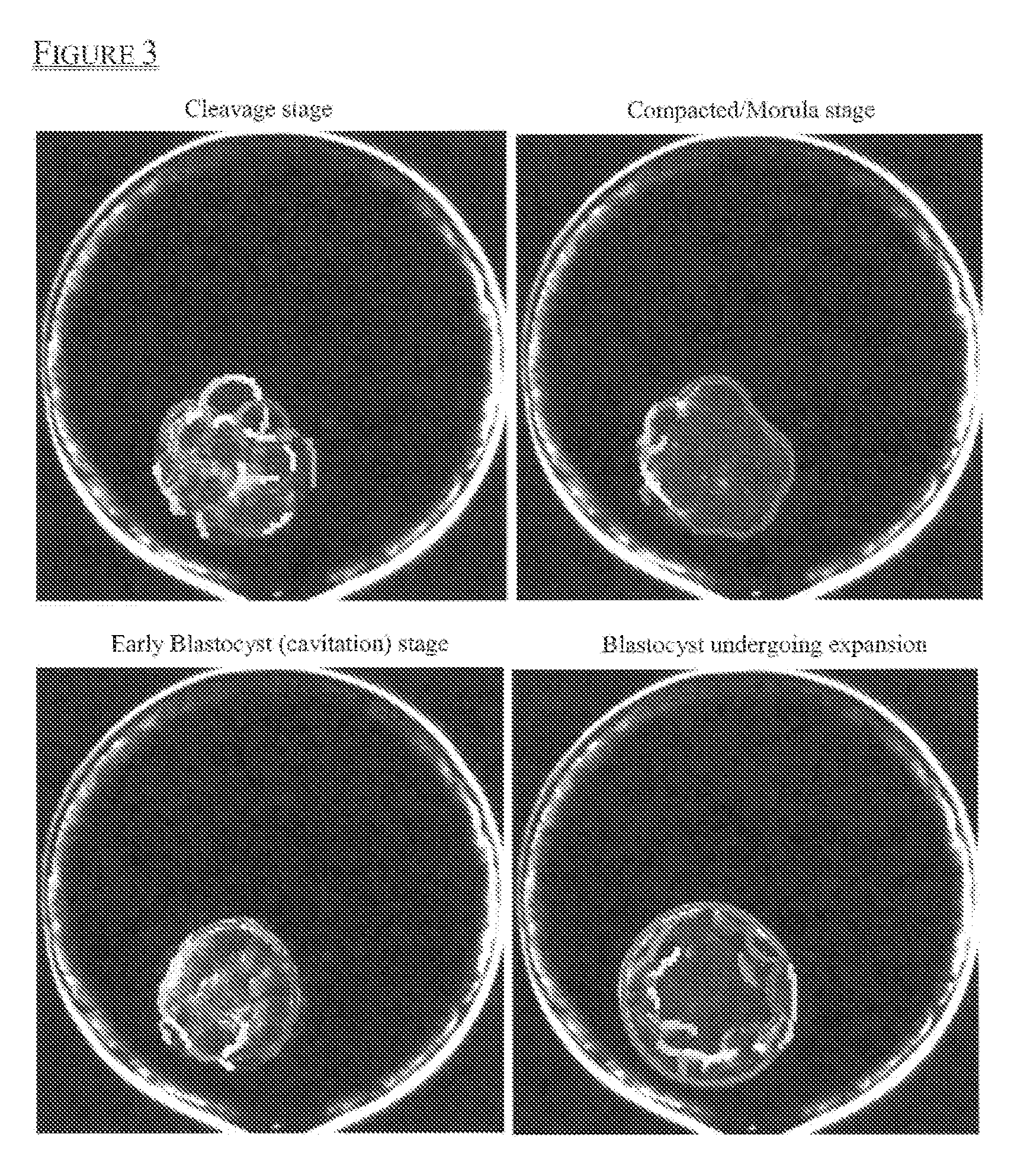
[0149]Compaction and cavitation are gradual transitioning processes, and it is challenging for human observers to identify a single time point when a transition occurs. In order to quantitatively measure compaction and cavitation events, embryos were imaged using a time-lapse imaging system through day 5 at an image frequency of 5 minutes per frame.

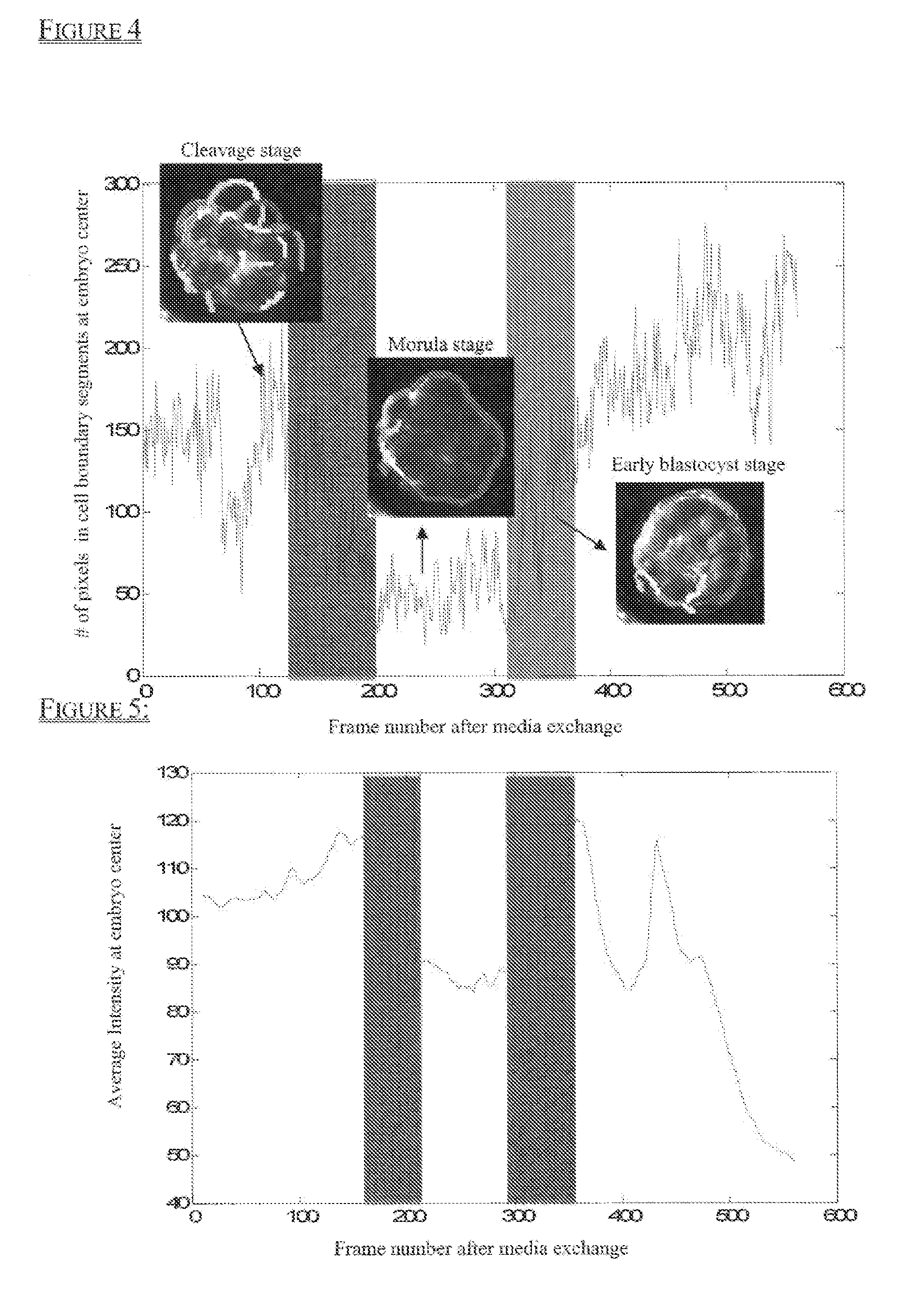
[0150]First, analysis was performed on all images to obtain coherent boundary features (i.e., segments) as shown in FIG. 3. The compaction process correlates to a decrease in edge-based segments located in the center of the embryo, while the cavitation process correlates to an increase in edge-based segments located in the center of the embryo. Therefore, segment distribution pattern changes over time were analyzed, and an example is shown in FIG. 4.

[0151]The second image feature analyzed was the average of integrated image intensity changes at the center of the embryo over tim...

example 3

Cavitation Parameters are Predictive of Aneuploidy and can be Combined with Other Parameters to Classify Risk of Embryonic Aneuploidy

[0156]It has been reported that more than 50% of the human embryos cultured in IVF clinics are aneuploid embryos that have an abnormal number of chromosomes. These aneuploid embryos have lower implantation rates and are not likely to result in live birth of healthy babies. Currently, pre-implantation genetic screening (PGS) is the most effective approach to select against aneuploidy embryos. However, due to its high cost, labor intensive requirements and invasive nature, less than 10% of patients that are treated for assisted reproduction undergo PGS prior to embryo transfer.

[0157]In order to develop a non-invasive time-lapse enabled assay to assess the risk of embryos being aneuploid, it was discovered that by assessing the tCav parameter, alone or in combination with other cellular parameters, embryo morphology and patient information, the likelihood...


PUM

PropertyMeasurementUnit
Exp Timeaaaaaaaaaa
widthaaaaaaaaaa
time lapse imagingaaaaaaaaaa

Abstract

Methods, compositions and kits for determining the developmental potential of one or more embryos are provided. These methods, compositions and kits find use in identifying embryos in vitro that are most useful in treating infertility in humans.

Description

CROSS REFERENT TO RELATED APPLICATIONS[0001]This application claims priority to U.S. Provisional Patent Application No. 61 / 968,260 filed Mar. 20, 2014, the contents of which are incorporated by reference herein in its entirety.FIELD OF THE INVENTION[0002]This invention relates to the field of biological and clinical testing, and particularly the imaging and evaluation of zygotes / embryos from both humans and animals.BACKGROUND OF THE INVENTION[0003]Infertility is a common health problem that affects 10-15% of couples of reproductive-age. In the United States alone in the year 2006, approximately 140,000 cycles of in vitro fertilization (IVF) were performed (cdc.gov / art). This resulted in the culture of more than a million embryos annually with variable, and often ill-defined, potential for implantation and development to term. The live birth rate, per cycle, following IVF was just 29%, while on average 30% of live births resulted in multiple gestations (cdc.gov / art). Multiple gestati...

Claims


Application Information

Patent Timeline
Patent Type & Authority Applications(United States)
IPC IPC(8): G01N33/50G01N15/14
CPCG01N33/5091G01N2015/1493G01N2015/1497G01N15/1463C12N5/0604G01N33/4833G06T7/0016G06T2207/30044G06T2207/10056G06V20/698
Inventor TAN, LEICHIAN, MARTINCHEN KIM, ALICELORENZEN, PETER
Owner ARES TRADING SA
Why Eureka
Try Eureka