Systems and methods for monitoring biological fluids

Inactive Publication Date: 2015-10-22
UNIV OSLO HF
View PDF1 Cites 3 Cited by
  • Summary
  • Abstract
  • Description
  • Claims
  • Application Information

AI Technical Summary

Benefits of technology

The present invention provides methods and compositions for detecting chemicals in biological fluids, such as methanol, ethanol, and ethylene glycol, using dehydrogenase enzymes and NAD+. The methods involve contacting a biological sample with a dehydrogenase enzyme and NAD+, and detecting the resulting NADH using a test strip or other means. The invention also provides kits for detecting these chemicals and monitoring their levels in a subject. The technical effects include improved diagnosis and screening for chemicals in biological samples, as well as the development of more efficient and reliable methods for detecting these substances.

Problems solved by technology

Diagnosis is difficult, and many never get a definitive diagnosis because methanol analyzes are seldom available.
In addition, samples must often be transmitted over great distances for analytical response, after which the patient may already have died or left the hospital without a diagnosis.

Method used


Image

  • Systems and methods for monitoring biological fluids
  • Systems and methods for monitoring biological fluids
  • Systems and methods for monitoring biological fluids

Examples


example 1

Reagents

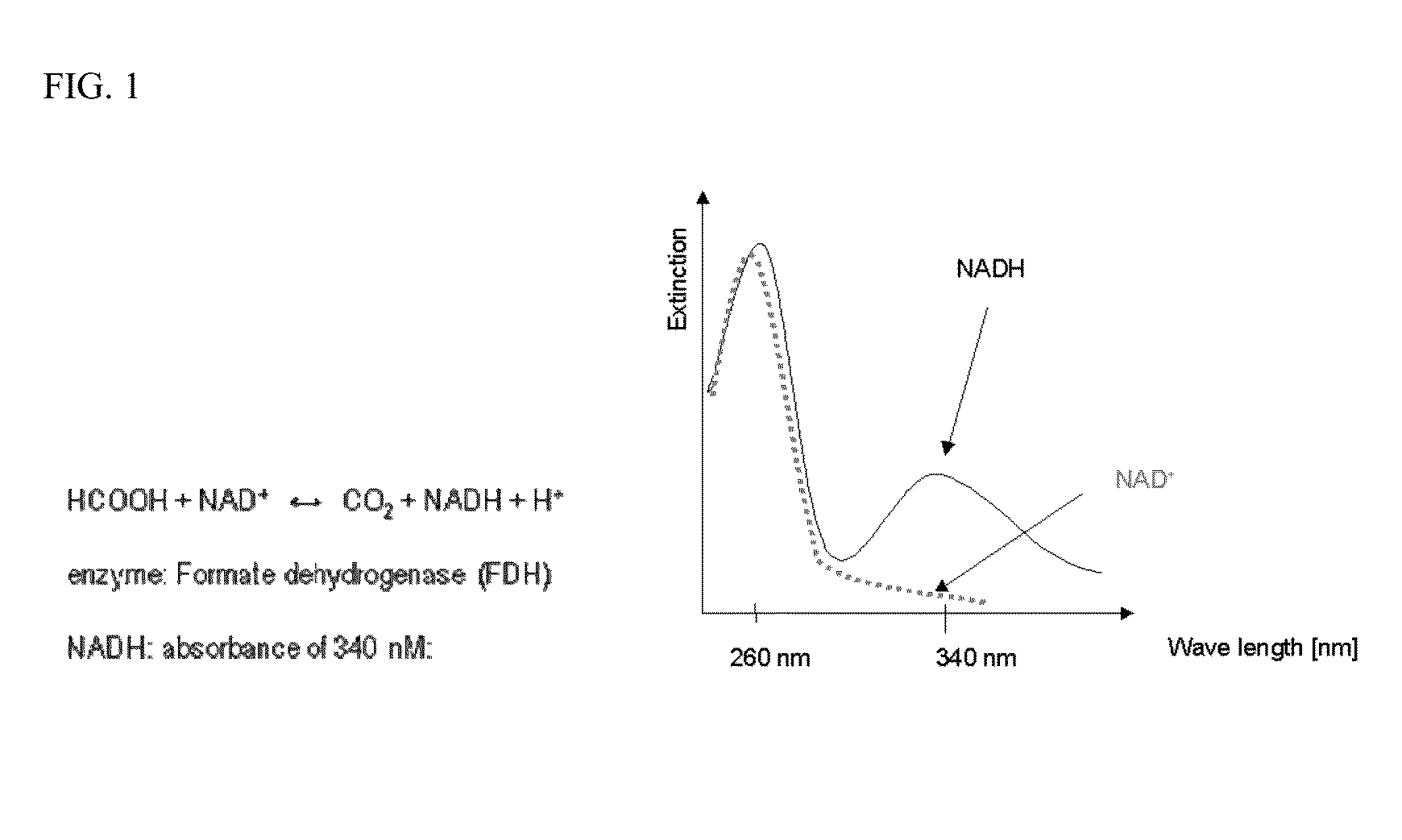
[0079]1. NAD (nicotinamide dinucleotide) commercially available from, for example, Sigma and many other companies).

2. Formate dehydrogenase from Roche (catalog no 244 678, contains 80 U or other concentration)

3. A phosphate buffer pH=7.5 such as 0.1M phosphate buffered saline (PBS) pH=7.5

Preparation of Reagents for Use:

[0080]Both NAD and formate dehydrogenase are sold in dry state and are stable for extended periods.

1. Dissolve approx 0.2 g of NAD in 30 ml buffer. (Reagent 1)

2. Dissolve the content of one bottle formate dehydrogenase (80 U) in 5 ml buffer (Reagent 2)

Both reagents are stable at 4-8° C. for approx 4-5 days and frozen reagents (preferentially at −40° C. or below) are stable for many months.

Analysis

[0081]1. Mix 1 part sample (serum or blood), 10 parts reagent 1 and 5 parts reagent 2.

2. Incubate for 5-10 minutes at 37° C. or at 20° C. for 7-10 min.

3. Measure absorbance in a photometer at 340 nm.

Evaluation of Result

[0082]1. The reagent itself (in the absence of sa...

example 2

[0084]This example describes the development of a colorimetric dry-reagent test strip for measuring formate in the range of 0-20 mM in buffer solutions with detection limit ≦2 mM by simple dip and read procedure. The strip also finds use in measuring formate in for example, whole blood and serum.

[0085]The developed formate test strip is a dry-reagent, self-dosing, “dip-and-read”, colorimetric test device, which contains in dry state all reagents needed for measuring formate. The following reagents were used: Formate Dehydrogenase (Roche Applied Sciences), Diaphorase (Sigma D2197), MTT (Sigma M2128), NAD (Sigma N1636). Other components were also included in the formate formula for better enzymes stability and strip performance.

[0086]The strip was made as follows:

[0087]a. Single reagent mixture was prepared by making HEPES buffer pH 7.9 first.

[0088]b. Enzymes were added from 100 U / mL stock solutions (in the same buffer).

[0089]c. All other ingredients were added to the mixture as dry p...

example 3

[0110]This example describes the detection of formate in serum and whole blood.

Formate in Serum

Materials Used:

[0111]For this study the early developed Formate test strip formula and pooled human serum from Cedarlane laboratories were used.

Preparation of Serum Samples:

[0112]Serum samples having 0, 1, 3, 5, 10 and 20 mM formate were prepared by addition of formate stock solutions (0.1 M and 0.5 M, pH 7.5) to the human serum.

Strip Activation:

[0113]20 μL of serum sample were put on the top surface of the formate reagent pad. After 5 seconds excess of liquid was removed from the strip surface. Color of the strip was measured in 2 minutes after strip activation.

Visual Evaluation:

[0114]Visual readings revealed clear color distinction between all the formate levels in serum −0, 1, 3, 5, 10 and 20 mM. Color of the strip changed from light yellow-greenish to green to blue in the 0-20 ppm formate range. The greenish color hue was due to the yellow serum color.

Instrumental Strip Readings:

[0115]...


PUM

PropertyMeasurementUnit
Timeaaaaaaaaaa
Concentrationaaaaaaaaaa
Levelaaaaaaaaaa

Abstract

The present disclosure relates to compositions and methods for diagnosis, research, and screening for chemicals in biological fluids (e.g., related to methanol poisoning, ethanol levels, and ethylene glycol poisoning). In particular, the present disclosure relates to point of care systems and methods for detecting formic acid or formate, ethanol, ethylene glycol, and other clinically relevant chemicals in biological fluids.

Description

FIELD OF THE INVENTION[0001]The present disclosure relates to compositions and methods for diagnosis, research, and screening for chemicals in biological fluids (e.g., related to methanol poisoning, ethanol levels, and ethylene glycol poisoning). In particular, the present disclosure relates to point of care systems and methods for detecting formic acid or formate, ethanol, ethylene glycol, and other clinically relevant chemicals in biological fluids.BACKGROUND OF THE INVENTION[0002]Methanol poisoning affects thousands each year, of which a large proportion (15-50%) die and many are left permanently blind or have brain damage. In a recent eruption in the Czech Republic, more than 40 dead and more than 120 methanol poisoned are this far reported.[0003]Early diagnosis is essential for successful treatment in methanol poisoning. Diagnosis is difficult, and many never get a definitive diagnosis because methanol analyzes are seldom available. In addition, samples must often be transmitte...

Claims


Application Information

Patent Timeline
IPC IPC(8): C12Q1/32A61K31/415A61K31/045
CPCC12Q1/32A61K31/045G01N2800/709G01N2333/90203G01N2333/904A61K31/415G01N33/5735
Inventor HOVDA, KNUT ERIKURDAL, PETTERGADEHOLT, GAUTJACOBSEN, DAG
Owner UNIV OSLO HF
Why Eureka
Try Eureka