Methods for Stabilizing Corneal Tissue

a corneal tissue and tissue technology, applied in the direction of cardiovascular disorders, drug compositions, peptide/protein ingredients, etc., can solve the problems of regression and corneal haze, corneal weakening, vision impairment, etc., to improve visual acuity and improve the outcome

Inactive Publication Date: 2016-01-07
THOMPSON VANCE +2
View PDF3 Cites 5 Cited by
  • Summary
  • Abstract
  • Description
  • Claims
  • Application Information

AI Technical Summary

Problems solved by technology

When the cornea is misshapen or injured, vision impairment can result.
Although this can correct the patient's vision, it also weakens the cornea, which may continue to change shape following the surgery.
However, regression and corneal haze can occur following PRK, and the greater the correction attempted, the greater the incidence and severity of the haze.
There is a risk that the flap created will later dislodge, however.
However, some patients with initially good results may experience a change in their refraction over the first 3 to 6 months (and possibly longer).
In addition, they reduce the eye's biomechanical rigidity, and postoperative keratectasia can result.
In some cases, the cornea thins and the resultant irregular astigmatism cannot be corrected, potentially requiring PRK to restore vision.
Keratoconus may involve a general weakening of the strength of the cornea, which eventually results in lesions in those areas of the cornea that are inherently less able to withstand the shear forces present within the cornea.
In keratoconus corneas, however, that orientation of collagen fibrils was lost within the diseased areas.
The slippage may be associated with loss of cohesive forces and mechanical failure in affected regions.
As discussed below, the methods of increasing corneal rigidity and compensating for corneal softness that currently exist suffer from drawbacks that include development of corneal haze and scarring, and the risk of endothelial cell damage.
These drawbacks are associated with the particular agents used in the methods.
The collagen fibrils in the scar tissue that forms following refractive surgery are disordered, resulting in corneal cloudiness.
Those authors also propose that part of this breakdown is triggered by a defect in the interfibrillar matrix that stabilizes the collagen fibrils, resulting in lamellar or fibrillar slippage.
In addition, the treatment also has the undesirable effect of inducing keratocyte apoptosis.
In application, however, aldehydes such as glutaraldehyde can lead to the development of corneal haze and scarring, while glyceraldehyde requires prolonged application times and its application is problematic.
Notably, the surgical techniques all involve at least some damage to the corneal structures and some tissue loss.

Method used


Image

  • Methods for Stabilizing Corneal Tissue
  • Methods for Stabilizing Corneal Tissue

Examples


example 2

Toxicity Evaluation of Decorin in the Feline Eye

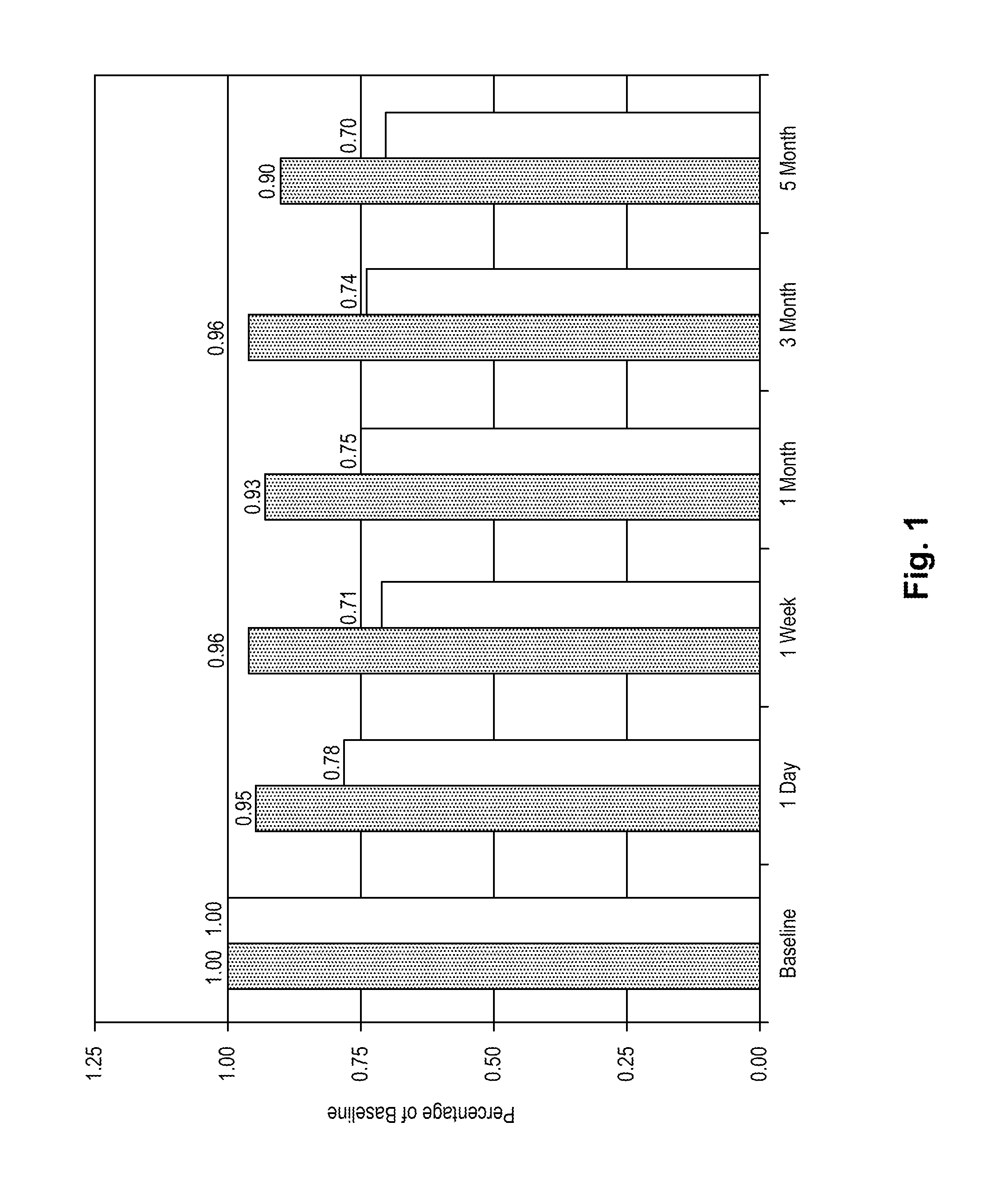
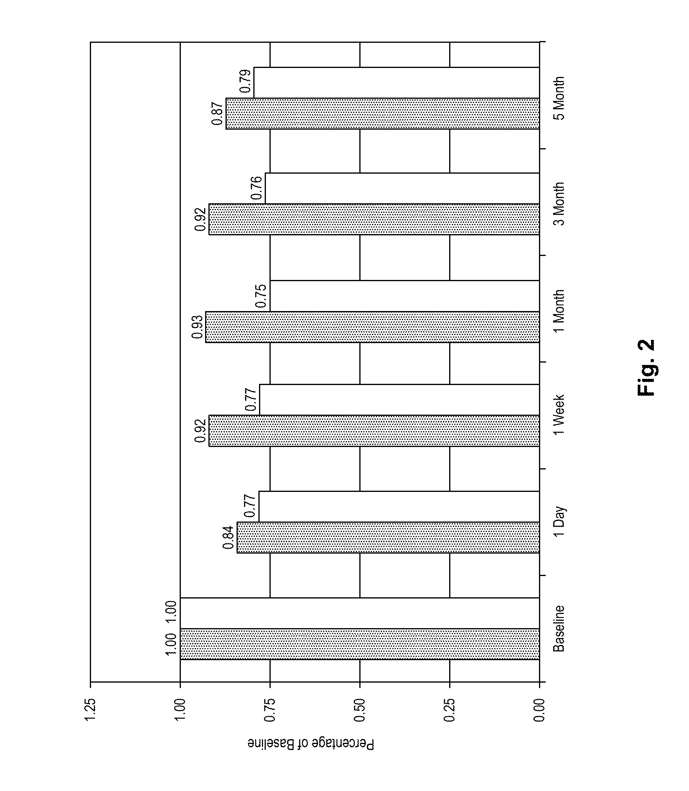
[0074]The purpose of the following evaluation was to determine if (1) there is toxicity associated with the use of decorin on the eye; (2) assess the penetration of decorin into the cornea; and (3) quantitate decorin in the cornea following exogenous application of decorin.

[0075]One, three, and five daily applications of decorin were assessed using female cats (6 months to 2 years of age) with normal corneas as the model system. The decorin was obtained as a dry powder (Sigma Chem. Co., Milwaukee, Wis.) and reconstituted in a 0.1 M phosphate buffer. In order to perform the microscopic evaluations, decorin was labeled with Oregon Green 514 using a commercially available kit from Molecular Probes.

[0076]Five cats were used in the study. Each cat was sedated prior to topical application of medication or photography of the eye. All animals received an ocular examination and photographs (whole eye, slit lamp, and endothelial cells) prior to ...

example 3

Measurement of Corneal Hysteresis in the Feline Model

[0083]The effects of decorin (human recombinant decorin provided by Catalent, Inc., Wisconsin) application on the biomechanical properties of the feline cornea were measured in five animals in a study performed at the Dartmouth-Hitchcock Surgical Research Center, Lebanon, N.H., Chemical agents were administered to the treated eyes to enhance decorin penetration and to dissociate proteoglycan bridges between collagen fibers, as referenced in paragraph [062]. The biomechanical integrity of the cornea was measured using the Reichert Ocular Response Analyzer (ORA). The ORA utilizes a dynamic by-directional applanation process to measure corneal hysteresis (CH).

[0084]Table 2 shows the results from this study.

TABLE 2Stabilization of Cornea Biomechanical propertiesfollowing Application of Decorin SolutionBeforeAfter decorinAnimal #Decorin (CH)Treatment (CH)After 21 DaysAHH35.507.437.50QJD43.906.306.90RAF63.135.186.20BEA43.654.806.20IRH67...

example 4

Ocular Irritation Studies in Humans

[0086]In safety trials with live human subjects in Shanghai, People's Republic of China in August 2004, 2.9 mg / ml of decorin in buffered saline solution was administered by the applicator method. To avoid discomfort from placing the applicator on the eye, proparican hydrochloride (0.5%) was first administered as an anesthetic. It appears that the clinician and the patient are most comfortable with a holding period of the applicator on the eye limited to about twenty (20) seconds, so that if the full dose cannot be delivered in that interval, repeat applications following one another over several minutes would be indicated. Work to date has been with a single application of a limited concentration of decorin only, however patients show no adverse effects and express no discomfort whatsoever. This type of safety study has been conducted on adolescent Chinese Ortho-K patients on three occasions. None of the safety tests suggest any adverse indications...


PUM

PropertyMeasurementUnit
diameteraaaaaaaaaa
diameteraaaaaaaaaa
pHaaaaaaaaaa

Abstract

Methods of stabilizing collagen fibrils in a cornea are disclosed. The stabilization may be effected by treating the cornea with a protein that crosslinks collagen fibrils, such as decorin. The stablization methods include treatment of corneas before, during, or after a surgical procedure, treatment of keratectasia, and treatment of keratoconus.

Description

RELATED APPLICATIONS [0001]This application is a continuation-in-part of application no. PCT / US2007 / 008049, filed Apr. 3, 2007, which claims benefit of provisional application No. 60 / 791,413, filed Apr. 13, 2006, the contents of each of which is incorporated by reference in its entirety.FIELD OF THE INVENTION [0002]The present invention relates to methods of stabilizing collagen fibrils in the cornea. These methods can be used to improve the outcome following refractive surgery, and to treat conditions of the cornea such as keratectasia and keratoconus.BACKGROUND OF THE INVENTION[0003]The cornea is the first and most powerful refracting surface of the optical system of the eye. It is made up of five layers, the outermost of which is the epithelium. The epithelium is only four to five cells thick, and renews itself continuously. Underneath the epithelium is the acellular Bowman's membrane. It is composed of collagen fibrils and normally transparent. Below Bowman's membrane is the str...

Claims


Application Information

Patent Timeline
Patent Type & Authority Applications(United States)
IPC IPC(8): A61K38/39A61K9/00
CPCA61K9/0048A61K38/39A61K38/1709A61K38/45C07K14/4725
Inventor THOMPSON, VANCEDEWOOLFSON, BRUCEDEVORE, DALE
Owner THOMPSON VANCE
Why Eureka
Try Eureka