Medicine for treating keratoconus

A keratoconus and pharmacological technology, applied in drug combinations, sensory diseases, pharmaceutical formulations, etc., can solve the problems of patients with great economic and physical and mental impact, cloudy implants, and missed treatment opportunities.

Active Publication Date: 2015-06-03
SHANDONG EYE INST
View PDF0 Cites 4 Cited by
  • Summary
  • Abstract
  • Description
  • Claims
  • Application Information

AI Technical Summary

Problems solved by technology

[0003] The etiology of keratoconus is still unknown, and it may be related to various reasons, such as mechanical friction of the ocular surface, long-term wearing of contact lenses, allergic inflammation of the ocular surface, etc. Early clinical intervention and treatment are still blank: even after early clinical diagnosis of keratoconus There is no method of treatment and intervention, only to wait for the natural development of the disease, wait for the cornea to expand to a certain stage, and rely on rigid conta

Method used


Image

  • Medicine for treating keratoconus
  • Medicine for treating keratoconus
  • Medicine for treating keratoconus

Examples


Embodiment 1

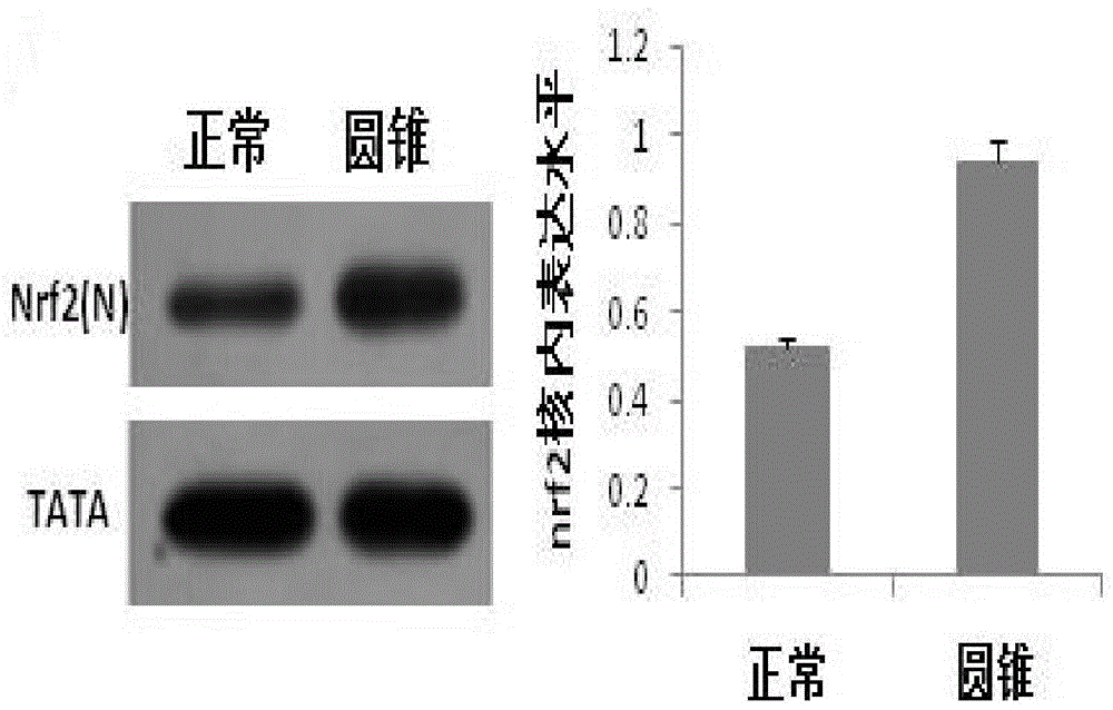
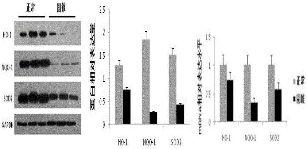
[0018] Example 1: Effects of adding different concentrations of 5'aza on antioxidant genes in keratoconus stromal cells

[0019] Corneal tissue was treated overnight at 4°C with Dispase (50mg / ml) to remove the corneal epithelium and endothelium. After the corneal stroma was cut into pieces, it was digested with collagenase (1.25mg / ml) at 37°C for 6-8 hours, separated by pipetting, centrifuged, and the supernatant was discarded. , add DMEM / F-12 medium containing 10% fetal bovine serum at 37°C CO 2 Cultured in an incubator, 0.25% trypsin + 0.02% EDTA for passage. During the isolation and culture of corneal stromal cells, the P3 generation cells were taken 1X10 6 The nuclear protein was extracted according to the steps of the nuclear protein-cytoplasmic protein extraction kit (Thermo, 78833), and the expression of Nrf2 protein in the nucleus of normal and keratoconus stromal cells was detected by wetern blot. The expression level of nrf2 gene in the nucleus of keratoconus sampl...

Embodiment 2

[0022] Example 2: Effect of adding different concentrations of 5'aza on the expression of matrix degrading enzymes in keratoconus stromal cells

[0023] During the isolation and culture of keratoconus stromal cells, different concentrations of methylation inhibitor 5’aza (0, 0.5, 1 μm) were added, and after 48 hours of culture, 5×10 cells were taken. 5 , conventional western blot detection of the protein expression of matrix degrading enzyme UPA in keratoconus stromal cells. After 5'aza treatment, the expression of uPA protein in keratoconus stromal cells was lower than that of the untreated group, such as Figure 5 . In addition, collagen degradation experiments were used to add culture solutions containing plasminogen (100 mg / L) and different concentrations (0, 0.5, 1 μm) of 5'aza to the surface of the three-dimensional collagen gel of keratoconus stroma, cultured for 48 hours, and determined The hydroxyproline content in the solution is used as the collagen degradation ac...

Embodiment 3

[0024] Example 3: The effect of adding different concentrations of SFN on the expression of matrix-degrading enzymes in keratoconus stromal cells

[0025] During the isolation and culture of keratoconus stromal cells, different concentrations of SFN (0, 5, 10 μM) were added, and after 24 hours of culture, 1X10 6 The nuclear protein was extracted according to the procedure of the nuclear protein-cytoplasmic protein extraction kit (Thermo, 78833), and the expression level of Nrf2 protein in the nucleus of keratoconus stromal cells after drug treatment was detected by wetern blot. In addition, take the above cells 5X10 5 , conventional western blot detection of the protein expression of matrix degrading enzyme UPA in keratoconus stromal cells. After SFN treatment, the expression of Nrf2 protein in the nucleus of keratoconus stromal cells increased; and after drug treatment, the expression of UPA protein in keratoconus stromal cells was lower than that in the untreated group. In...


PUM


Abstract

The invention aims at providing a medicine for treating keratoconus, which takes an Nrf2-ARE signal path as a target. The medicine can activate the signal path or improve the expression of downstream antioxidant genes NQO-1 and SOD2, so as to reduce the expression of matrix degrading enzymes; therefore, the medicine is suitable for the treatment of keratoconus. The medicine disclosed by the invention, which is used for treating the keratoconus by virtue of a methylation inhibitor 5-aza, can improve the expression of antioxidant genes of keratoconus matrix cells and can reduce the content of the matrix degrading enzymes as well.

Description

technical field [0001] The invention belongs to the technical field of ophthalmic disease treatment, and in particular relates to the application of a drug targeting the Nrf2-ARE signaling pathway in the treatment of keratoconus. Background technique [0002] Keratoconus (KC) is a corneal lesion characterized by dilatation of the cornea, causing the central part of the cornea to protrude forward, thinning into a conical shape, and producing highly irregular astigmatism. Histopathologically, corneal epithelial cell layer thinning, tearing of Bowman's membrane and Descemet's layer, corneal stroma thinning, decreased collagen content and scarring. The disease usually begins in adolescence, and patients can often use frame lenses to correct their vision in the early stage; as the disease progresses, they can wear rigid contact lenses or perform corneal surface lens surgery; in the late stage, the vision declines significantly, and corneal transplantation is required to restore p...

Claims


Application Information

Patent Timeline
IPC IPC(8): A61K45/00A61K31/12A61K31/26A61P27/02
Inventor 曲明俐周庆军边江王瑶杨玲玲
Owner SHANDONG EYE INST
Why Eureka
Try Eureka