Automatic measurement of lesions on medical images

a technology of medical images and lesions, applied in the field of analysis of medical images, can solve the problems of poor inter- or intra-observer reproducibility between single experts, increased mortality, and single experts not reaching excellent reproducibility

Active Publication Date: 2016-01-14
UNIV DANGERS +1
View PDF6 Cites 26 Cited by
  • Summary
  • Abstract
  • Description
  • Claims
  • Application Information

AI Technical Summary

Benefits of technology

[0111]In one embodiment, the step (a) of method of the invention comprises or consists in measuring the Area and fractal dimension of porto-septal and peri-sinusoidal fibrosis. Fibrosis can start in the centro-lobular region (alcoholic liver disease) and / or in the periphery of the liver lobule or around the portal tracts (viral liver disease). This fibrosis grows gradually to form bands of fibrosis, or septa, called septal fibrosis. This septal fibrosis is extended between the portal tracts or between the portal tracts and the centro-lobular region. The cirrhosis stage occurs when these different septa entirely surround hepatocytes. When pathologists are assessing the Metavir stage, their judgment is based on the porto-septal fibrosis. The other form of fibrosis is located between the rows of hepatocytes and sinusoids, the equivalent of the capillary in the liver, that is to say, in the intercellular space where there are many liver metabolic exchanges. This is called the peri-sinusoidal fibrosis. Although it is not taken into account in the fibrosis scores in clinical use, this fibrosis is important because it has a key role in the genesis of liver failure and portal hypertension. In one embodiment, the method of the invention thus comprises measuring descriptors associated with porto-septal and / or peri-sinusoidal fibrosis, such as, for example, area and fractal dimension of porto-septal fibrosis, area and fractal dimension of peri-sinusoidal fibrosis, area of lobular peri-sinusoidal fibrosis and ratio of peri-sinusoidal fibrosis among the whole fibrosis. The present invention also relates to a method for measuring descriptors associated with porto-septal and / or peri-sinusoidal fibrosis such as, for example, area and fractal dimension of porto-septal fibrosis, area and fractal dimension of peri-sinusoidal fibrosis, area of lobular peri-sinusoidal fibrosis and ratio of peri-sinusoidal fibrosis among the whole fibrosis. A non-limiting example of such a method is shown hereinafter. The Inventors thus developed a mask (MASKPORT) which allows distinguishing porto-septal and peri-sinusoidal fibrosis. In one embodiment, as the creation of this mask requires a large number of morphometric treatments (regarding erosions, dilations, the size of fibrosis areas . . . ) the image dimensions may be reduced.
[0196]In one embodiment of the invention, the method of the invention is for diagnosing a disease characterized by the presence of lesions in an organ or tissue. According to this embodiment, the score obtained at step (c) of the method of the invention is a diagnostic score, which means that it is indicative of the presence and / or of the severity of a specific disease, defined as the “diagnostic target”. In one embodiment of the invention, the score ranges from 0 to 1, and the higher the value of the score, the higher the probability for the subject to be diagnosed with the diagnostic target.
[0198]In one embodiment of the invention, the method of the invention allows a direct assessment of the presence and / or severity of a lesion, or a direct diagnostic of the diagnostic target, i.e. the method leads to a binary diagnostic. In other words, according to this embodiment, the method of the invention usually leads to a dual type response “yes or no”: according to the value of the score obtained at step (c), a direct conclusion about the presence and / or severity of a lesion or a disease may be given. For example, according to this embodiment, when the diagnostic target is clinically significant fibrosis, the method of the invention allows determining if the subject is affected or not by clinically significant fibrosis.
[0361]The method of the present invention thus presents the following advantages. First, the method of the invention involves the automatic measurement and assessment of the severity of lesions in an organ. Consequently, the method of the invention allows reducing or eliminating subjectivity from diagnosis. Second, the method of the invention leads to a score allowing a direct diagnosis or prognosis, without additional step of comparison with a reference database.

Problems solved by technology

This liver fibrosis induces liver architecture modifications which are the source of major complications responsible for increased mortality.
Indeed, these semi-quantitative scoring systems of fibrosis are limited by poor inter- or intra-observer reproducibility between pathologists (Rousselet et al, Hepatology, 2005, 41(2):257-264).
The most important issue regarding the classification system by pathologists is that it is subjective.
However, a recent study showed that single experts did not reach excellent reproducibility whereas an expert panel seems to be the best reading reference (Boursier et al, BMC Gastroenterol, 2011, 11:132).
Thus, the challenging issue in pathology is to work with expert centers, with the probable risk of saturation.
In addition, there is a rarefaction or increasing unavailability of pathologists.
However, these published techniques give quantitative information but are not well correlated with the diagnostic reference system of pathologists, i.e. for example in the field of hepatology, the METAVIR fibrosis score with 5 classes (F0: no fibrosis; F1: portal fibrosis without septa; F2: portal fibrosis with rare septa; F3: numerous septa without cirrhosis; F4: cirrhosis) and the Ishak staging system with 7 classes based on portal and septal fibrosis and the degree of completeness for cirrhosis.

Method used


Image

  • Automatic measurement of lesions on medical images
  • Automatic measurement of lesions on medical images
  • Automatic measurement of lesions on medical images

Examples


example 1

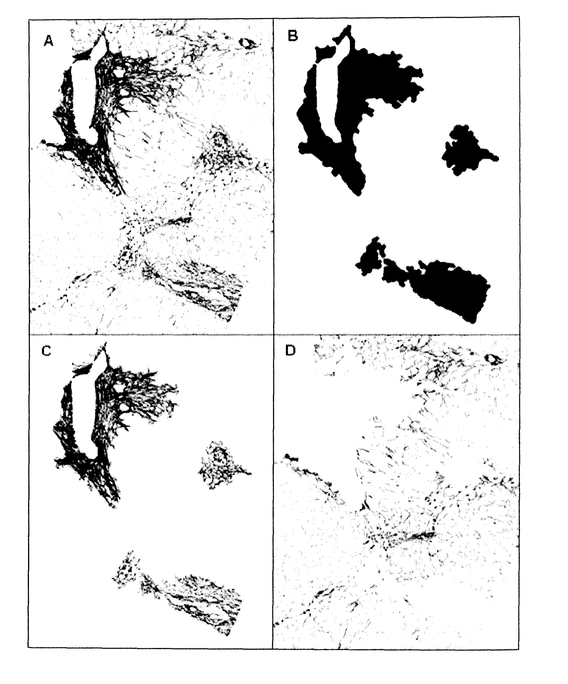
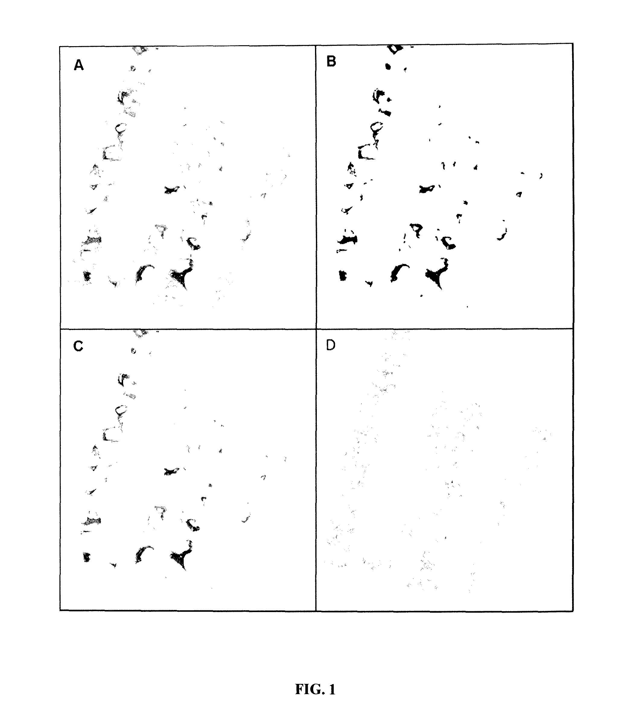
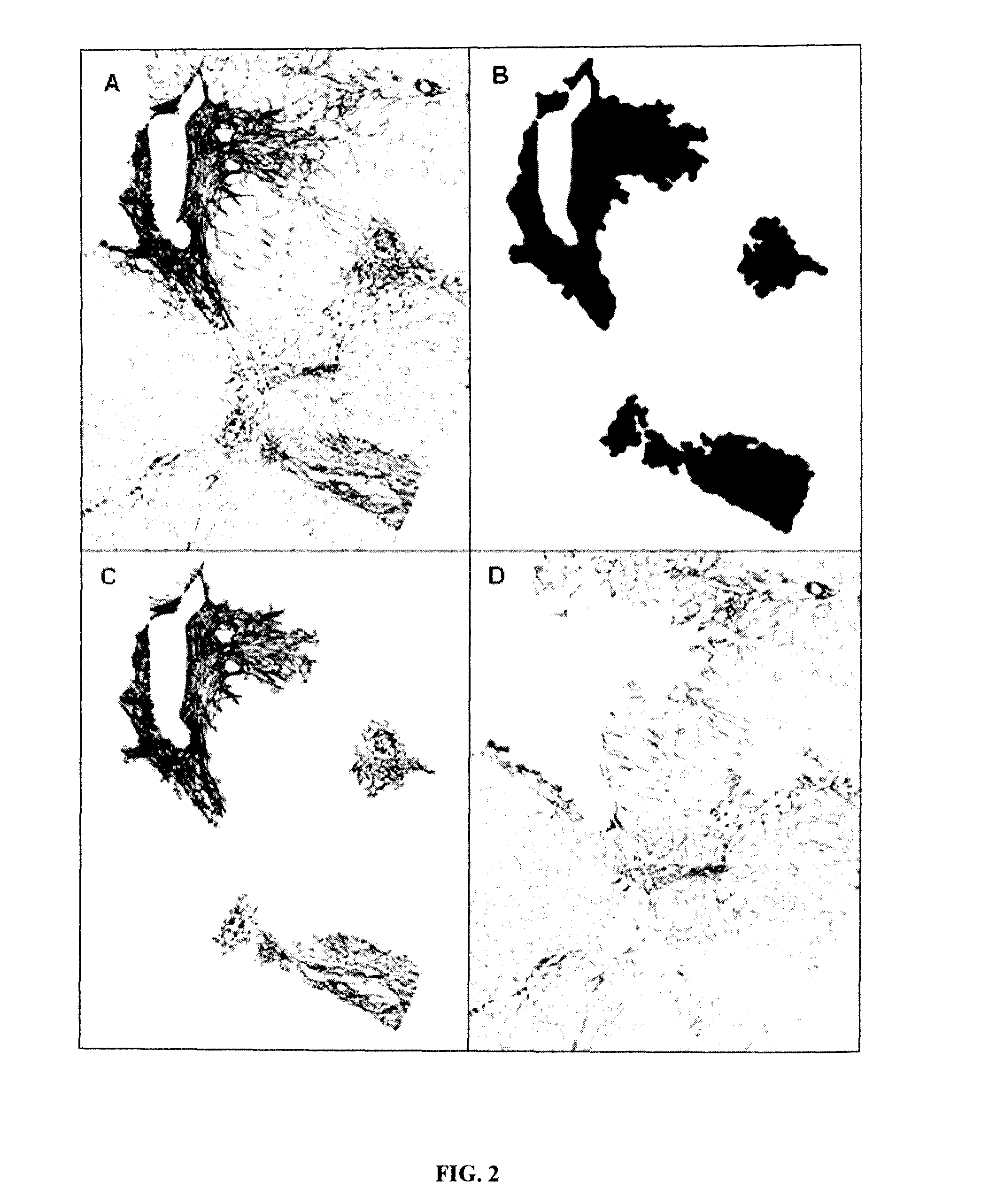
Automatic Measurement of Lesions in a Histological Image

Methods

Study Design

[0387]The morphometric diagnosis by automated morphometry was designed to diagnose clinically significant fibrosis (CSF), cirrhosis and Metavir F stages. The diagnostic models were developed in the derivation population. These results were validated by using two kinds of population:[0388]Populations with reference Metavir staging by expert to validate the diagnostic accuracy of diagnostic models based on automated morphometry.[0389]Population with Metavir staging performed by a first line pathologist to compare the diagnosis made by automated morphometry with that performed in real life conditions.

The conditions of liver biopsy specimens were the same in all populations. Especially, specimen lengths were the same and staining and morphometry performed by the same engineer were made centrally in the HIFIH laboratory.

Populations

Derivation Population

[0390]416 patients with chronic viral hepatitis C and a length ...

example 2

Reliability

[0436]The accuracy of prediction is imperfect. These inaccurate results are not due to chance and can be predicted by statistical analysis providing significant independent predictors of accuracy. This defines the reliability analysis. Thus, one can calculate reliability classes where the accuracy varies and predictors can be different.

[0437]For example, a perfect result has 100% accuracy. A reliability analysis can determine reliability classes from 0 to 10, 20, 30, 40, 50, 60, 70, 80, 90% accuracy with specific predictors for each reliability class.

[0438]Predictors are issued from the list of descriptors of the invention.

[0439]For example, we provide here different predictive models obtained in different populations by binary logistic regression.

Tables of the Coefficients of the Binary Logistic Regression for Prediction of Accuracy for Clinically Significant Fibrosis

[0440]

Model # 1, AUROC = 0.656VariableLowerCoefficientUpperStd. errorz-valueProb(Intercept)1.93884.21356....

example 3

Diagnosis of NASH

[0442]Non-alcoholic fatty liver disease (NAFLD) is a frequent pathology. It encompasses the liver lesions linked to metabolic syndrome. It evolves from pure steatosis to non-alcoholic steato-hepatitis (NASH) and liver fibrosis. NASH includes several lesions: steatosis, hepatocyte ballooning, and lobular inflammation (Sanyal Hepatology 2011). An international expert team has described a NAFLD activity score (NAS) (Kleiner Hepatology 2005).

[0443]We have measured several lesions in a cohort of 235 patients with NAFLD. Optical microscopy included classical lesions (steatosis degree, NAS, NASH and fibrosis). Morphometry with image analysis included automatic measurement of area of steatosis (AOS) and fractal dimension of steatosis (DS) as well as area of fibrosis (AOF) and fractal dimension of fibrosis (DF). We also defined the relative Area of Steatosis (rAOS) as the area of steatosis in the non-fibrotic area.

[0444]Whatever the fibrosis stage, NAS was globally well corr...


PUM


Abstract

The present invention relates to a method for assessing the presence and / or the severity of a lesion in an organ or tissue of a subject through automated analysis of at least one image of said organ or tissue, wherein said organ or tissue is preferably a liver organ or liver tissue, comprising the calculation of a score combining descriptors of said image, wherein said method comprises the steps of: a. measuring on said at least one image at least two descriptors of said at least one image; b. mathematically combining said at least two descriptors in a score; and c. assessing the presence and / or the severity of a lesion in the organ or tissue based on the value of the score calculated at step (b).

Description

FIELD OF INVENTION[0001]The present invention relates to the analysis of medical images. More specifically, the present invention relates to an automatic method for assessing the presence and / or the severity of lesions in an organ, based on the computerized analysis of a medical image of this organ.BACKGROUND OF INVENTION[0002]All chronic liver diseases (causes: viral, alcohol, metabolic steatosis . . . ) are characterized by the development of liver lesions and the development of liver fibrosis. This liver fibrosis induces liver architecture modifications which are the source of major complications responsible for increased mortality. Fibrosis is progressive and usually reversible.[0003]Quantification of hepatic fibrosis is important to determine the severity of liver disease, its prognosis and treatment indication. The quantification reference of hepatic fibrosis is a microscopical examination by an expert liver pathologist of a liver specimen usually obtained by liver biopsy (LB)...

Claims


Application Information

Patent Timeline
Patent Type & Authority Applications(United States)
IPC IPC(8): G06T7/00G06T7/40
CPCG06T7/0012G06T7/408G06T2207/30096G06T2207/30056G06T7/0081G06T2207/10056G06T7/11G06T7/90
Inventor CALES, PAULCHAIGNEAU, JULIENHUNAULT, GILLESROUSSELET, MARIE-CHRISTINEAUBE, CHRISTOPHE
Owner UNIV DANGERS
Why Eureka
Try Eureka