Microscopy of a tissue sampling using structured illumination

a tissue sample and structured illumination technology, applied in the field of medical imaging systems, can solve the problems of large amount of unwanted background fluorescence, limited intraoperative tools available to assist surgeons in tumor margin, and high cost and time-consuming intraoperative pathology for surgical tumor resection

Active Publication Date: 2016-03-03
THE ADMINISTRATORS OF THE TULANE EDUCATIONAL FUND
View PDF3 Cites 18 Cited by
  • Summary
  • Abstract
  • Description
  • Claims
  • Application Information

AI Technical Summary

Problems solved by technology

Currently, there are very few intra-operative tools available to assist surgeons in tumor margin diagnosis to ensure complete removal of the tumor.
Intraoperative pathology for surgical tumor resections is expensive and time-consuming and consequently is only available at a handful of surgery centers in the US.
This creates a problem as fluorophores outside the plane of focus are also excited and emit photons, generating a significant amount of unwanted background fluorescence.
This in turn significantly degrades contrast of features of interest in the focal plane.
This is a common issue in wide-field fluorescence microscopy and several specialized techniques exist to reject background fluorescence, such as fluorescence confocal microscopy.
While effective in rejecting background fluorescence, confocal microscopy requires specialized optics in the form of rapid scanning mirrors and adjustable pinhole collection, which increases the cost and decreases the speed, which hinders the intra-operative intraoperative clinical feasibility.
In addition, the amount of photons collected at each beam scan position is limited, so the system requires a sensitive detector, such as a photomultiplier tube.

Method used


Image

  • Microscopy of a tissue sampling using structured illumination
  • Microscopy of a tissue sampling using structured illumination
  • Microscopy of a tissue sampling using structured illumination

Examples


Embodiment Construction

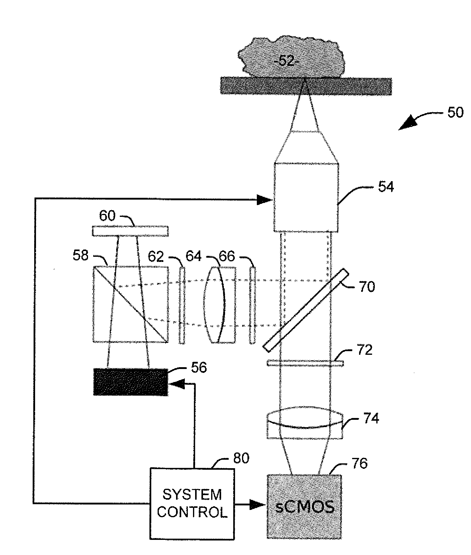
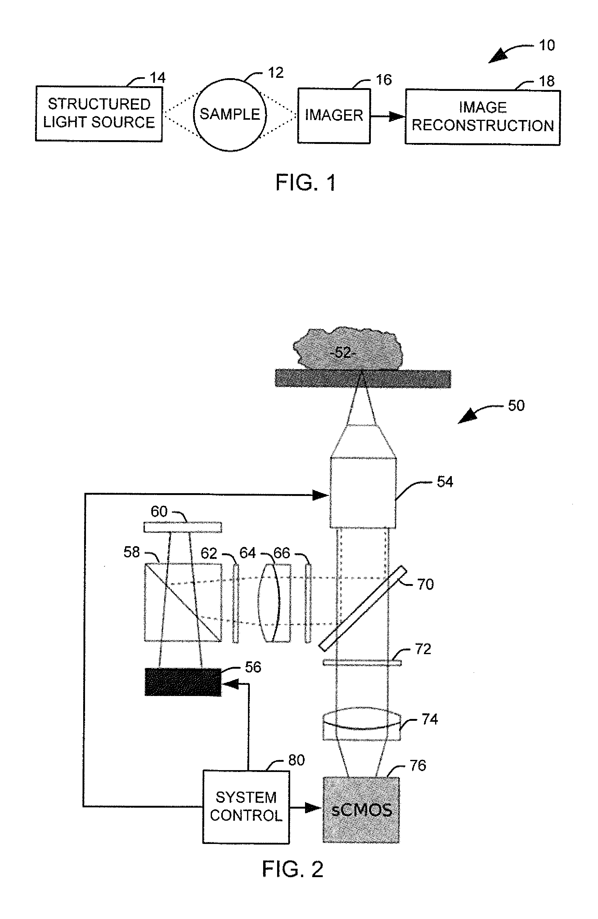
[0017]The present invention discloses the development of a next-generation Rapid Optical Sectioning Specimen Scanner based on video-rate incoherent structured illumination microscopy and developed with consideration of the design requirements for rapid, high-area throughput fluorescence microscopy of intact surgical and biopsy specimens. An imaging system in accordance with an aspect of the present invention provides the ability to image fresh tissue immediately after removal from the subject to provide a clinically useful sample. By “fresh,” it is meant that the tissue has been removed or exposed during a medical procedure occurring at a same time as the imaging of the sample. In other words, a fresh sample can be tissue removed from a patient during the procedure or tissue that is still within the patient, but made accessible as part of the medical procedure. It will be appreciated that a fresh sample will be significantly thicker than a traditionally prepared sample, increasing t...


PUM

PropertyMeasurementUnit
diameteraaaaaaaaaa
fluorescenceaaaaaaaaaa
fluorescentaaaaaaaaaa

Abstract

Systems and methods are provided for evaluating a fresh tissue sample, prepared as to fluoresce under illumination, during a medical procedure. A structured light source is configured to project a spatially patterned light beam onto the fresh tissue sample. An imaging system is configured to produce an image from fluorescence emitted from the illuminated fresh tissue sample. A system control is configured to provide a human-comprehensible clinically useful output associated with the medical procedure.

Description

RELATED APPLICATIONS[0001]This application claims priority to U.S. Provisional Patent Application Ser. No. 61 / 823,817, filed May 15, 2013, and U.S. Provisional Patent Application Ser. No. 61 / 919,434, filed Dec. 20, 2013. The entire contents of both applications are hereby incorporated by reference, such that where an element or a part of the description, claims or drawings of this application is not otherwise contained in the text of the application but is completely contained in either or both of the '817 and the '434 applications, that element or part is hereby incorporated by reference for the purposes of Rule 20.6.TECHNICAL FIELD[0002]This invention relates to medical imaging systems, and more particularly, to microscopy of a tissue sample using structured illumination.BACKGROUND[0003]A number of clinical situations could be benefited by a more rapid histological processing method, which reduces the number of steps between obtaining a fresh tissue specimen and rendering a diagno...

Claims


Application Information

Patent Timeline
Patent Type & Authority Applications(United States)
IPC IPC(8): G02B21/16A61B5/00A61B10/02G01N1/30G02B21/36G06K9/52G06K9/62H04N5/225
CPCA61B5/0071G06K9/6267G06K9/52G02B21/367H04N5/2256G02B21/16G01N2201/0635G01N1/30A61B10/02G02B21/365G01N21/6458G02B21/06G02B21/36G01N2001/302G01N2021/6419G01N2021/6421G01N2021/6441G01N2201/0675G01N2201/129G02B21/0076A61B2090/3941A61B2090/366G06V10/42H04N23/56G06F18/24
Inventor BROWN, JONATHON, QUINCY
Owner THE ADMINISTRATORS OF THE TULANE EDUCATIONAL FUND
Why Eureka
Try Eureka