Application Device for Inducing Cytotoxicity to Tumor Cells Via Coated Cerium Oxide Nanoparticles

a cerium oxide nanoparticle and tumor cell technology, applied in the field of human and animal cell treatment, can solve the problems of nanomaterials and living systems that can be potentially harmful, silver nanoparticles have been found to display size-dependent toxicity, and the greatest liability of nanomaterials, etc., and achieve the effect of no toxicity

Inactive Publication Date: 2016-03-17
UNIV OF CENT FLORIDA RES FOUND INC
View PDF1 Cites 4 Cited by
  • Summary
  • Abstract
  • Description
  • Claims
  • Application Information

AI Technical Summary

Benefits of technology

[0045]Another preferred surface coated nanoceria particle with a neutral surface charge that is administered to a plurality of isolated normal and a plurality of isolated transformed c...

Problems solved by technology

However, the greatest strength of nanomaterial, which relies primarily on the enhanced physical and chemical characteristics that matter exhibits at this scale, has the potential to be its greatest liability.
Potentially harmful interactions can occur between nanomaterials and living systems, including systems ...

Method used


Image

  • Application Device for Inducing Cytotoxicity to Tumor Cells Via Coated Cerium Oxide Nanoparticles
  • Application Device for Inducing Cytotoxicity to Tumor Cells Via Coated Cerium Oxide Nanoparticles
  • Application Device for Inducing Cytotoxicity to Tumor Cells Via Coated Cerium Oxide Nanoparticles

Examples


example 1

Synthesis and Characterization of Polymer-Coated Cerium Oxide Nanoparticles

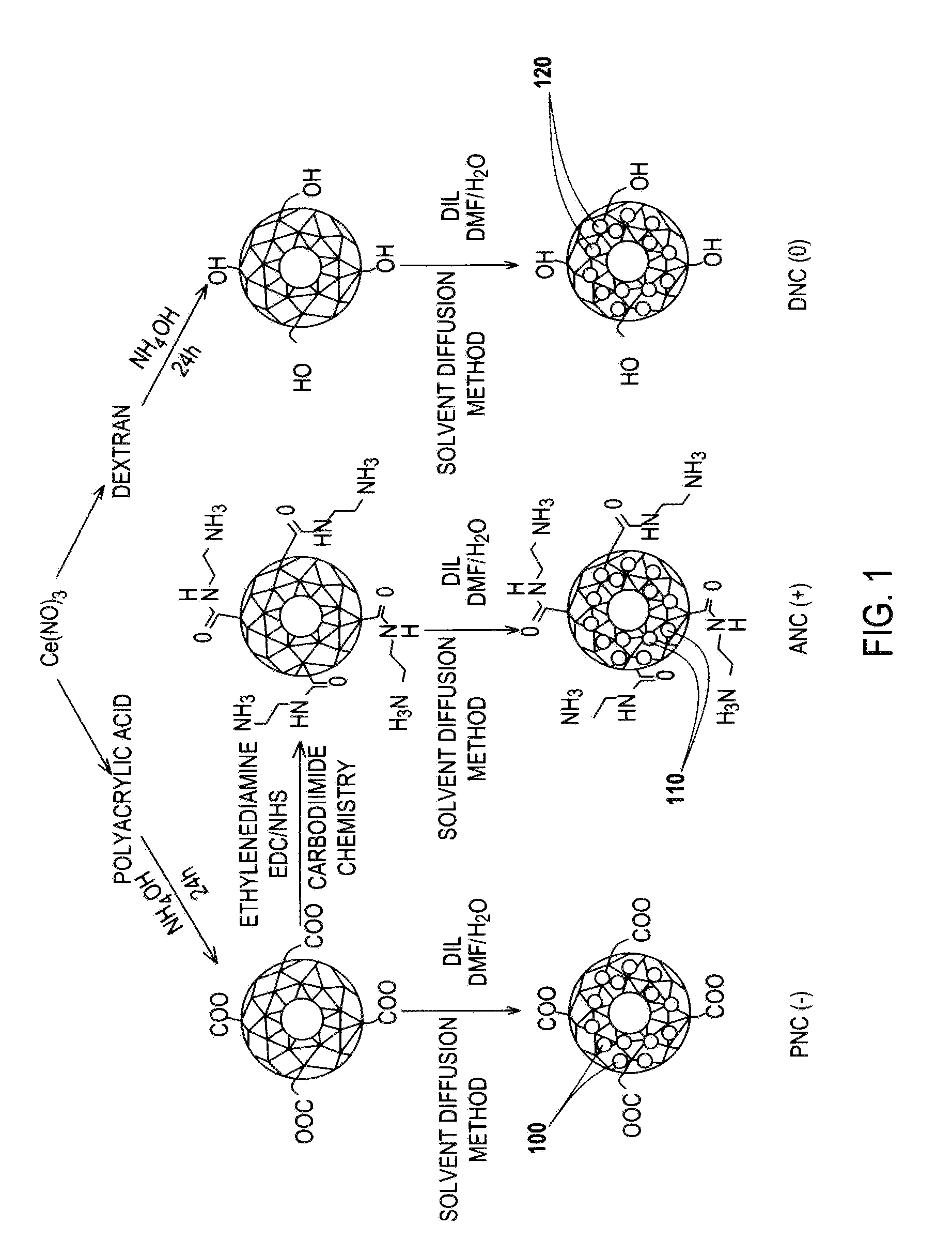
[0092]FIG. 1 is an illustration of various nanoceria preparations coated with either polyacrylic acid (PNC), aminated polyacrylic acid (ANC), or dextran (DNC) that endow nanoparticles with a negative (−), positive (+) or neutral (0) surface charge, respectively.

[0093]During synthesis, the nanoparticles were labeled fluorescently, by encapsulating a dye (DiI) 100, 110, 120 within the hydrophobic microdomains of the polymeric coatings of each nanoceria preparation, following a previously reported solvent diffusion methodology disclosed by S. Santra in Drug / dye-loaded, Multifunctional Iron oxide Nanoparticles for Combined Targeted Cancer Therapy and Dual Optical / Magnetic Resonance Imaging, Small 2009, 5, 1862-1868.

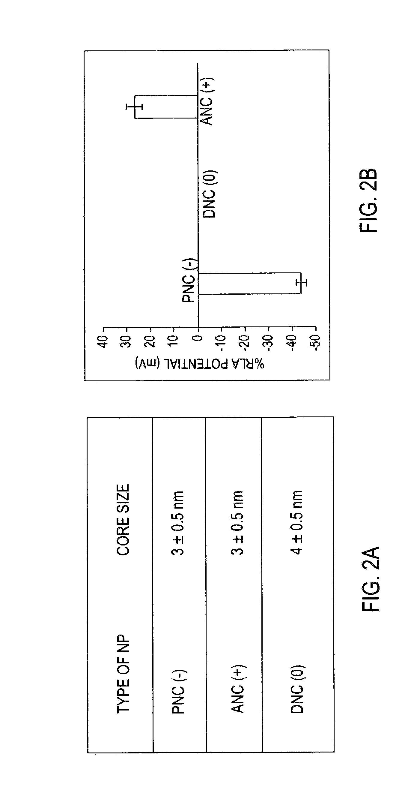
[0094]Transmission electron microscopy (TEM) studies reported in FIG. 2A show the presence of nanoparticles of similar core size (3 to 4 nm) in all preparations as reported by A. Asati, et al., in Ange...

example 3

Surface-Charge-Dependent Cellular Interaction of Nanoceria

[0098]Confocal microscopy experiments were performed in order to study the cellular uptake and intracellular localizaton of the DiI-labeled polymer-coated nanoceria. In these experiments, PNC (−), ANC (+) and DNC (0) (1.0 mM) were incubated with two transformed carcinoma cell lines (A549 lung and MCF-7 breast carcinomas), and two non-transformed (normal) cell lines (H9c2 cardiac myocytes and HEK293 human embryonic kidney cells). These cells lines were selected in order to investigate whether there is a difference in uptake by ANC(+), PNC(−) and DNC(0) polymer-coated nanoparticles, thus potentially displaying different toxicity profiles.

[0099]First, the internalization pattern of ANC(+), PNC(−) and DNC(0) was studied in normal cardiac myocyte cells (H9c2), using 10,000 cells as shown in FIGS. 3A-3C. After a 3-hour incubation, results showed that the negatively charged carboxylated nanoparticles [PNC(−)] were minimally uptaken ...

example 4

Internalization Experiments with Cancer Cells

[0102]FIGS. 4A-4C show results of internalization experiments performed with cancer cells (10,000 cells) derived from lung (A549) carcinoma cells. PNC(−) uptake by the A549 cells is minimal or slight as shown in FIG. 4A. In FIG. 4B, lung (A549) carcinoma cells were able to uptake ANC (+); interestingly, a higher degree of internalization and pronounced punctuated fluorescence was observed with the ANC(+) in the A549 cells. This might indicate that in these cells the majority of the ANC(+) localized into endosomal compartments. In contrast, in FIG. 4C, minimal and diffused intracellular fluorescence was observed with the DNC (0) in the A549 cells, suggesting that these nanoparticles are mostly localized in the cytosol with a small fraction confined within the endosomal compartments. Taken together, the above results suggest that the surface charge on polymer-coated nanoceria dictates their differential internalization and localization in n...


PUM

PropertyMeasurementUnit
Cytotoxicityaaaaaaaaaa
Toxicityaaaaaaaaaa
Surface chargeaaaaaaaaaa

Abstract

Differential surface-charge-dependent localization of nanoceria in normal cells and cancer cells plays a critical role in the toxicity profile of a nanoceria particle. Engineered surface-coated cerium oxide nanoparticles with different surface charges that are positive, negative and neutral provide therapeutic results for normal and cancer cell lines. Results show that nanoceria with a positive or neutral charge enters most of the cell lines studied, while nanoceria with a negative charge internalizes mostly in the cancer cell lines. Moreover, upon entry into the cells, nanoceria is localized to different cell compartments (e.g. cytoplasm and lysosomes) depending on the nanoparticle surface charge. The internalization and subcellular localization of nanoceria plays a key role in the nanoparticle cytotoxicity profile, exhibiting significant toxicity when they localize in the lysosomes of the cancer cell lines. In contrast, minimal toxicity is observed when they localize into the cytoplasm or do not enter the cells.

Description

[0001]This application is a divisional of U.S. Ser. No. 13 / 188,695 filed on Jul. 22, 2011 which claims priority based on U.S. Provisional Patent Application Ser. No. 61 / 366,697 filed on Jul. 22, 2010, both of which are incorporated herein by reference.FIELD OF THE INVENTION[0002]This invention relates to polymer coated nanoceria particles and more particularly to treatment of human and animal cells wherein cell internalization and cytotoxicity of polymer-coated nanoceria plays a key role in the nanoparticle cytotoxicity profile, exhibiting significant toxicity when localized in the lysosomes of cancer cell lines. The research herein is partially funded by the National Institutes of Health (NIH) under contract number CA10178.BACKGROUND AND PRIOR ART[0003]Nanomaterials with unique magnetic, luminescent and catalytic properties are being engineered for numerous biomedical applications, ranging from imaging, diagnostics and therapy, as reported in the following references: R. Weissleder...

Claims


Application Information

Patent Timeline
IPC IPC(8): A61K33/244A61K9/50
CPCA61K9/5026A61K33/24A61K9/5138A61K9/5161A61K33/244
Inventor PEREZ, JESUS MANUELASATI, ATULSANTRA, SANTIMUKULKAITTANIS, CHARALAMBOS
Owner UNIV OF CENT FLORIDA RES FOUND INC
Why Eureka
Try Eureka