Use of tenascin-c as an extracellular marker of tumor-derived microparticles

a tumor-derived microparticle and tenascin-c technology, applied in the field of tumor-derived microparticle isolation and analysis, can solve the problems of wasting time and expense, unable to develop robust methods and tools for early detection and monitoring, and patients are exposed to unpleasant and dangerous side effects

Inactive Publication Date: 2016-04-07
NX PHARMAGEN
View PDF2 Cites 3 Cited by
  • Summary
  • Abstract
  • Description
  • Claims
  • Application Information

AI Technical Summary

Benefits of technology

[0006]In one aspect, the present disclosure provides methods of isolating tumor-derived microparticles from a subject for analysis, including: providing a sample including bodily fluid from a subject; isolating one or more microparticles from the sample; and isolating one or more Tenascin-C positive microparticles from the one or more microparticles in the sample to obtain tumor-derived microparticles. In some embodiments, the tumor-derived microparticles are analyzed to determine the expression status of one or more biomarkers. In some embodiments, the tumor-derived micrparticles are analyzed to determine the status of one or more tumor-derived microparticles. In some embodiments, one or more of the tumor-derived microparticles are derived from a tumor arising from a cancer selected from brain cancer, breast cancer, ovarian cancer, lung cancer, and gastrointestinal cancer.
[0007]In another aspect, the present disclosure provides methods of identifying tumor-derived microparticles from a subject for analysis, including: providing a sample including bodily fluid from a subject; identifying one or more microparticles from the sample; and identifying one or more Tenascin-C positive microparticles from the one or more microparticles in the sample to identify tumor-derived microparticles. In some embodiments, the tumor-derived microparticles are analyzed to determine the expression status of one or more biomarkers. In some embodiments, the tumor-derived microparticles are analyzed to determine the status of one or more tumor-derived microparticles. In some embodiments, one or more of the tumor-derived microparticles are derived from a tumor arising from a cancer selected from brain cancer, breast cancer, ovarian cancer, lung cancer, and gastrointestinal cancer.
[0008]In another aspect, the present disclosure provides methods of isolating brain tumor-derived microparticles from a subject for analysis, including: providing a sample including bodily fluid from a subject; isolating one or more microparticles from the sample; and isolating one or more Tenascin-C positive microparticles from the one or more microparticles in the sample via immunopurification to obtain brain tumor-derived microparticles. In some embodiments, the brain tumor-derived microparticles are analyzed to determine the expression status of one or more biomarkers selected from EGFRvIII, 14-3-3, H2AX, PPIX, and glycoporin A, where the presence of EGFRvIII, the presence of 14-3-3, the presence of H2AX, the presence of PPIX, and the absence of glycoporin A are indicative of brain-tumor derived microparticles.
[0009]In another aspect, the present disclosure provides methods of diagnosing brain cancer in a subject, including: providing a sample including bodily fluid from a subject; isolating one or more microparticles from the sample; isolating one or more Tenascin-C positive microparticles from the one or more microparticles in the sample to obtain tumor-derived microparticles; determining the expression status of one or more biomarkers in the tumor-derived microparticles; and diagnosing brain cancer in the subject based on the expression status of the one or more biomarkers. In some embodiments, the expression status of the one or more biomarkers is selected from EGFRvIII, 14-3-3, H2AX, PPIX, and glycoporin A, where the presence of EGFRvIII, the presence of 14-3-3, the presence of H2AX, the presence of PPIX, and the absence of glycoporin A are indicative of brain tumor-derived microparticles.
[0010]In some embodiments that may be combined with any of the preceding embodiments, the bodily fluid is selected from blood, serum, plasma, urine, saliva, and cerebrospinal fluid. In some embodiments that may be combined with any of the preceding embodiments, the microparticles are isolated using a solid support. In some embodiments, the solid support is a column. In some embodiments, the column is a Sepharose 2B column. In some embodiments that may be combined with any of the preceding embodiments, a mobile phase is added to the solid support. In some embodiments that may be combined with any of the preceding embodiments, the mobile phase is water. In some embodiments that may be combined with any of the preceding embodiments, the mobile phase includes an amino acid. In some embodiments, the mobile phase includes arginine. In some embodiments that may be combined with any of the preceding embodiments, the solid support includes a HDN peptide. In some embodiments that may be combined with any of the preceding embodiments, the solid support includes a Vn96 peptide. In some embodiments that may be combined with any of the preceding embodiments, the Tenascin-C positive microparticles are isolated using a solid support. In some embodiments that may be combined with any of the preceding embodiments, the solid support includes an anti-Tenascin-C antibody. In some embodiments that may be combined with any of the preceding embodiments, the biomarkers are selected from EGFRvIII, HER-2, MET, K-ras, c-myc, PI3K, Akt, BRCA, PTEN, and VEGFR. In some embodiments that may be combined with any of the preceding embodiments, the expression status of the biomarkers is determined by Western blot or mass spectrometry. In some embodiments that may be combined with any of the preceding embodiments, the microparticle characteristics are the number of Tenascin-C positive microparticles relative to the total microparticles isolated or the concentration of Ten-C positive microparticles. In some embodiments that may be combined with any of the preceding embodiments, analyzing the tumor-derived microparticles is used to diagnose cancer, prognose cancer, and / or monitor the progression or recurrence of cancer. In some embodiments that may be combined with any of the preceding embodiments, the Tenascin-C positive microparticles are isolated by direct affinity pulldown. In some embodiments, the Tenascin-C positive microparticles are contacted with an anti-Tenascin-C antibody. In some embodiments, the anti-Tenascin-C antibody is 81C6. In some embodiments that may be combined with any of the preceding embodiments, one or more microparticles isolated from the sample are incubated with a solution including LaurA. In some embodiments, the solution includes about 5% LaurA. In some embodiments that may be combined with any of the preceding embodiments, one or more microparticles isolated from the sample are contacted with a solution including arginine and glutamine. In some embodiments, the arginine and glutamine are present in the solution at substantially equal concentrations. In some embodiments, the concentration of arginine and glutamine is 1 M. In some embodiments that may be combined with any of the preceding embodiments, one or more microparticles isolated from the sample are contacted with a solution comprising arginine and ethanol. In some embodiments that may be combined with any of the preceding embodiments, one or more microparticles isolated from the sample are contacted with a solution comprising 1 M arginine buffer.

Problems solved by technology

Cancer is a leading cause of death worldwide.
Early detection of cancer and monitoring of the progression of cancer are critical to reducing the worldwide cancer burden, yet the development of robust methods and tools for early detection and monitoring remain major challenges in the field.
Furthermore, a large proportion of patients with cancer are over-treated, resulting in wasted time and expense and unnecessary exposure of patients to unpleasant and dangerous side effects (Hartmann et al., Lancet 2010, 11: 383-390).
These processes are often expensive and inaccurate due to subjective interpretation by different technicians and clinicians.
Classification and diagnosis do not, however, lead to successful treatment since typically only 25-50% of patients derive benefit from any given drug therapy.

Method used


Image

  • Use of tenascin-c as an extracellular marker of tumor-derived microparticles
  • Use of tenascin-c as an extracellular marker of tumor-derived microparticles
  • Use of tenascin-c as an extracellular marker of tumor-derived microparticles

Examples


example 1

Tenascin-C as an Extracellular Marker for Tumor-Derived Microparticles

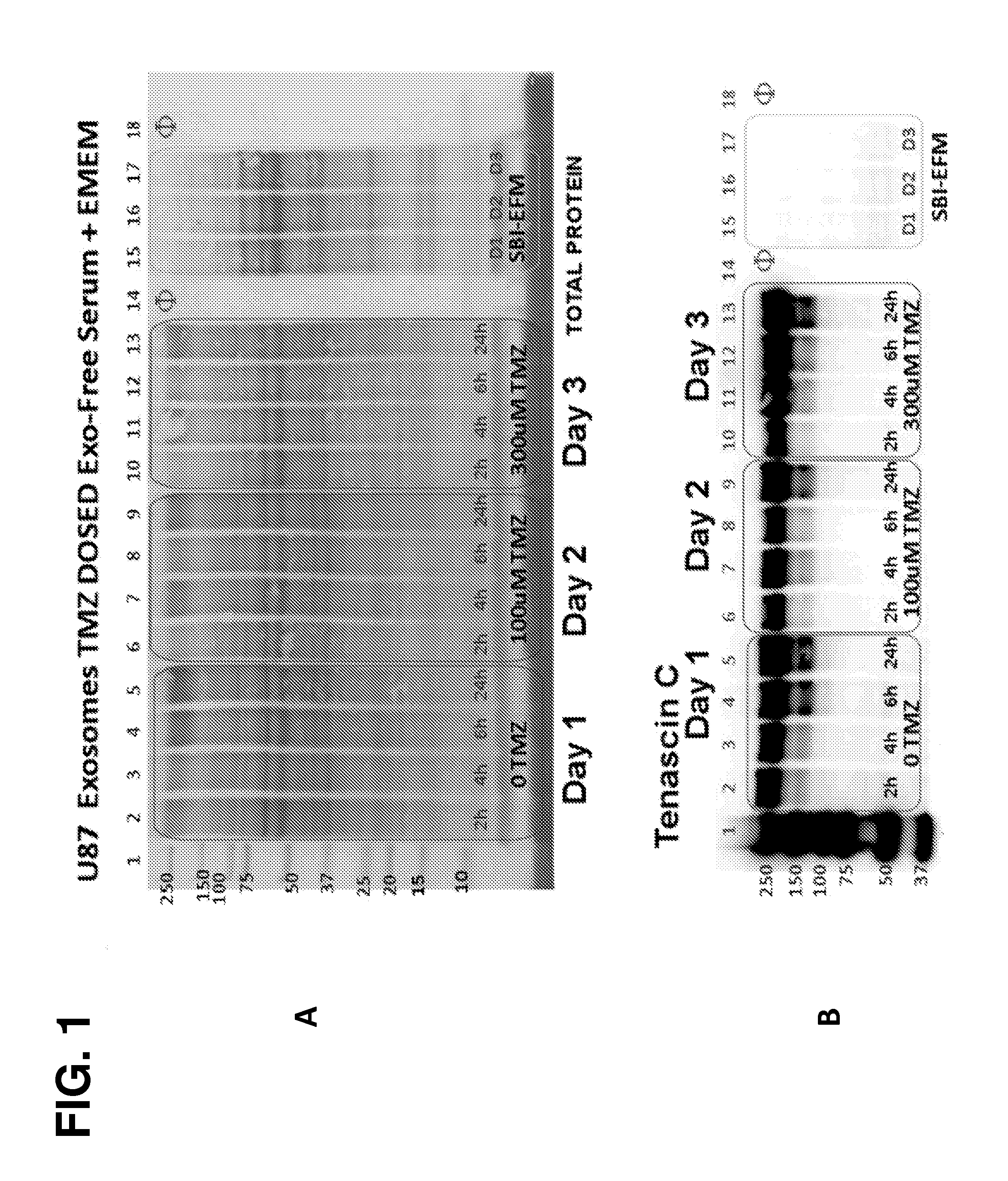
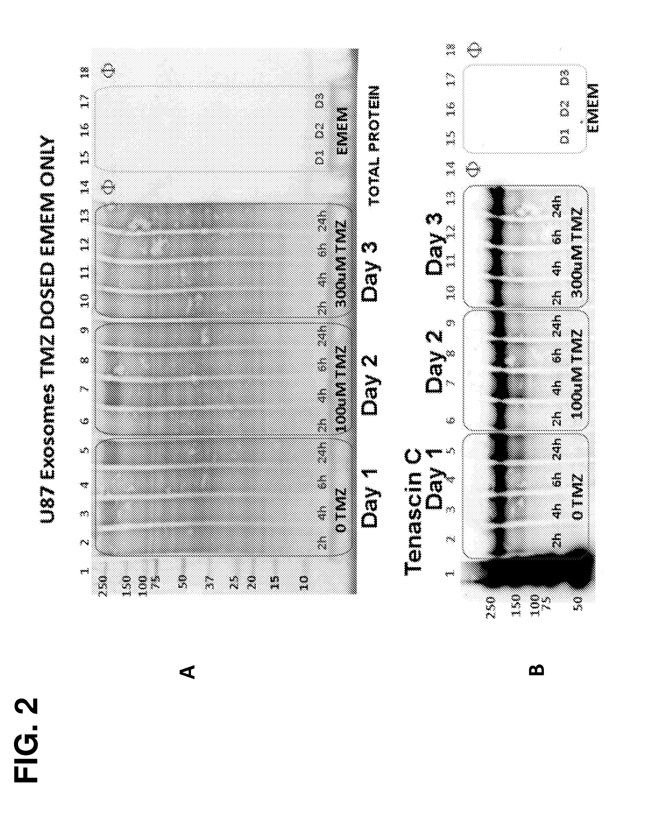
[0178]The following example demonstrates that Tenascin-C is a marker for tumor-derived microparticles. Isolation and analysis of microparticles shed from U87 brain cancer cells under normal conditions, serum-deprived conditions, or treated with the DNA alkylating agent temozolomide (TMZ) revealed robust abundance of Tenascin-C in these tumor-derived microparticles. Robust abundance of Tenascin-C persisted even in the presence of a genotoxic agent (TMZ) and growth of the U87 cells in a nutrient-deprived media.

Materials and Methods

[0179]Overview of Microparticles Harvested from Tumor Cell Lines-Experimental Outline

[0180]U87 MG (ATCC® HTB 14™) cell cultures represent glioblastoma multiforme (GBM). Two Integra CELLine Bioreactors (Hudson, N.H.) were seeded from T-75 cultures of U87 into the 15 ml lower chamber, separated by a 10 kDa MWCO filter from a 500 ml upper reservoir chamber. After growth was established in the...

example 2

Two-Step Recovery of Microparticles Bound to Size Exclusion Chromatography (SEC) Columns and Identification of Tenascin-C

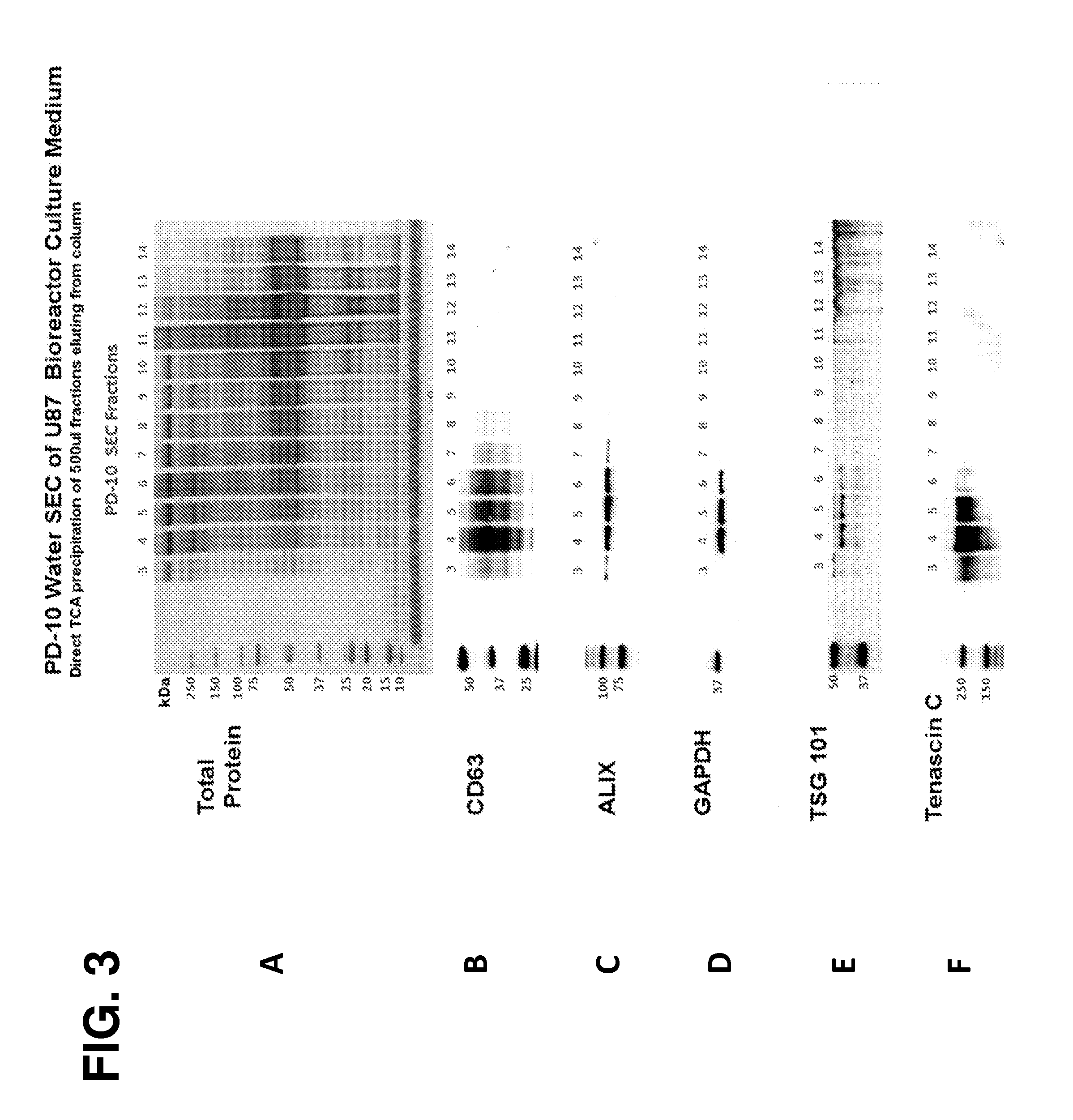
[0218]The following example demonstrates that microparticles may be effectively recovered from a serum-containing sample using a two-step purification method in a size exclusion chromatography (SEC) column. The use of water as a first mobile phase, followed by an arginine-containing solution as a second mobile phase, resulted in controlled elution and separation of microparticles from other sample components. Further, Tenascin-C was identified in the U87-derived microparticle-containing fractions eluted from the column.

Materials and Methods

[0219]PD-10 Fractionation of Serum

[0220]Microparticles to be peptide recovered were prepared by retrieving two separate, 1 ml volumes of glioblastoma U87 bioreactor culture medium from refrigerated storage (containing 0.04% sodium azide and 5 μl / ml of Protease Inhibitor Cocktail III, EMD Millipore). 10 μl of Hdn peptide stock so...

example 3

Direct Affinity Pulldown of Tenascin-C Positive Microparticles from U87 Culture Medium Incubated with Protein Solubilizing Buffer “LaurA”

[0239]The following Example demonstrates the influence of increasing concentrations of a protein solubilizing buffer (“LaurA”) on the protein profile of Tenascin-C and other antigens from affinity captured microparticles from U87 cell culture medium by either the peptide recovered microparticle (PREX) reagent Vn96, or an anti-Tenascin C monoclonal antibody (81C6).

Introduction

[0240]It was seen from Examples 1 and 2 that Tenascin-C protein was robustly associated with U87-derived microparticles, suggesting that Tenascin-C may be used as a robust extracellular marker of tumor-derived microparticles. Without wishing to be bound by theory, it is thought that exposure of Tenascin-C (TNC) on microparticles may be able to be improved by removing background protein contamination. To this end, Applicants explored the use of a protein solubilizing buffer, cal...


PUM

PropertyMeasurementUnit
pore diameteraaaaaaaaaa
densityaaaaaaaaaa

Abstract

The present disclosure provides methods for isolating tumor-derived microparticles from a subject for analysis, specifically by isolating Tenascin-C positive microparticles from a sample from the subject to obtain tumor-derived microparticles. Methods for determining the expression status of biomarkers in the tumor-derived microparticles and methods for determining additional characteristics of the tumor-derived microparticles are also provided.

Description

CROSS-REFERENCE TO RELATED APPLICATION(S)[0001]This application claims the benefit of U.S. Provisional Application No. 61 / 825,951 filed May 21, 2013, which is incorporated herein by reference in its entirety.FIELD[0002]The present disclosure relates generally to the isolation and analysis of microparticles, and more specifically to the isolation and analysis of tumor-derived microparticles.BACKGROUND[0003]Cancer is a leading cause of death worldwide. In 2004, it accounted for 7.4 million deaths (around 13% of all deaths). Deaths from cancer are expected to continue to rise, with a predicted 12 million deaths in 2030 (WHO, February 2009). Early detection of cancer and monitoring of the progression of cancer are critical to reducing the worldwide cancer burden, yet the development of robust methods and tools for early detection and monitoring remain major challenges in the field. Furthermore, a large proportion of patients with cancer are over-treated, resulting in wasted time and exp...

Claims


Application Information

Patent Timeline
Patent Type & Authority Applications(United States)
IPC IPC(8): G01N33/574
CPCG01N33/57488G01N2333/4703G01N33/57407G01N2333/78
Inventor EZRIN, ALAN M.GRIFFITHS, STEVEN G.
Owner NX PHARMAGEN
Why Eureka
Try Eureka