Methods for phenotyping of intact bones by tissue clearing and staining

a technology of intact bones and tissue, applied in the field of tissue preparation and characterization, can solve the problems of large damage to tissue samples, physical challenges, and inability to easily access three-dimensional information in most mammalian osseous tissue,

Inactive Publication Date: 2016-05-12
CALIFORNIA INST OF TECH
View PDF1 Cites 7 Cited by
  • Summary
  • Abstract
  • Description
  • Claims
  • Application Information

AI Technical Summary

Benefits of technology

[0006]In various embodiments, the invention teaches a method, including: applying a fixing solution to a tissue including a bone of a subject; applying a hydrogel monomer solution to the tissue including the bone of the subject; applying a detergent solution to the tissue including the bone of the subject; and applying a solution including a calcium chelating agent to the tissue including the bone of the subject. In some embodiments, the method further includes applying a solution including an amino alcohol to the tissue the bone of the subject. In some embodiments, the solution including the amino alcohol includes N,N,N′,N′-Tetrakis(2-Hydroxypropyl)ethylenediamine. In some embodiments, the calcium chelating agent includes ethylenediaminetetraacetic acid (EDTA) and/or ethylene glycol tetraacetic acid (EGTA). In some embodiments, the hydrogel monomer solution includes 2-8% acrylamide. In some embodiments, the method further includes applying a photoinitiator solution including 2,2′-Azobis[2-(2-imidazolin-2-yl)propane]dihydrochloride to the tissue including the bone of the subject. In some embodiments, the method further includes placing the tissue including the bone of the subject into a substantially air-tight chamber, and introducing nitrogen into the substantially air tight chamber, thereby forming a de-gassed tissue including bone. In certain embodiments, the detergent solution includes 6-15% sodium dodecyl sulfate (SDS). In certain embodiments, the method further includes serially incubating the tissue including the bone of the subject in refractive index matching solutions (RIMS) with progressively higher refractive indexes (RIs), wherein the final RIMS in which the tissue including the bone of the subject is incubated has an RI of 1.46-1.52 or 1.38-1.46. In some embodiments, the tissue including the bone of the subject is first incubated in a RIMS of a first RI, thereby forming a primary RIMS treated tissue including bone, and the primary RIMS treated tissue including bone is subsequently incubated in a second RIMS with a second RI, wherein the second RI is higher than the first RI, thereby forming a secondary RIMS treated tissue including bone. In certain embodiments, the method further includes incubating the secondary RIMS treated tissue including bone in a third RIMS, thereby forming a tertiary RIMS treated tissue including bone, wherein the third RIMS has a higher RI than the first and second RIMS. In some embodiments, the first RIMS in which the tissue including the bone of the subject is incubated has an RI of 1.38-1.44. In some embodiments, the second RIMS in which the tissue including bone is incubated has an RI of 1.44-1.48. In certain embodiments, the tissue including bone is incubated in a third RIMS that has an RI of 1.47-1.52. In

Problems solved by technology

Due to its calcified nature, most mammalian osseous tissue does not afford easy access to three-dimensional information.
Bone-sectioning, howev

Method used


Image

  • Methods for phenotyping of intact bones by tissue clearing and staining
  • Methods for phenotyping of intact bones by tissue clearing and staining
  • Methods for phenotyping of intact bones by tissue clearing and staining

Examples


example 1

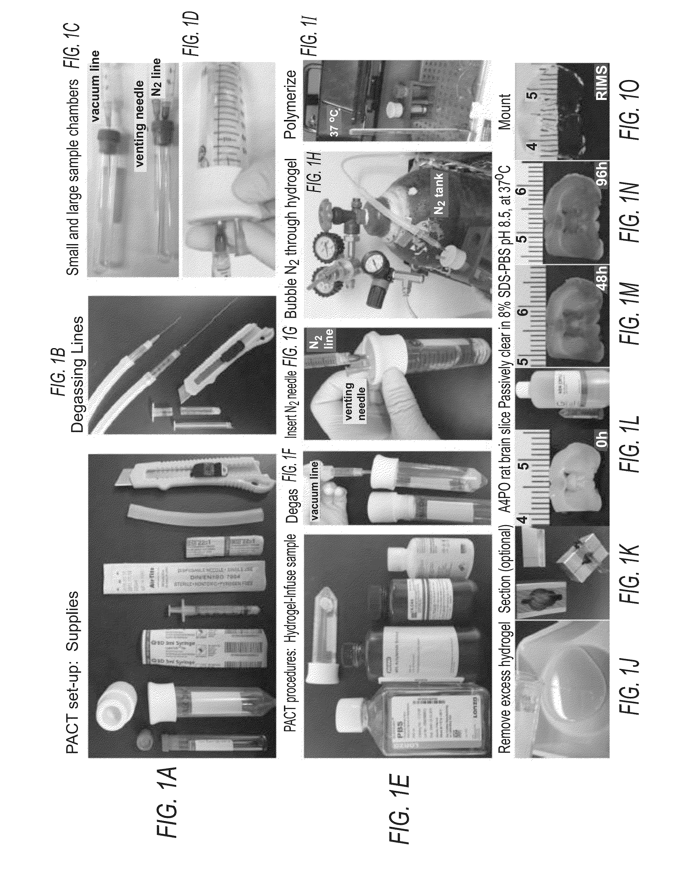
Tissue Preparation for PACT

[0057]A bone containing sample (“bone sample”) to be PACT processed is excised. The bone sample is post-fixed in 4% PFA for 1-2 hours at RT with gentle agitation on a rocking platform shaker. If desired, the sample can be post-fixed overnight at 4° C. Fixing samples for extended periods of time may result in over-fixation and antigen masking.

Formation of a Tissue-Hydrogel Matrix

[0058]The polymerization of tissue components with hydrogel monomers is important as it ensures that SDS micelles preferentially solubilize and remove tissue lipids during clearing. It was previously determined that a minimal acrylamide-based network, which supports more rapid clearing, was nevertheless sufficient for stabilizing proteins and nucleic acids. To increase the level of crosslinking without the addition of bis-acrylamide or PFA to the hydrogel monomer solution, the hydrogel-infused tissue can be carried through rigorous degassing steps.

Hydrogel-Embedding of PACT Samples

[...

example 2

Reagents and Setup

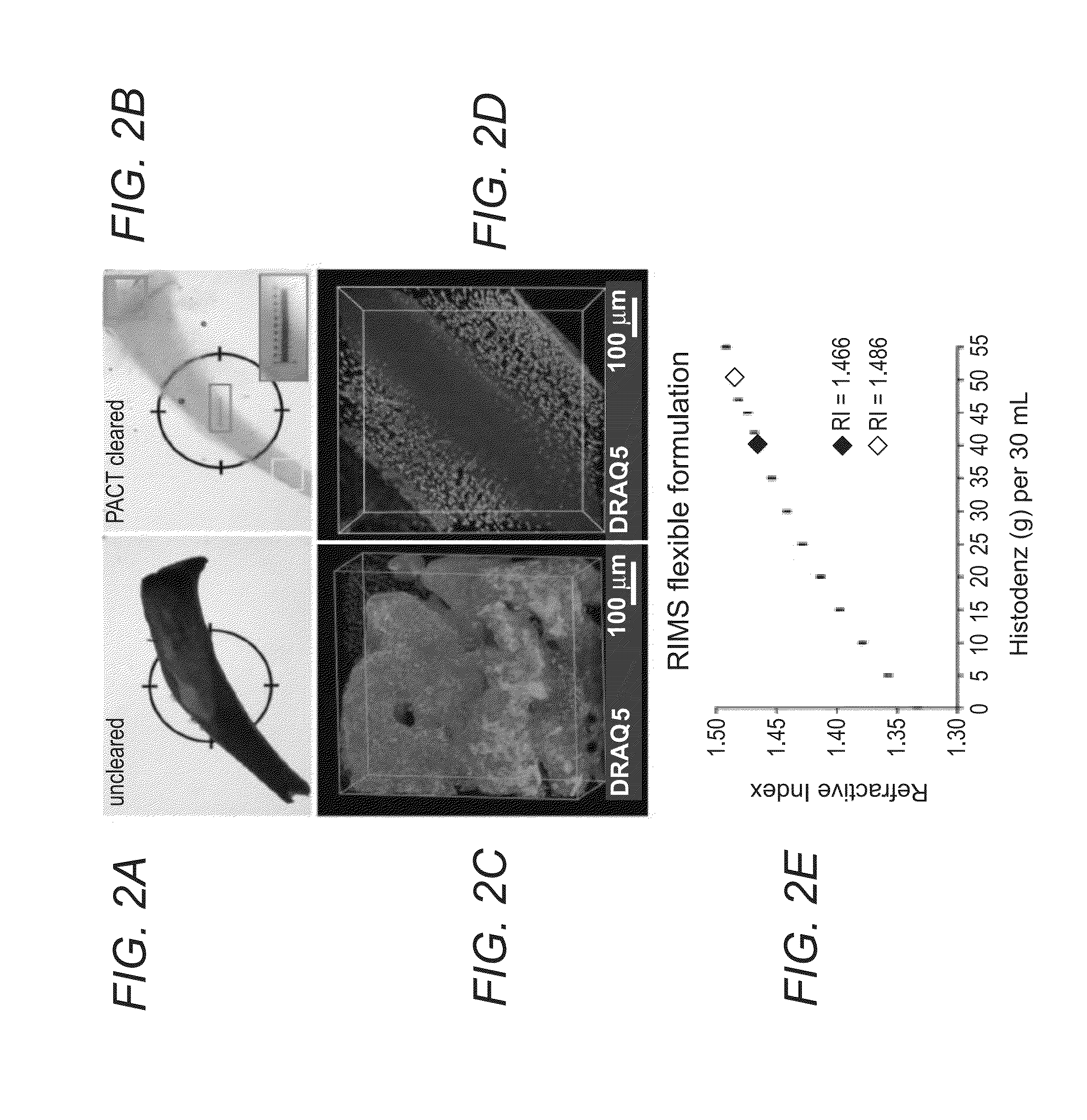
Refractive Index Matching Solution (RIMS)

[0071]Histodenz™ (Sigma-Aldrich, cat. no. D2158)[0072]0.02 M Phosphate buffer[0073]Sodium azide (Fisher Scientific, cat. no. 71448-16)

[0074]To prevent microbial growth, sodium azide may be added to all mounting medias (RIMS and sRIMS), as well as to all immunostaining dilutions and wash buffers that are used in extended incubations.

Sorbitol-Based Refractive Index Matching Solution (sRIMS)[0075]70% Sorbitol (Sigma-Aldrich, cat. no. 309532)[0076]0.02 M Phosphate buffer[0077]Sodium azide (Fisher Scientific, cat. no. 71448-16)

Refractive Index Matching Solution for Cold Storage (cRIMS)[0078]Histodenz™ (Sigma-Aldrich, cat. no. D2158)[0079]0.005 M Phosphate buffer[0080]Sodium azide (Fisher Scientific, cat. no. 71448-16)

Immersion Media and Alternative Mounting Media

[0081]There are numerous commercial and home-made RIMS alternatives, including FocusClear, Cargille Labs optical liquids, 2,2′-thiodiethanol, and diluted glycerol.

Glycero...


PUM


Abstract

In various embodiments, the present application teaches methods and kits for clearing and optionally subsequently visualizing tissue containing bone. In some embodiments, the method includes serially incubating cleared bone in refractive index matching solutions with progressively higher refractive indexes. In some embodiments, the methods teach immunolabeling and/or staining tissue containing bone and optionally visualizing the immunolabeled and/or stained tissue containing bone.

Description

CROSS REFERENCE TO RELATED APPLICATION[0001]This application claims priority under 35 U.S.C. §119(e) from U.S. Provisional Patent Application No. 62 / 076,705, filed Nov. 7, 2014, the content of which is hereby incorporated herein by reference in its entirety.STATEMENT REGARDING FEDERALLY-SPONSORED RESEARCH[0002]This invention was made with government support under Grant Nos. IDP20D017782-01 and 1R01AG047664-01 awarded by the National Institutes of Health. The government has certain rights in the invention.FIELD OF THE INVENTION[0003]The present invention generally relates to the field of tissue preparation and characterization.BACKGROUND[0004]The following description includes information that may be useful in understanding the present invention. It is not an admission that any of the information provided herein is prior art or relevant to the presently claimed invention.[0005]Due to its calcified nature, most mammalian osseous tissue does not afford easy access to three-dimensional ...

Claims


Application Information

Patent Timeline
IPC IPC(8): G01N1/30
CPCG01N2001/302G01N1/30
Inventor GRADINARU, VIVIANAYANG, BINTREWEEK, JENNIFERCHAN, KEN
Owner CALIFORNIA INST OF TECH
Why Eureka
Try Eureka