
Methods of treating pain using protein solutions

a protein solution and pain technology, applied in the field of pain treatment, can solve the problems of affecting the quality of life, affecting the emotional well-being and ability, and limiting the perception of pain, so as to reduce the pain experienced, prevent, reduce or eliminate the effect of pain

Active Publication Date: 2016-05-19
BIOMET BIOLOGICS
View PDF0 Cites 15 Cited by
  • Summary
  • Abstract
  • Description
  • Claims
  • Application Information

AI Technical Summary

Benefits of technology

The patent describes a method for treating pain by injecting an anti-inflammatory composition into the subject. The method involves measuring a therapeutic indicator in the subject's blood or a fraction of blood, and administering the anti-inflammatory composition if the therapeutic indicator is equal to or above a threshold level. The anti-inflammatory composition includes a protein solution with interleukin-1 receptor antagonist (IL-1ra) and interleukin-1 (IL-1), which can prevent, reduce, or eliminate pain. The method can also involve producing an autologous protein solution (APS) by contacting a volume of blood or a fraction of blood obtained from the subject with a solid extraction material, and administering the APS to the subject if the therapeutic indicator is equal to or above a threshold level of the select therapeutic indicator. The APS is effective for preventing, reducing, or eliminating pain.

Problems solved by technology

However, pain—particularly chronic pain—can significantly interfere with quality of life, including emotional well-being and ability to work.
Thus acute pain is typically transient, and self-limiting.
Many treatments focus on removing the underlying pain stimulus, while others block the perception of pain.
However, many such treatments may present side effects, and may have limited long term utility as underlying conditions become worse.
However, as with any therapy, such treatments may not always be effective for specific patients.
Some patients experiencing pain may undergo a medical procedure that fails to alleviate their pain.
Such unsuccessful procedures are discouraging to the patients, and may create unnecessary costs, delays in pursuing alternative therapies, and other potential risks than can be inherent in any medical procedure.
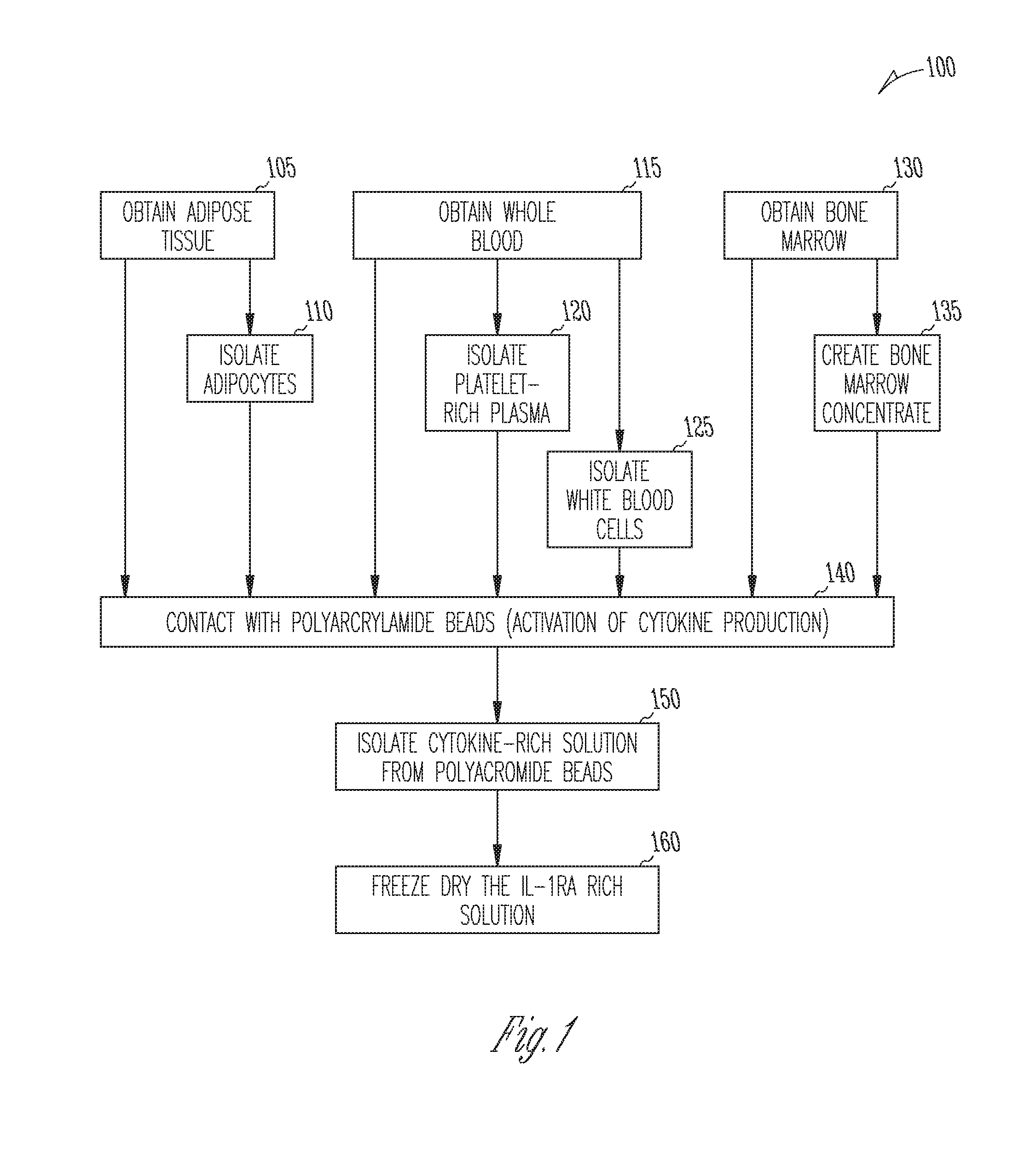
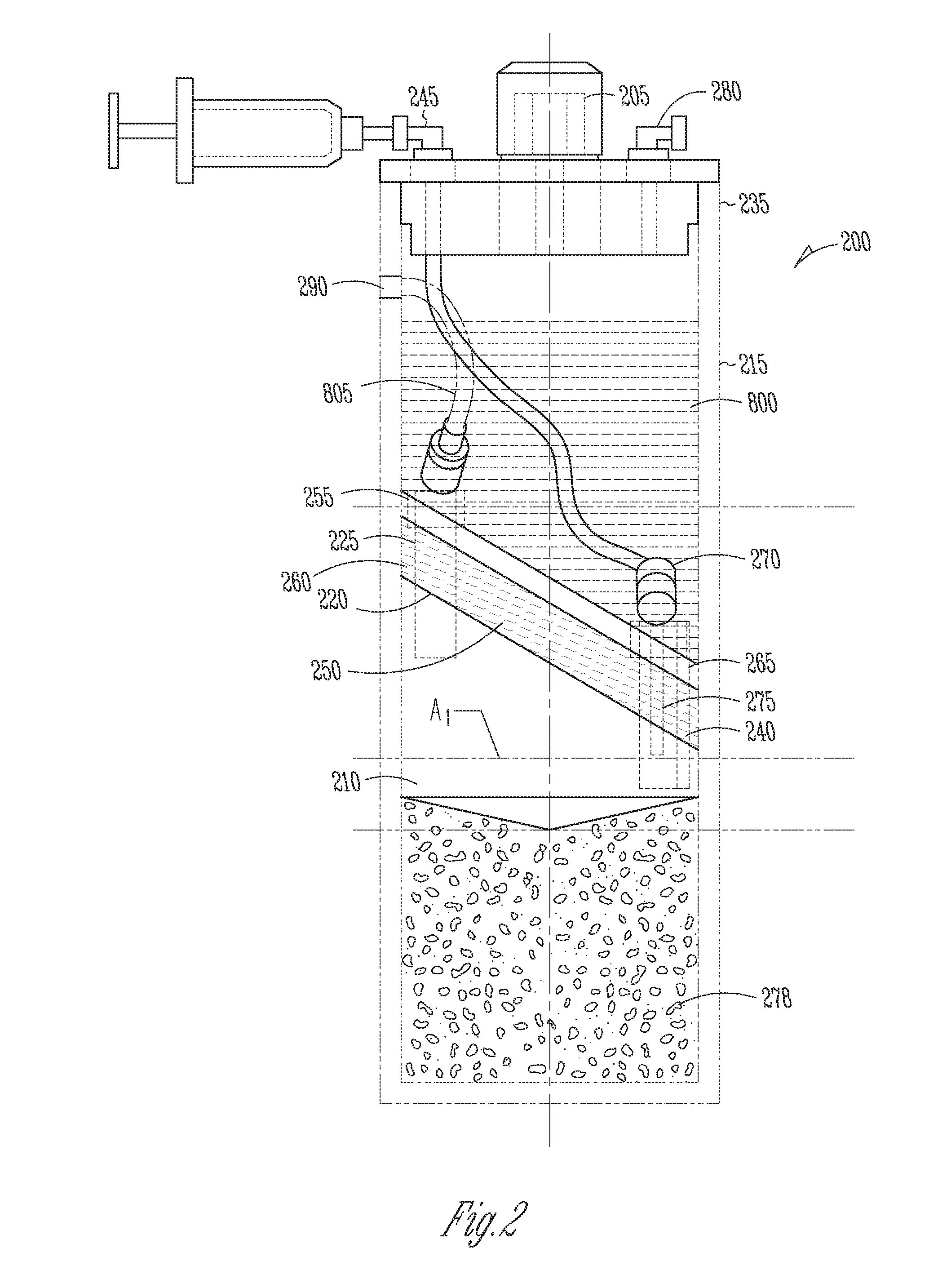
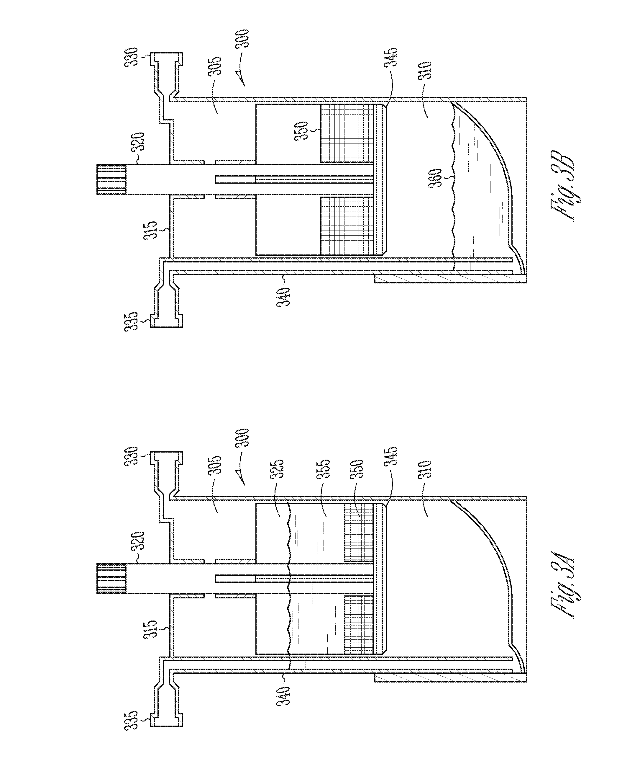
These figures may not precisely reflect the characteristics of any given embodiment, and are not necessarily intended to fully define or limit specific embodiments within the scope of this technology.

Method used


Image

  • Methods of treating pain using protein solutions
  • Methods of treating pain using protein solutions
  • Methods of treating pain using protein solutions

Examples


example 1

Preparing and Characterizing an Anti-Inflammatory Composition

[0224]An anti-inflammatory protein solution composition rich in interleukin-1 receptor antagonist is prepared from seven consented human providers. Blood (55 mL) is drawn into a 60 cc syringe with 5 mL of anticoagulant citrate dextrose solution A (ACD-A, Citra Labs, Braintree, Mass.). Platelet-rich plasma (PRP) is created using the GPS® III platelet concentration system (800-1003A, Biomet Biologics, Warsaw, Ind.) according to the instructions for use. The solution is generated by adding 6 mL of PRP to a modified Plasmax® device containing 1 gram of polyacrylamide beads (Biomet Biologics, Warsaw, Ind.). The IL-1ra solution is removed from the Plasmax® devices and frozen at minus 50° C. for the assay. Cytokine content is assayed on a 16-plex ELISA (Searchlight Protein Array, Aushon Biosystems, Billerica, Mass.). The analytes included IL-4, IL-10, IL-11, IL-13, IL-1ra, IFN-γ, sTNF-RI, sTNF-RII, IL-1α, IL-1β, TNF-α, IL-17, IL-...

example 2

Generation of IL-1ra from Platelet-Rich Plasma

[0226]An IL-1ra-rich solution is created as follows. Whole blood (70 mL) anticoagulated (10%) with ACD-A (Braintree, Mass., USA) is drawn from 5 healthy volunteers. A portion (10 mL) is reserved for a whole blood measurement. Platelet-rich plasma (PRP) (6 mL) is produced using the GPS® II System (Biomet Biologics, LLC, Warsaw, Ind., USA). Complete blood counts are collected for the whole blood and PRP samples following a validated procedure, as described in Woodell-May J E, Ridderman D N, Swift M J, Higgins J. “Producing Accurate Platelet Counts for Platelet Rich Plasma: Validation of a Hematology Analyzer and Preparation Techniques for Counting” J. Craniofac. Surg. (2005) September 16(5):749-56.

[0227]Following the PRP production, 5 mL of the PRP is added to a modified plasma concentration device containing 1 gram of polyacrylamide beads (Plasmax® device, Biomet Biologics LLC, Warsaw, Ind., USA) and incubated with polyacrylamide desiccat...

example 3

Production of Protein Solution from PRP

[0231]Anticoagulated blood (120 cc) is collected from 5 human donors. Platelet-rich plasma (PRP) is prepared using GPS® III disposables (Biomet Biologics LLC, Warsaw, Ind., USA). PRP is loaded into modified plasma concentration devices containing 1 gram of polyacrylamide beads (Plasmax® device, Biomet Biologics LLC, Warsaw, Ind., USA) and processed. The output is divided into 4 groups: IL-1ra in concentrated plasma with and without thrombin activation (1000 U / mL in 1M CaCl2), or cell-free IL-1ra with and without thrombin activation. IL-1ra is measured using ELISA (R&D Systems) over time.

[0232]The PRP contacts polyacrylamide beads in the Plasmax® device while electromagnetic field stimulation is provided using a capacitively coupled electromagnetic field.

[0233]Unclotted PRP produces an average of about 50 ng over 24 hrs. The cell-free samples produce about 34 ng without changing over 24 hrs. Once clotted, the elution of IL-1ra is slowed, with on...


PUM

PropertyMeasurementUnit
Concentrationaaaaaaaaaa
Densityaaaaaaaaaa
Concentrationaaaaaaaaaa

Abstract

Methods and compositions for treating pain in mammalian subjects are provided. The methods include obtaining blood or a fraction of blood from the subject, measuring a therapeutic indicator in the blood or in the fraction blood, and administering an anti-inflammatory composition to the subject if the therapeutic indicator is equal to or above a threshold level. The anti-inflammatory composition comprises a protein solution including interleukin-1 receptor antagonist. The protein solution may be prepared from the blood or blood fraction obtained from the subject.

Description

CLAIM OF PRIORITY[0001]This application claims the benefit of U.S. Provisional Patent Application Ser. No. 62 / 077,396, filed on Nov. 10, 2014, the benefit of priority of which is claimed hereby, and which is incorporated by reference herein in its entirety.INTRODUCTION[0002]The present technology relates to methods of treating pain, including pain associated with injury and inflammatory disorders. In particular, methods comprise determining whether a subject experiencing pain is likely to experience less pain after administration of a solution comprising anti-inflammatory cytokines, including such solutions derived from blood and other tissues.[0003]Pain can be described as an unpleasant sensation associated with actual or potential tissue damage or disease, and is the most common reason for consultations with physicians in the United States. On a basic level, pain serves as a warning mechanism to the body, to avoid or minimize exposure to potentially harmful environmental or physio...

Claims


Application Information

Patent Timeline
IPC IPC(8): A61K38/20G01N33/50G01N33/68A61K38/17A61K38/18
CPCA61K38/2006A61K38/1793A61K38/1841A61K38/18G01N2800/2842A61K38/1858G01N33/6869G01N33/5094G01N2333/545A61K38/1866A61K35/15A61K2300/00
Inventor TOLER, KRISTAKING, WILLIAMWOODELL-MAY, JENNIFER E.
Owner BIOMET BIOLOGICS
Learn More