Novel hemostatic patch and uses thereof

a technology of hemostatic patch and patch, which is applied in the field of new hemostatic patch and hemostatic patch, can solve the problems of difficult to locate wounds, unique problems of internal tissue wounds, and time lost from treating the actual point of trauma, and achieve the effect of controlling or arresting bleeding in patients

Inactive Publication Date: 2016-06-02
RUBIN LEO +1
View PDF3 Cites 2 Cited by
  • Summary
  • Abstract
  • Description
  • Claims
  • Application Information

AI Technical Summary

Benefits of technology

[0075]In one aspect, the present invention provides for methods of using the hemostatic patch, e.g. as described throughout, e.g., any Hemostatic Patch of A-L, to control or arrest bleeding internally. For example, one advantage of the present invention described herein, e.g., any Hemostatic Patch of A-L, is that it can be used to control or arrest internal organ or tissue bleeding directly at the site of bleeding or trauma to the organ or tissue. In addition, the present invention also contemplates that the hemostatic patch may be used to control or arrest bleeding caused by damage to arteries. It is contemplated by the present invention that the arterial damage could have been caused by various catheterization procedures.
[0076]In one aspect the present invention described herein, e.g., any Hemostatic Patch of A-L, may be placed directly at the source of bleeding. In one aspect of the present invention the internal bleeding can be detected via endoscopic means. It is an advantage of the present invention that the hemostatic patch may be endoscopically placed on a wound in order to control or arrest bleeding in a patient.
[0077]In one aspect, the present invention described herein, e.g., any Hemostatic Patch of A-L, may be in the form of a spray or gel, or any other scaffold known in the art, and may be used during open, endoscopic or catheter-based procedures or surgeries. Patch compositions may be in the form of a spray or gel and include polyethylene glycol-based systems. It is contemplated that the vasocontrictant compound may be crosslinked to the gel. In one aspect the patch may be applied as a composition that forms a crosslinked gel when placed on the intended application site.
[0078]The present invention also provides methods for treating bleeding, e.g. arterial damage, internal organ damage, comprising placing a hemostatic patch as described herein, e.g., any Hemostatic Patch of A-L, over the injured area wherein said hemostatic patch comprises an effective amount of a mucoadhesive and an effective amount of a compound that causes vasoconstriction.
[0079]In one aspect, the present invention provides for a method for treating bleeding, e.g. arterial damage, internal organ damage, comprising placing a hemostatic patch as described herein, e.g., Hemostatic Patch A-L, over the injured area. In one aspect, hemostatic patch as described herein, e.g., any Hemostatic Patch of A-L, comprises nanoparticles which comprise chitosan and Neuropeptide Y. In another aspect the hemostatic patch comprises chitosan and epinephrine.
[0080]In one aspect the present invention described herein, e.g., any Hemostatic Patch of A-L, may be used to treat external patient bleeding as well.

Problems solved by technology

Despite the products that have been made available, and the techniques now known, the control or arrest of patient bleeding, by virtue of the most minimally invasive means achievable, still remains a unique and salient issue for many physicians.
For example, internal tissue wounds present unique problems that must be addressed when attempting to close such wounds.
Where there is bleeding within the field of injury around the wound, consequently this can cause the wound to be difficult to locate.
The time lost from treating the actual point of trauma can, in some instances, be life-threatening depending upon the amount of blood loss a patient may incur as a result of lost time.
Moreover, because access to an internal wound sometimes requires open surgery this can necessarily cause risk to the patient.
However, consistent and reliable closure of a wound using only a single instrument is difficult to achieve.
Furthermore, localized vascular complications associated with catheterization of a vein or an artery is often times the common side effect of many medical procedures.
Such complications including hemorrhaging of blood vessels, delayed homeostasis time, hematoma, pseudoaneurysm, and arteriovenous (AV) fistula formation can be life threatening Since cardiac catheterization, for example, remains the primary technique for diagnosing coronary artery disease and is used to help delineate coronary anatomy, the information gained is often a medical necessity, despite the risks.
Traditionally, the application of pressure to a vein or artery after cardiac catheterization can be very painful for the patients.
However, patients are then at risk to develop back pain and urinary retention in addition to the localized vascular complications.
One problem with certain presently available hemostatic patches occurs because, while they may be effective at the interface of the wound and the patch, hemostatic patches have not been demonstrated to be effective at a distance in treating, for example, punctures caused by catheterization that may arise due to damage or trauma that arise from deeper internal puncture in the vein or artery.
However, sutures may cause additional trauma to the wound site and are time consuming to replace.
However, surgical staples may also impose additional wound trauma and require the use of ancillary and often expensive devices for positioning and applying the staples.
In addition, it may be the case where serious bleeding exists, for example, that endoscopically stapling a wound does not function to arrest arterial bleeding.
Therefore, because the wound continues to bleed even after the endoscopic insertion of a staple, open surgery may still be required.
Such surgery is necessarily more invasive, and may be more painful and entail a greater risk of infection than if a physician were able to simply arrest bleeding at the point of injury through some other less invasive means.

Method used


Image

  • Novel hemostatic patch and uses thereof
  • Novel hemostatic patch and uses thereof
  • Novel hemostatic patch and uses thereof

Examples


example 1

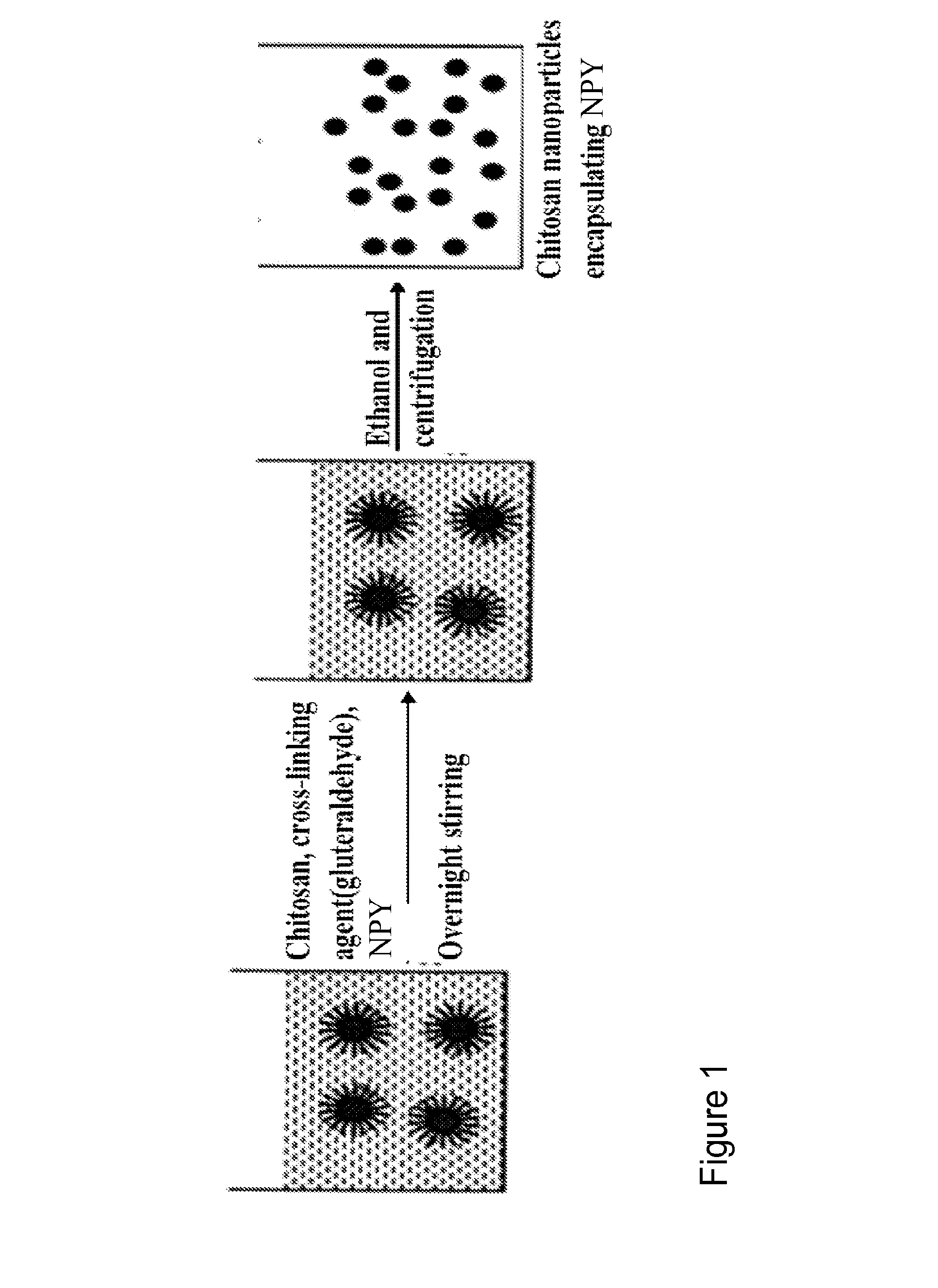
Synthesis of NPY Encapsulated Nanoparticles

[0120]Chitosan nanoparticles encapsulating NPY are produced using a reverse micellar method. Chitosan polymer and NPY are added to 0.1M AOT / hexane (AOT-Aerosol OT is used as a surfactant) solution to form reverse micelles. Bifunctional reagent gluteraldehyde is added to this reverse micelles system as a cross-linking agent. The chemical cross-linking of chitosan polymers with gluteraldehyde occurs by Schiff's reaction of aldehyde groups on gluteraldehyde and amino groups on the chitosan chain. Finally nanoparticles are separated out by high speed centrifugation.

[0121]In these examples, nanoparticles are optimized as to size, and entrapment efficiency to get an optimum formulation with maximum loading.

example 2

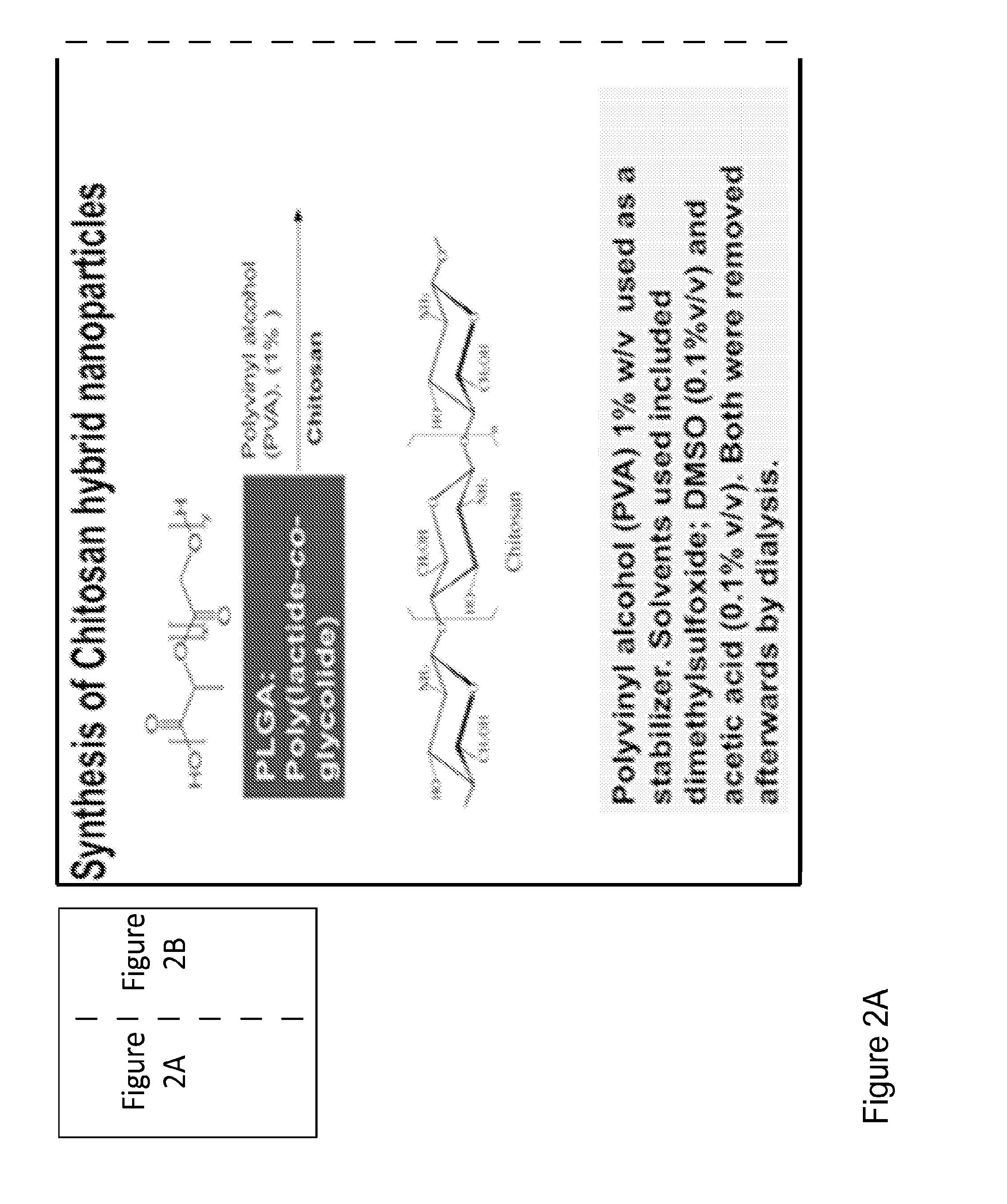
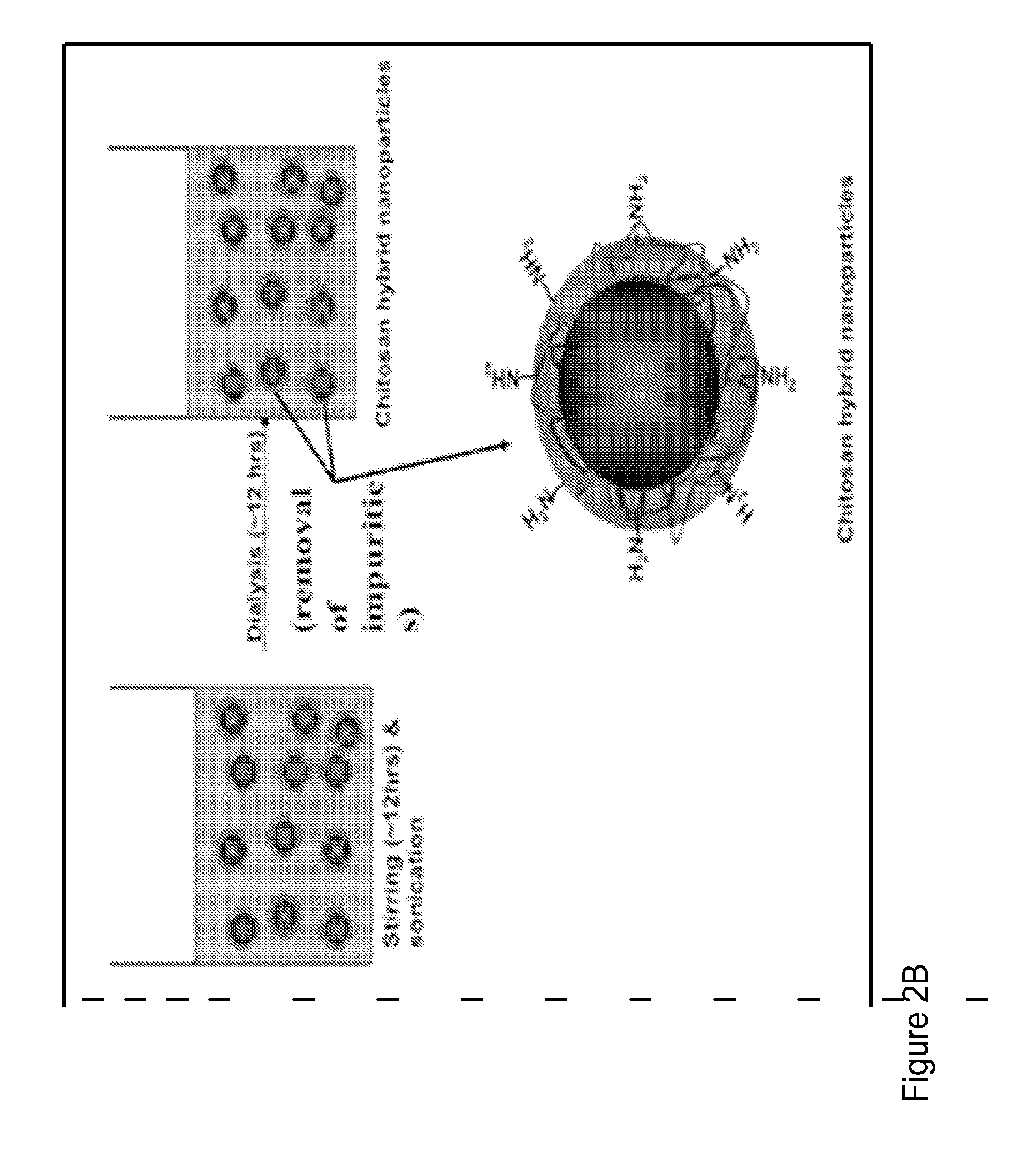
Synthesis of Chitosan-PLGA Nanoparticles

[0122]FIGS. 3A and 3B depict the synthesis and preparation of chitosan-PLGA hybrid nanoparticles with and without VIP. In FIG. 3A, PLGA is mixed with chitosan and PVA(1%) in an overnight stirring and sonication step. Subsequently the mixture undergoes a dialysis step to remove impurities. PVA is used as a stabilizer, while DMSO (0.1% v / v) and acetic acid (0.1% v / v) were incorporated as solvents. These may be removed by the subsequent dialysis step. FIG. 3B, PLGA, NPY, chitosan, and gluteraldehyde are mixed together, for approximately twenty-four hours, in a stirring and sonication step. Subsequently the mixture undergoes a dialysis step to remove impurities. The result is a PLGA-chitosan nanoparticle, wherein the chitosan layer is cross-linked with gluteraldehyde.

[0123]NPY encapsulated in nanoparticles with different degrees of cross-linking is tested for optimal pharmacokinetics. The formulation is optimized for loading efficiency. The ratios...

example 3

Synthesis of Epinephrine Encapsulated Nanoparticles

[0124]Chitosan nanoparticles encapsulating epinepherine are produced using a reverse micellar method. Chitosan polymer and epinephrine are added to 0.1M AOT / hexane (AOT-Aerosol OT is used as a surfactant) solution to form reverse micelles. Bifunctional reagent gluteraldehyde is added to this reverse micelles system as a cross-linking agent. The chemical cross-linking of chitosan polymers with gluteraldehyde occurs by Schiff's reaction of aldehyde groups on gluteraldehyde and amino groups on the chitosan chain. Finally nanoparticles are separated out by high speed centrifugation.

[0125]In these examples, nanoparticles are optimized as to size, and entrapment efficiency to get an optimum formulation with maximum loading.


PUM

PropertyMeasurementUnit
apparent viscosityaaaaaaaaaa
diameteraaaaaaaaaa
diameteraaaaaaaaaa

Abstract

Disclosed herein is a novel hemostatic patch that may be used to control and / or arrest bleeding in patients. The patch offers an effective but also minimally invasive way to control and / or arrest bleeding in a patient. The patch comprises a mucoadhesive and a compound that causes vasoconstriction. In a preferred aspect, the patch comprises chitosan and Neuropeptide Y. Also disclosed are methods of using the novel hemostatic patch.

Description

CROSS REFERENCE TO RELATED APPLICATIONS[0001]This application claims the benefit of U.S. Provisional Application 61 / 514,587 filed Aug. 3, 2011, the contents of which are incorporated herein by reference.TECHNICAL FIELD[0002]The field relates generally to a hemostatic device. The field further relates to methods for using said hemostatic device. The field further relates to a hemostatic patch that comprises both a mucoadhesive and a compound that causes vasoconstriction. Specifically, the field relates to a hemostatic patch that comprises chitosan and a vasoconstrictant, e.g., neuropeptide Y, epinephrine.BACKGROUND OF THE INVENTION[0003]Despite the products that have been made available, and the techniques now known, the control or arrest of patient bleeding, by virtue of the most minimally invasive means achievable, still remains a unique and salient issue for many physicians. In particular, physicians still seek some comprehensive, effective, and minimally invasive means in order t...

Claims


Application Information

Patent Timeline
Patent Type & Authority Applications(United States)
IPC IPC(8): A61L15/28A61L15/44A61L15/20A61L15/58A61L15/64A61L15/26
CPCA61L15/28A61L15/64A61L15/26A61L15/20A61L2300/204A61L15/44A61L2400/04A61L2300/62A61L15/58A61K9/0024A61K47/36A61K9/5153A61K9/5161A61K9/7007A61K31/137A61L2300/418A61K38/2271C08L5/08A61K9/1647A61K9/1652A61K9/703A61L15/225A61L2300/252C08B37/003
Inventor RUBIN, LEOMOUSA, SHAKER
Owner RUBIN LEO
Why Eureka
Try Eureka