Method of distinguishing mesenchymal stem cells

Active Publication Date: 2016-06-09
MERIDIGEN BIOTECH
View PDF0 Cites 1 Cited by
  • Summary
  • Abstract
  • Description
  • Claims
  • Application Information

AI Technical Summary

Benefits of technology

This patent provides a way to get a purer population of cells from a placenta-related tissue. This is done by using a surface marker called EphA2. This can help to improve the quality of the research being done with these cells.

Problems solved by technology

However, the isolation procedure is extremely invasive.
Due to the heterogeneous nature and the absence of known biomarkers specific for mesenchymal stem cells, it is a challenging task to define MSC phenotypes and characteristics [52-54].
In addition, lack of specific cell surface markers renders the cell culture at potential contamination risk with other cell types, in particular, those mature stromal cells such as fibroblasts, which, conversely, are abundant in mesenchymal tissue [52-54].
In particular, fibroblast is the main source of contamination.
Not only is it difficult to apply techniques which successfully eliminate fibroblasts from a culture, it is also particularly complex to distinguish MSCs from fibroblasts in the same culture.
The current definition suggested by ISCT is thus incapable of distinguishing MSC from generic fibroblasts.
However, the mechanism of Eph in stem cell niche maintenance and its role in regulating stem cells are not well understood.
However, in view of that Vescovi's study entirely and only focused on human glioblastomas (hGBMs) and the facts that EphA2[Low] hGBM still possess significant tumor-initiating capability, one skilled in the art would have in no way recognized that EphA2 is a specific marker competent in identifying multipotent stem cells.

Method used


Image

  • Method of distinguishing mesenchymal stem cells
  • Method of distinguishing mesenchymal stem cells
  • Method of distinguishing mesenchymal stem cells

Examples


example 1

The Immunophenotypic Characterization of the Placenta-Derived Mesenchymal Stem Cells (MSCs) and Fibroblasts

[0042]Full-term placentas (n=8) were collected after obtaining written informed consent from donors. MSCs were derived from amniotic membrane (AM), chorionic disk (CD), chorionic membrane (CM), and umbilical cord (UC). Placenta-derived cells were cultured, expanded and maintained in α-MEM with FBS and basic FGF at 37° C., saturating humidity and 5% CO2, and were sub-cultured when cells reached 80% confluence, later phenotypically characterized by flow cytometry. In the process of immunostaining subject to flow cytometry, cells were incubated with the antibodies following manufacturer's instructions. Nonspecific IgG of the corresponding class served as the negative control. Cell suspensions were analyzed on a flow cytometer (BD Biosciences FACSCanto II) with Flowjo 7.6.1 software.

[0043]We assessed expression of CD11b, CD19, CD34, CD45, CD73, CD90, CD105, HLA-DR, and EphA2. Flow ...

example 2

The Immunophenotypic Characterization of the EphA2-Sorted Placenta-Derived Mesenchymal Stem Cells (MSCs)

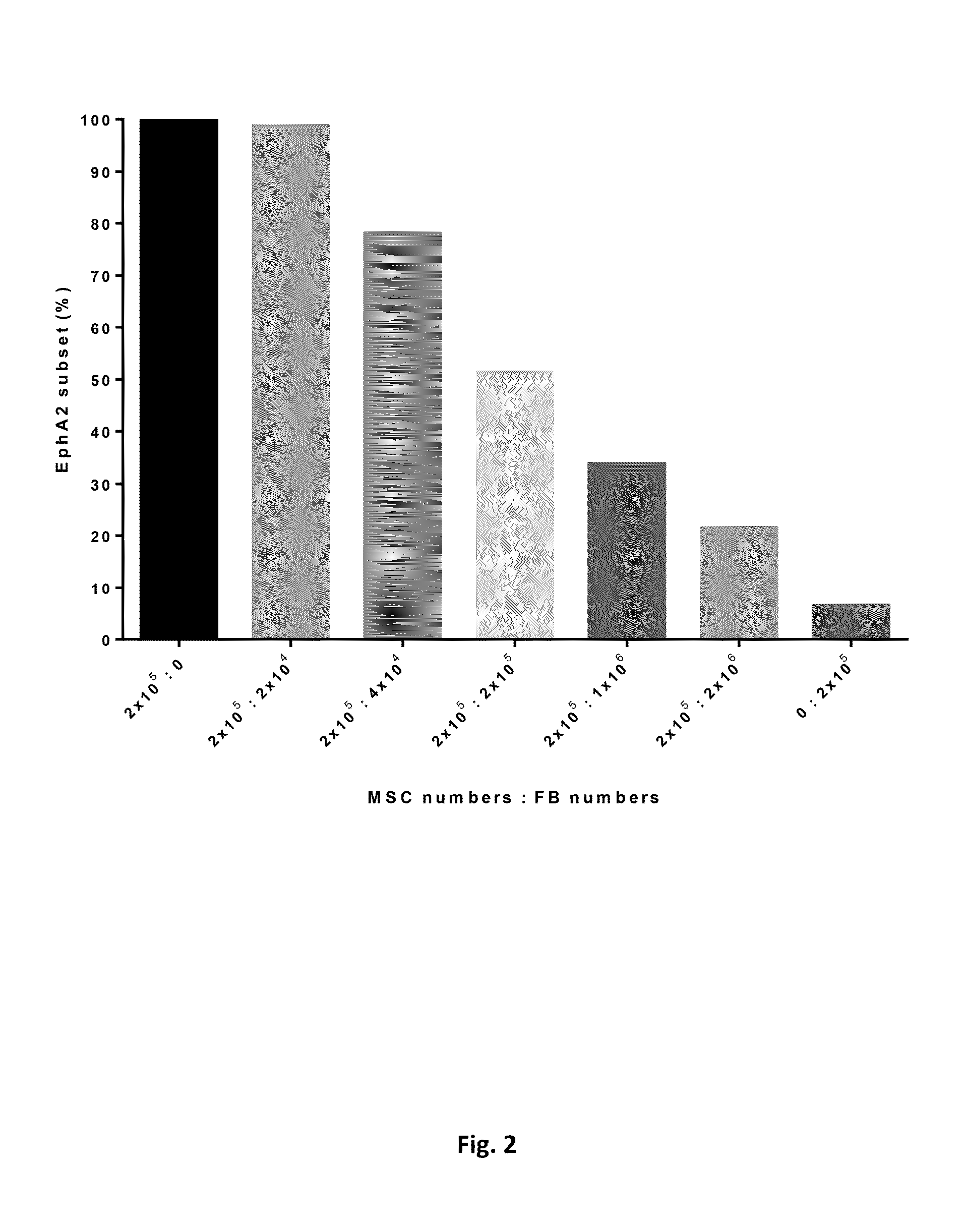
[0044]a. Flow Cytometry Analysis of EphA2-Enriched MSCs Sorted by Magnetic-Activated Cell Sorting (MACS)

[0045]The MACS method (MACS® Technology, Miltenyi Biotec) allows cells to be separated by incubating with magnetic nanoparticles coated with antibodies against EphA2 surface antigen. Primary culture of MSCs derived from placenta were incubated with the fluorescence conjugated anti-human antibodies against EphA2 and sorted by R-Phycoerythrin (PE) Magnetic Particles according to manufacturer's instructions. Flow cytometry analysis of MACS sorted MSCs at P0 revealed that cell population could become homogeneous in 100% CD73 positive, 97.2˜99.5% CD90 positive, 96.0˜99.9% CD 105 positive and 96.6˜100% EphA2 positive expression since passage 0 (see Table 2 below), demonstrating that EphA2 sorting via antibodies conjugated magnetic beads could dramatically improve the MSC purity from P...

example 3

Quantitative Real-Time PCR Evaluation of EphA2 Transcript in Placenta-Derived Mesenchymal Stem Cells (MSCs) and Fibroblasts

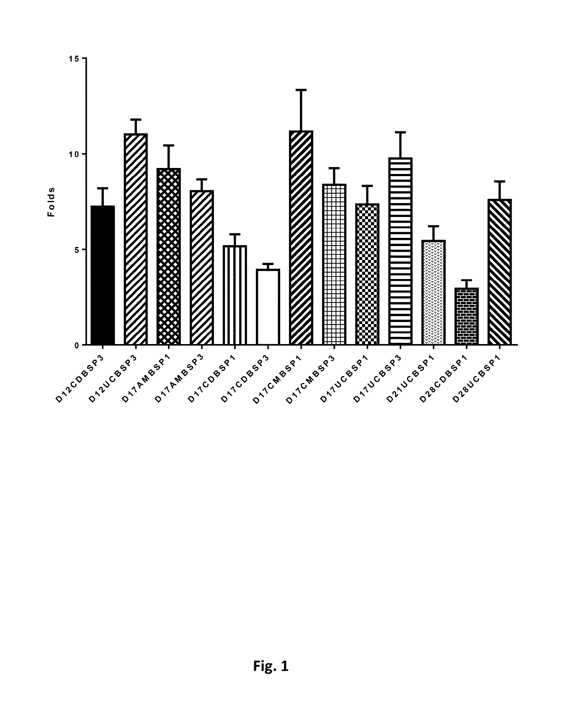
[0048]Total RNA from 64 populations of placenta-derived cells (n=8, including passage 1 and passage 3, from AM, CD, CM, UC, 4 different parts of a placenta) and Human Foreskin Fibroblasts (neonatal, PC501A-HFF, SBI) were isolated using the Direct-zol miniprep Kit (Zymo Research Corporation, CA, USA). The complementary DNA (cDNA) was synthesized with Transcriptor First Strand cDNA Synthesis Kit (Roche, Basel, Switzerland). Then Quantitative RT-PCR was performed using the Roche Universal ProbeLibrary System with a LightCycler480 II (Roche, Basel, Switzerland) according to the manufacturer's instructions.

[0049]We assessed expression of EphA2 by quantitative real-time PCR in order to compare placenta-derived multipotent MSCs and fibroblasts. Gene expression was normalized to the endogenous gene glyceraldehyde-3-phosphate dehydrogenase expression in the different cel...


PUM

PropertyMeasurementUnit
Fractionaaaaaaaaaa
Responsivityaaaaaaaaaa
Purityaaaaaaaaaa

Abstract

A method of distinguishing mesenchymal stem cells (MSCs) in a primary culture of cells derived from a placenta-related tissue is provided. Also provided is a method of increasing the purity of MSC population in a primary culture of cells derived from a placenta-related tissue. Further provided is a method of isolating a MSC population which is more responsive in an inflammatory environment. Said methods each comprise a step of sorting the cells by a marker of EphA2.

Description

FIELD OF THE INVENTION[0001]The present invention pertains to a method of distinguishing mesenchymal stem cells (MSCs) in a primary culture of cells derived from a placenta-related tissue. The present invention also relates to a method of increasing the purity of MSC population in a primary culture of cells derived from a placenta-related tissue. In another aspect, the invention pertains to a method of isolating a MSC population which is more responsive in an inflammatory environment.BACKGROUND OF THE INVENTION[0002]Mesenchymal stem or stromal cells (MSCs) are multipotent cells of embryonic mesodermal origin, with a fibroblast-like morphology. These cells can differentiate into adipocytes, osteocytes, chondrocytes, neural lineage cells, and myocytes among other cell types depending on the stimuli and culture conditions. Although the plasticity of hMSCs and their role in tissue repair and regeneration have been extensively studied, it is their immunological trophic property that has ...

Claims


Application Information

Patent Timeline
IPC IPC(8): G01N33/569C12Q1/68
CPCG01N33/56966C12Q2600/158G01N2333/71C12Q1/6881G01N2333/91205
Inventor HSUAN, CHANG-YOLIU, WEI-TINGSHEN, SHIH-PEILIN, YUNLI, YUAN-TSUNGCHANG, CHIA-CHI
Owner MERIDIGEN BIOTECH
Why Eureka
Try Eureka