Therapeutic and Diagnostic Method for Ataxia-Telangiectasia

a diagnostic method and ataxia technology, applied in the field of ataxia-telangiectasia therapy and diagnostic method, can solve the problems of severe consequences of atm damage in dna damaged cells, and achieve the effects of reducing neurodegeneration in a-t, and regulating proteasome-mediated protein turnover

Active Publication Date: 2016-07-28
BOARD OF SUPERVISORS OF LOUISIANA STATE UNIV & AGRI & MECHANICAL COLLEGE
View PDF3 Cites 0 Cited by
  • Summary
  • Abstract
  • Description
  • Claims
  • Application Information

AI Technical Summary

Problems solved by technology

Therefore, a defect in ATM has severe consequences in DNA damaged cells, especially in terminally differentiated cells such as neurons (11).

Method used


Image

  • Therapeutic and Diagnostic Method for Ataxia-Telangiectasia
  • Therapeutic and Diagnostic Method for Ataxia-Telangiectasia
  • Therapeutic and Diagnostic Method for Ataxia-Telangiectasia

Examples


example 1

Materials and Methods

[0073]Human tissues and Animal Studies.

[0074]Frozen human mid-brain tissues containing specifically substantia nigra were obtained postmortem from patients with confirmed A-T disease and control individuals (without any known disease). Slides with paraffin-embedded sections of the midbrain tissues were used in immunoflorescence study. Human brain tissues and tissue sections were obtained from the NICHD Brain and Tissue Bank for Developmental Disorders at the University of Maryland under protocols approved by the University of Maryland Institutional Review Board.

[0075]Animal study was carried out in strict accordance with the recommendations in the Guide for the Care and Use of Laboratory Animals of the National Institutes of Health. The protocol was approved by the Louisiana State University Health Sciences Center—NO Institutional Animal Care and Use Committee under its assurance with the Office of Laboratory Animal Welfare of the National Institutes of Health.

[...

example 2

Protein Polyubiquitylation and Degradation is Reduced in Cells Deficient in ATM

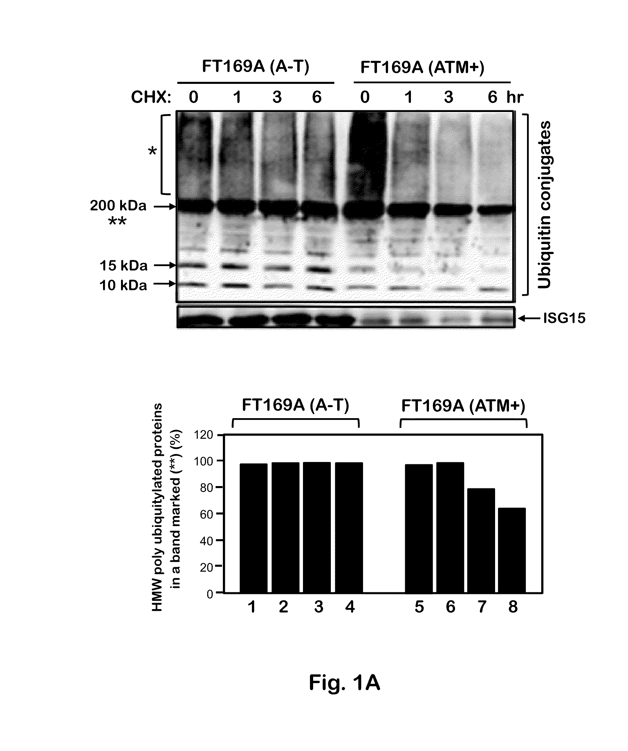
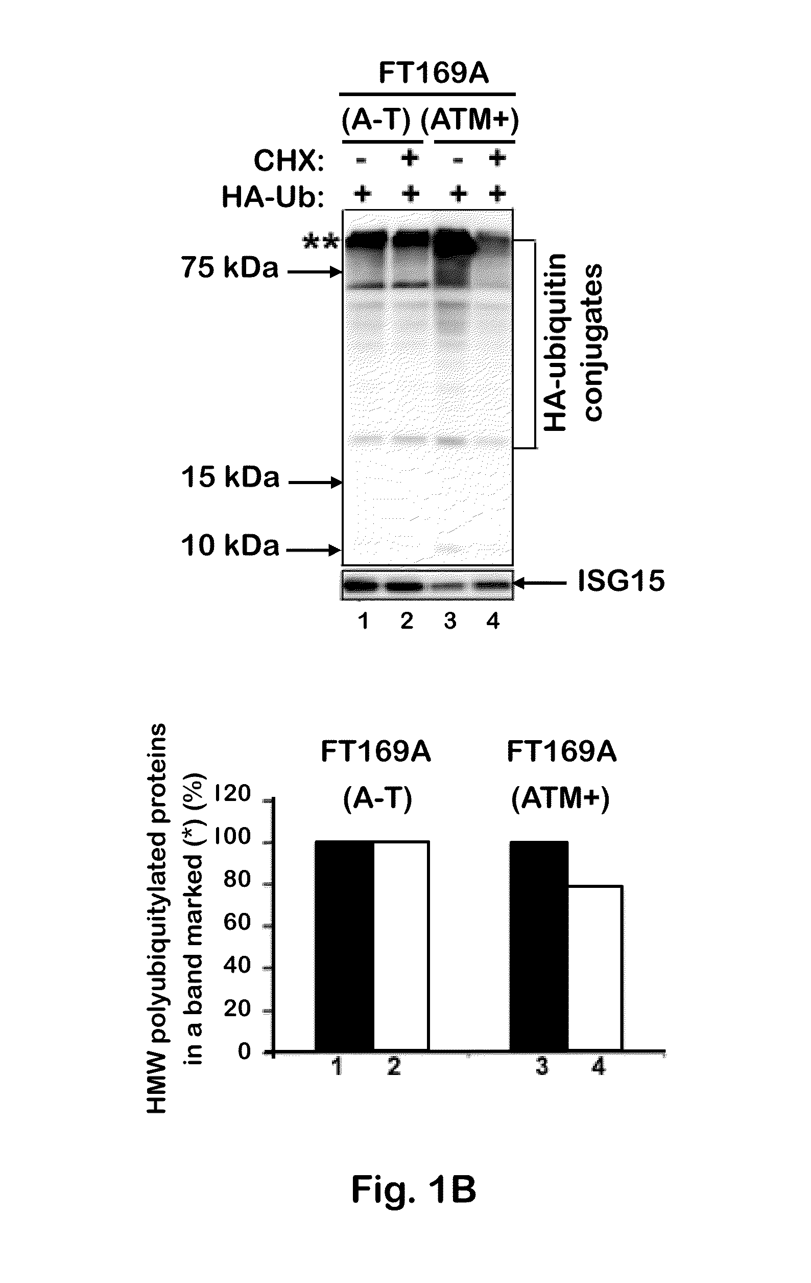
[0092]To test whether the defective ubiquitin-mediated degradation of cellular proteins contributes to neurodegeneration in A-T, the rate of degradation of overall cellular polyubiquitylated proteins was monitored in FT169A (A-T) (ATM null) and FT169A (ATM+) (ATM reconstituted FT169A) isogenic cells as described (71) using the protein synthesis inhibitor cycloheximide (CHX) (Sigma-Aldrich). In FIG. 1A, FT169A (A-T) (lanes 1-4) and FT169A (ATM+) (lanes 5-8) cells were treated with the protein synthesis inhibitor CHX (10 μg / ml) for 0, 1, 3, and 6 hours. Cell lysates were analyzed using discontinuous (15%) SDS-PAGE followed by immunoblotting with anti-ubiquitin antibody. The symbols * and ** mark the position of high-molecular-weight (HMW) polyubiquitylated proteins. Quantitation of the high-molecular-weight (HMW) polyubiquitylated proteins (shown as **) is shown in the bar graph.

[0093]As shown in FIG. 1A, t...

example 3

ATM Negatively Regulates the ISG15 Pathway

[0106]Previous studies have shown that ISG15 is increased in A-T lymphoblasts (70). Overexpression of ISG15 in tumor cells has been linked to reduced protein polyubiquitylation and turnover (24). To determine whether overexpression of the ISG15 pathway is responsible for reduced protein polyubiquitylation in FT169A (A-T) cells as shown above, the levels of ISG15 and its conjugates were measured in ATM null FT169A (A-T) and ATM-reconstituted FT169A (ATM+) cells using anti-ISG15 antibodies by Western analysis. In FIGS. 3A-3C, extracts of FT169A (A-T) and FT169A (ATM+) cells were analyzed by 5% (FIG. 3A) or 15% (FIG. 3B) SDS-PAGE, followed by immunoblotting using either anti-ATM (FIG. 3A) or anti-ISG15 antibody (FIG. 3B). The same membrane shown in FIG. 3B was stripped and re-probed with anti-tubulin antibody to assure equal protein loading. The average band intensity of the free ISG15 protein (error bar represents SEM) from three independent e...


PUM

PropertyMeasurementUnit
molecular weightaaaaaaaaaa
thickaaaaaaaaaa
concentrationaaaaaaaaaa

Abstract

ATM kinase is shown to regulate proteasome-mediated protein turnover through suppression of the expression of the ubiquitin-like protein ISG15 (Interferon Stimulated Gene 15). Silencing of the ISG15 pathway restored both the ubiquitin and autophagy pathways, and the UV-mediated degradation of their substrates in A-T cells. The ATM kinase negatively regulates the ISG15 pathway, and the constitutively elevated ISG15 pathway induces proteinopathy in A-T cells, and in A-T patients. These findings indicate that proteasome-mediated protein degradation is impaired in A-T cells due to elevated expression of the ISG15 conjugation pathway, which contributes to progressive neurodegeneration in A-T patients. The ISG15 pathway is a new target for both detection and treatment of A-T Inhibitors if ISG15 expression can be used to inhibit or attenuate neurodegeneration in A-T patients. In addition, an inhibitor of the early phase of autophagy, 3-MA, was shown to be effective in decreasing the impaired proteasome-mediated protein degradation in A-T cells, and thus would be effective in decreasing the neurodegeneration in A-T patients.

Description

[0001]This application claims priority under 35 U.S.C. §119(e) from U.S. Provisional Application Ser. No. 61 / 565,715, filed 1 Dec. 2011, entitled “Therapeutic and Diagnostic Method for Ataxia-Telangiectasia,” and from U.S. Provisional Application Ser. No. 61 / 706,863, filed 28 Sep. 2012, entitled “An Improvement to Targeting the ISG15 Pathway in Ataxia-Telangiectasia: A Novel Therapeutic Approach for Treating A-T;” the contents of both provisional applications are fully incorporated by reference herein.[0002]This invention was made with government support under grant number R21NS060960 awarded by the National Institutes of Health / National Institute of Neurological Disorders and Stroke. The government has certain rights in the invention.[0003]Ataxia-telangiectasia (A-T) is a childhood disease with an incident of 1 in 40,000 children in the U.S. and 1 in 200,000 worldwide each year (1-4). Ataxia refers to uncoordinated movements, such as walking, and telangiectasia is the enlargement o...

Claims


Application Information

Patent Timeline
Patent Type & Authority Applications(United States)
IPC IPC(8): G01N33/68A61K31/52A61K31/713
CPCG01N33/6896A61K31/52A61K31/713G01N2500/00G01N2800/2835
Inventor DESAI, SHYAMAL D.
Owner BOARD OF SUPERVISORS OF LOUISIANA STATE UNIV & AGRI & MECHANICAL COLLEGE
Why Eureka
Try Eureka