Method of processing x-ray images of a breast

a breast and x-ray technology, applied in the field of x-ray mammography, can solve the problems of significantly increasing the duration of the biopsy, time-consuming, and difficult to detect micro calcifications with such techniques, and achieve the effects of less time for the procedure, improved accuracy, and reduced time consumed in determining the target position of the needl

Active Publication Date: 2016-07-28
GENERAL ELECTRIC CO
View PDF10 Cites 6 Cited by
  • Summary
  • Abstract
  • Description
  • Claims
  • Application Information

AI Technical Summary

Benefits of technology

[0016]This method allows the operator to be offered needle target position where to perform a biopsy rather than deducing such needle target position after browsing slices images. As a consequence, the time consumed in determining a needle target position is reduced.
[0017]As patient's breast is maintained under compression during examination of the slice images, the less time the procedure takes the better and painless it is for the patient.

Problems solved by technology

Ultrasound guided biopsy is preferentially performed, as it is a non-irradiating modality, but micro calcifications are often hard to detect with such techniques.
However, such navigation is very time consuming for the radiologist and thus may significantly increase the duration of the biopsy.

Method used


Image

  • Method of processing x-ray images of a breast
  • Method of processing x-ray images of a breast
  • Method of processing x-ray images of a breast

Examples


Embodiment Construction

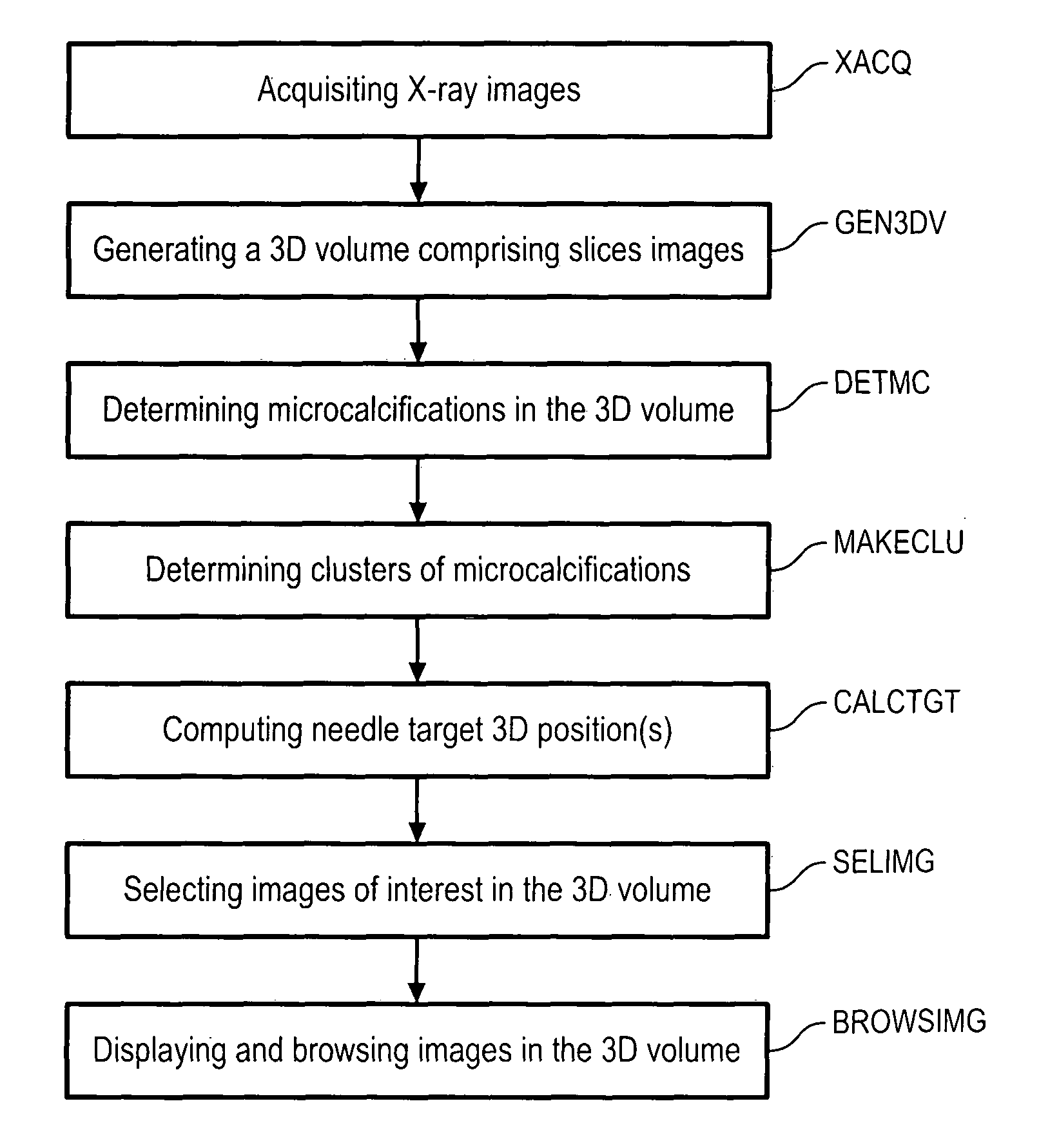
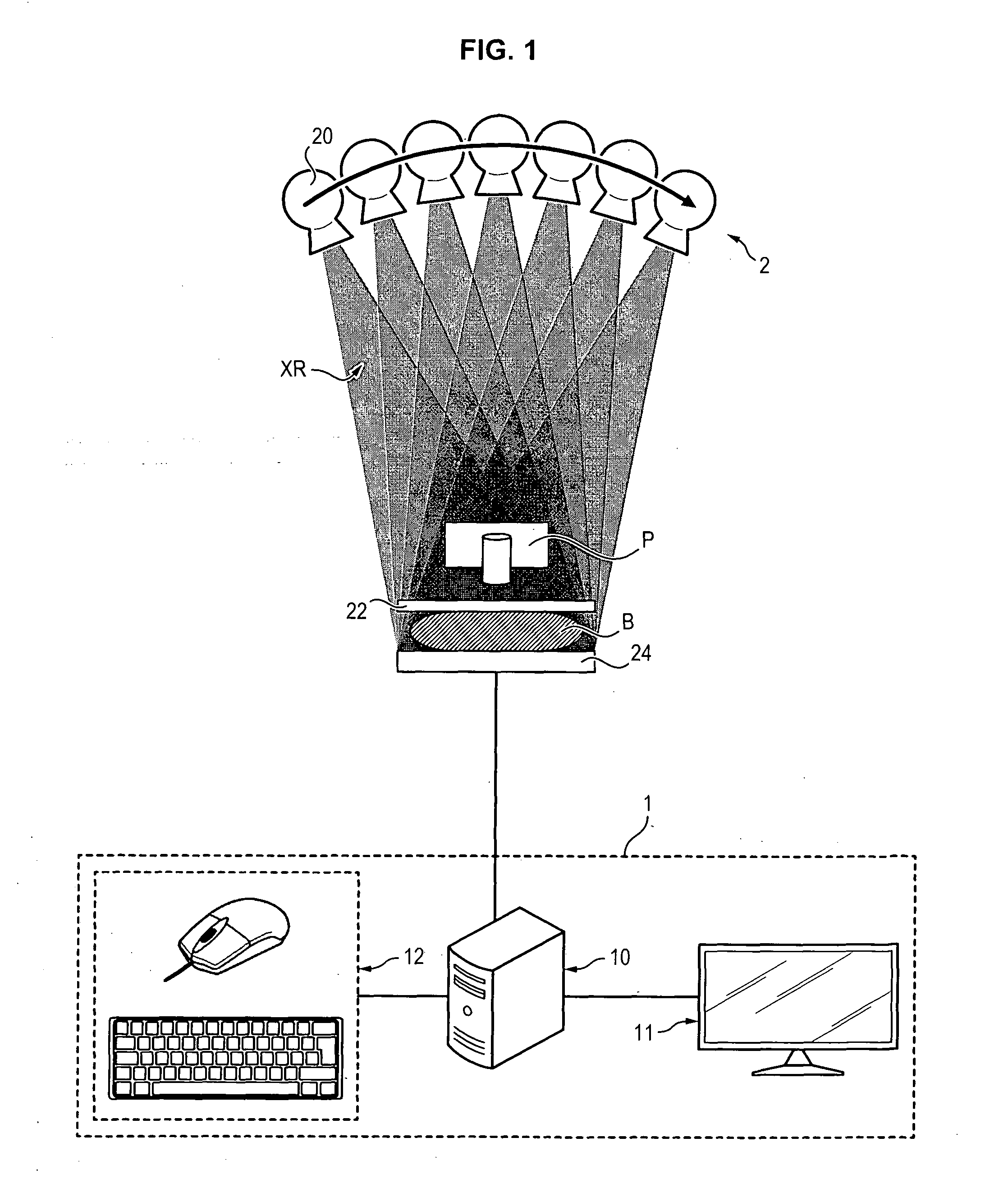
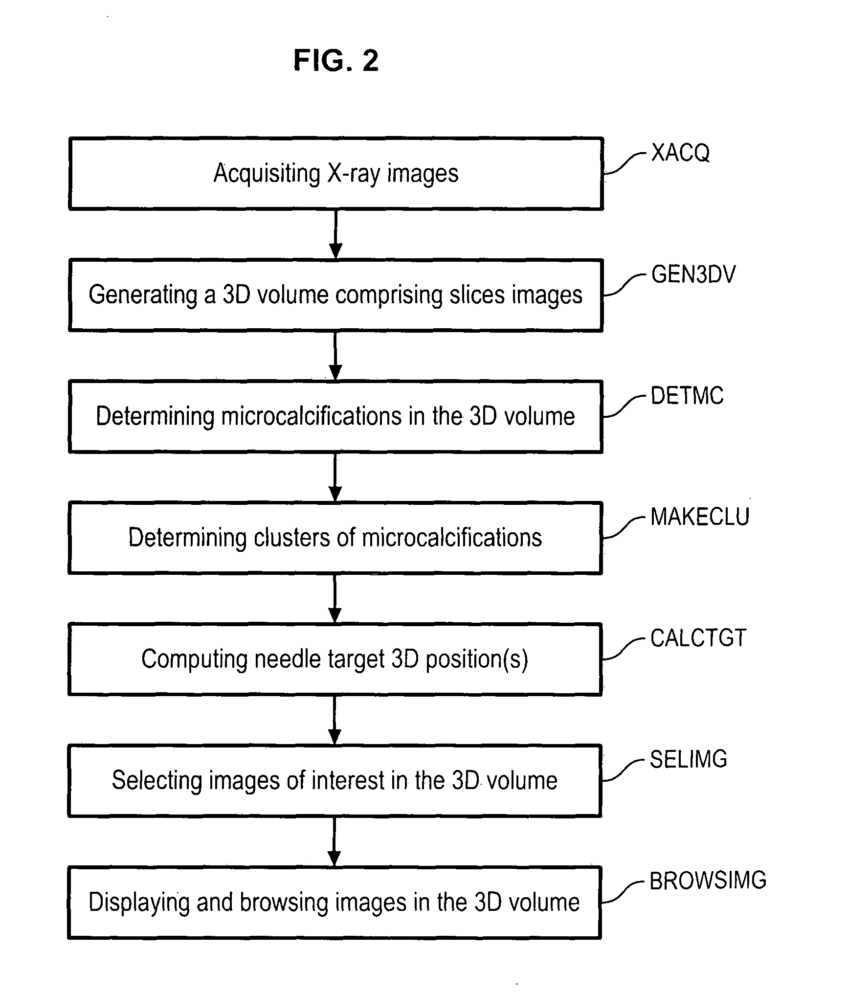
[0026]Referring to FIG. 1, a device 1 for processing X-ray images comprises an image processing unit 10, a graphical interface 11 and an input interface 12.

[0027]The image processing unit 10 is adapted to process X-ray images and display data on the graphical interface 11. It may comprise one or more processors and a memory storage unit adapted for storing X-ray images.

[0028]The graphical interface 11 is adapted to display images processed by the image processing unit: it may comprise one or many screens.

[0029]The input interface 12 is adapted for capturing event triggered by the operator: it may comprise a keyboard, a mouse, a touchscreen, or a combination of those. In the following, data inputted by an operator through the input interface 12 will be named “event”. Events may be for instance: clicking on a displayed item, entering one or more keys through a keyboard, etc.

[0030]The image processing unit is typically coupled with an X-ray device 2 adapted for acquiring X-ray images o...


PUM


Abstract

A method of processing X-ray images of a breast (B), the method comprising the steps of —generating (GEN3DV) a 3D volume from a plurality of X-ray images, —processing (DETMC) the 3D volume and/or the plurality of X-ray images, —computing (DETMC-CTR), for each region of interest in the 3D volume, a 3D characteristic position of the region of interest, —computing (CALCTGT) at least one needle target 3D position from a plurality of said computed 3D characteristic positions, —associating (SELIMG-TGT) each needle target position with a target image, —displaying (DISP-IMG) a slice image on a graphical interface, and —if the current slice image is a target image, displaying (DISP-TGT) on the graphical interface a target marker indicating each needle target position associated with the displayed current slice image.

Description

BACKGROUND[0001]1. Technical Field[0002]Embodiments of the present invention relate to X-ray mammography and more particularly to assistance tools for carrying out an X-ray-guided breast biopsy.[0003]2. Description of Related Art[0004]Microcalcifications are tiny specks of mineral deposits (calcium) that can be scattered throughout the mammary gland or occur in clusters. Commonly, they simply indicate the presence of tiny benign cysts, but can signify the presence of early breast cancer.[0005]Suspicious microcalcifications are commonly punctured thanks to biopsy, which is a medical test commonly performed by a surgeon or an interventional radiologist involving sampling of cells or tissues for examination.[0006]Ultrasound guided biopsy is preferentially performed, as it is a non-irradiating modality, but micro calcifications are often hard to detect with such techniques. On the contrary, micro-calcifications are well detected with X-rays, and that is why mammography is used to punctu...

Claims


Application Information

Patent Timeline
Patent Type & Authority Applications(United States)
IPC IPC(8): G06T7/00G06T19/00
CPCG06T7/0012G06T7/0081G06T19/003G06T2207/30204G06T2207/30068G06T2207/30096G06T2207/10112G06T7/11G06T11/008G06T2211/436
Inventor VANCAMBERG, LAURENCEPALMA, JR., GIOVANNI JOHN JACQUES
Owner GENERAL ELECTRIC CO
Why Eureka
Try Eureka