

Immune competent models of human disease
a human disease, immune-competent technology, applied in the field of immune-competent animal models, can solve the problems of difficult transgenic animals, difficult to initiate, and many preclinical therapies that are tested in rodents fail miserably when they reach human trials
- Summary
- Abstract
- Description
- Claims
- Application Information
AI Technical Summary
Benefits of technology
Problems solved by technology
Method used
Image

Examples
example 1
General Procedure for Porcine Model
[0049]Tumor injection in fetuses. A pregnant sow at approximately 50 days gestation is anesthetized and given a dose of banamine. After induction of general anesthesia, a paramedian incision is made in the lower abdomen in order to externalize the uterus. The uterus is externalized, one horn at a time, and fetuses are palpated, isolated and immobilized. The fetuses are injected with human tumor cells (1-4 million cells) into the peritoneal cavity and / or the fetal liver using either ultrasound guided injection or landmark guided injection. Alternatively, cells are transplanted into the amnion or amniotic sac of the fetus. After all fetuses are injected, the uterus is replaced and the incision was sutured. The sow is watched closely for 24 hours. At 24 hours, another dose of banamine is given, as well as a broad spectrum antibiotic.
[0050]Remainder of gestation and post-farrowing: The sow is monitored closely during the remainder of gestation. At farr...
example 2
Porcine Model
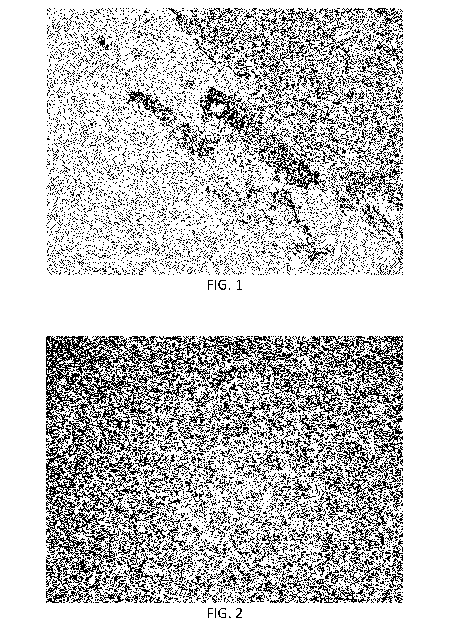
[0053]An experiment was carried out following the protocol described above in Example 1. Before farrowing, the sow developed sepsis and lost most of the fetuses. At farrowing, one live piglet was recovered. To determine the effectiveness of the procedure, this piglet was euthanized and tissues were collected. Liver was fixed and sectioned and probed with anti-human nuclear antibodies. IHC demonstrated the presence of small pockets of human cells in the liver. This indicates the fetal injections were successful and desensitized the developing fetus to human cells. In FIG. 1, brown staining indicates detection of human cells by anti-human antibodies at the surface of the liver.
example 3
Porcine Model
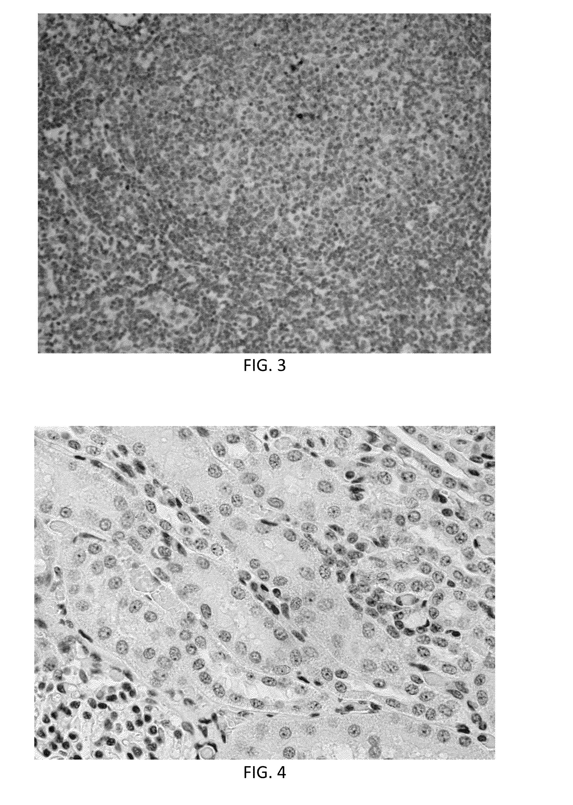
[0054]A second experiment was carried out according to the protocol for Example 1, except that human triple negative breast cancer cells (MDA-MB-231) were transplanted into the pre-immune fetuses. In this case, nine live piglets were born. Two days after weaning, cells from the same cell culture were transplanted in a subcutaneous location in an ear of each piglet.
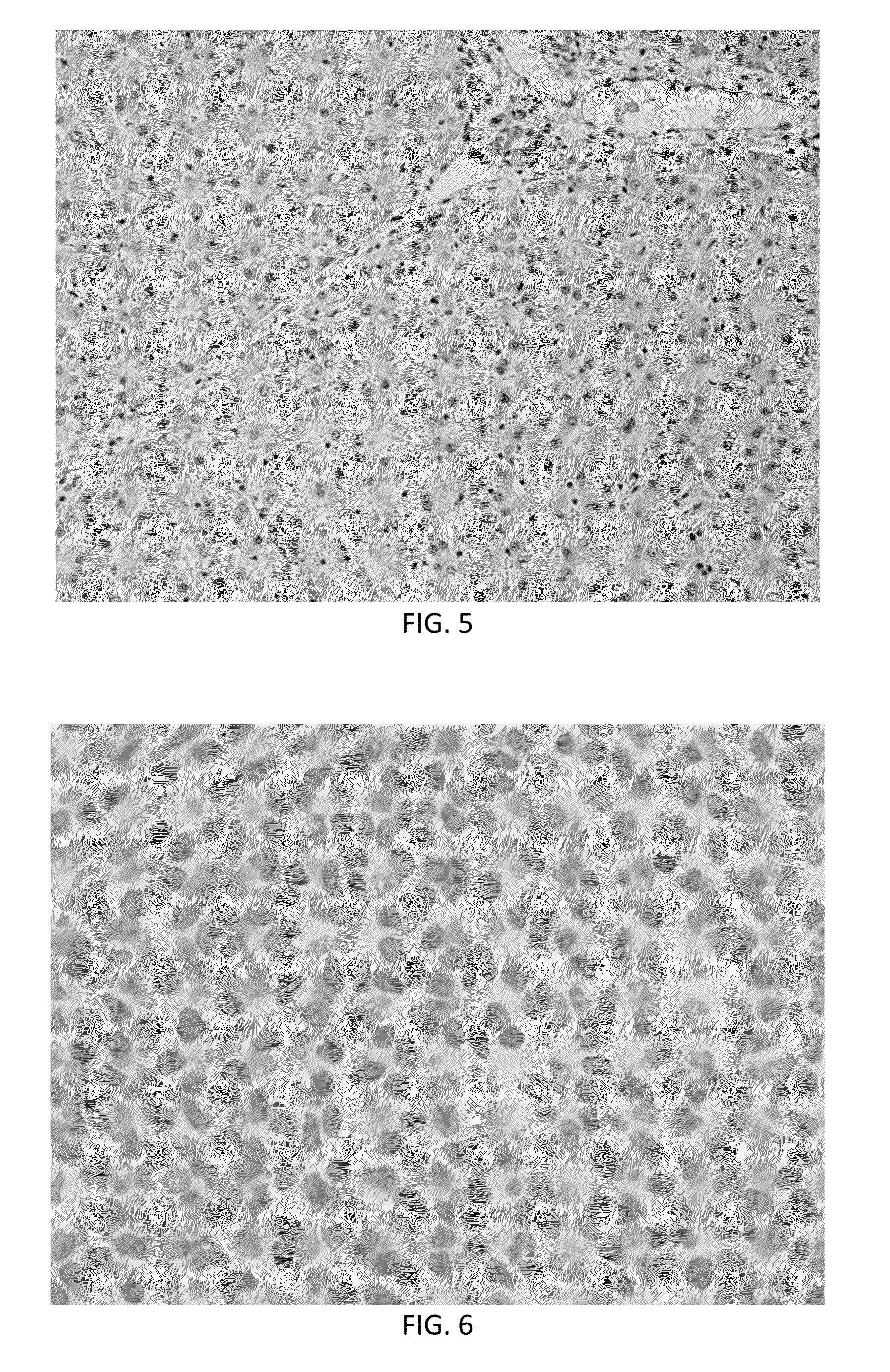
[0055]Analysis of tissues from this experiment showed many human cells incorporated in many pig tissues (identified by immunohistochemistry using a human specific anti-nuclear antibody) from the first in utero injection. Cells (brown in color) were found in retropharyngeal nodes (FIG. 2), mesenteric lymph nodes (FIG. 3), thymus, adrenal cortex, lung, kidney (FIG. 4) and liver (FIG. 5). Note that nearly 50% of liver cells have brown staining nuclei in FIG. 5 indicating that the breast cancer stem cell population responded to developmental cues of the developing liver. No positive cells were found in pigs not tr...
PUM
 Abstract
Description
Claims
Application Information