Integrated Ultrasound, OCT, PA and/or Florescence Imaging Endoscope for Diagnosing Cancers in Gastrointestinal, Respiratory, and Urogenital Tracts

a cancer and endoscope technology, applied in the field of endoscopic probes, can solve the problems of poor prognosis, crude approach, invasiveness and harm to patients, and exacerbate the problem of early detection of dysplasia in the gut, etc., and achieve the effect of poor prognosis

Active Publication Date: 2016-08-25
UNIV OF SOUTHERN CALIFORNIA +1
View PDF5 Cites 17 Cited by
  • Summary
  • Abstract
  • Description
  • Claims
  • Application Information

AI Technical Summary

Benefits of technology

[0011]The illustrated embodiments of the invention include an integrated ultrasound-optical coherence tomography (OCT) imaging system and endoscopic catheter for imaging inside a bile duct and pancreatic duct to diagnose a cholangiocarcinoma and cystic neoplasm of the pancreas. The medical imaging endoscope is intended for clinical practice. The imaging device includes a multiple-function imaging catheter and an imaging system. During the diagnostic procedure, known as ERCP (endoscopic retrograde cholangiopancreatography), the catheter is advanced either into the bile ducts or pancreas of patients, providing real time cross sectional ultrasound. OCT high resolution imaging of duct walls, photoacoustic imaging and / or florescence imaging with molecular contrasts for the cancerous tissue or abnormal tissue, to diagnose the etiology of stricture / obstruction, vascular compression, tumor staging, and other lesions. As tumors of the bile duct and pancreas have a particularly poor prognosis when detected in their advanced forms, the early diagnosis made possible by the invention has a profound effect on the medical management and outcome of the disease.

Problems solved by technology

Besides inadequate sampling, the problem of early detection of dysplasia in the gut is exacerbated in the presence of chronic inflammatory conditions such as esophagitis, since early-stage lesions are difficult to distinguish from inflamed gastrointestinal mucosa by the endoscopist.
This crude approach is very invasive and harmful to the patients and the accuracy is still below 50%.
Moreover, the biopsy approach only allows the clinician to harvest cells on the inner surface of bile duct, and has no diagnosing ability for tissues several millimeters beyond the epithelium of the bile duet.
Another specific problem to be solved is the diagnosis of cystic neoplasms of the pancreas.

Method used


Image

  • Integrated Ultrasound, OCT, PA and/or Florescence Imaging Endoscope for Diagnosing Cancers in Gastrointestinal, Respiratory, and Urogenital Tracts
  • Integrated Ultrasound, OCT, PA and/or Florescence Imaging Endoscope for Diagnosing Cancers in Gastrointestinal, Respiratory, and Urogenital Tracts
  • Integrated Ultrasound, OCT, PA and/or Florescence Imaging Endoscope for Diagnosing Cancers in Gastrointestinal, Respiratory, and Urogenital Tracts

Examples


Embodiment Construction

[0045]Imaging System and Catheter Design

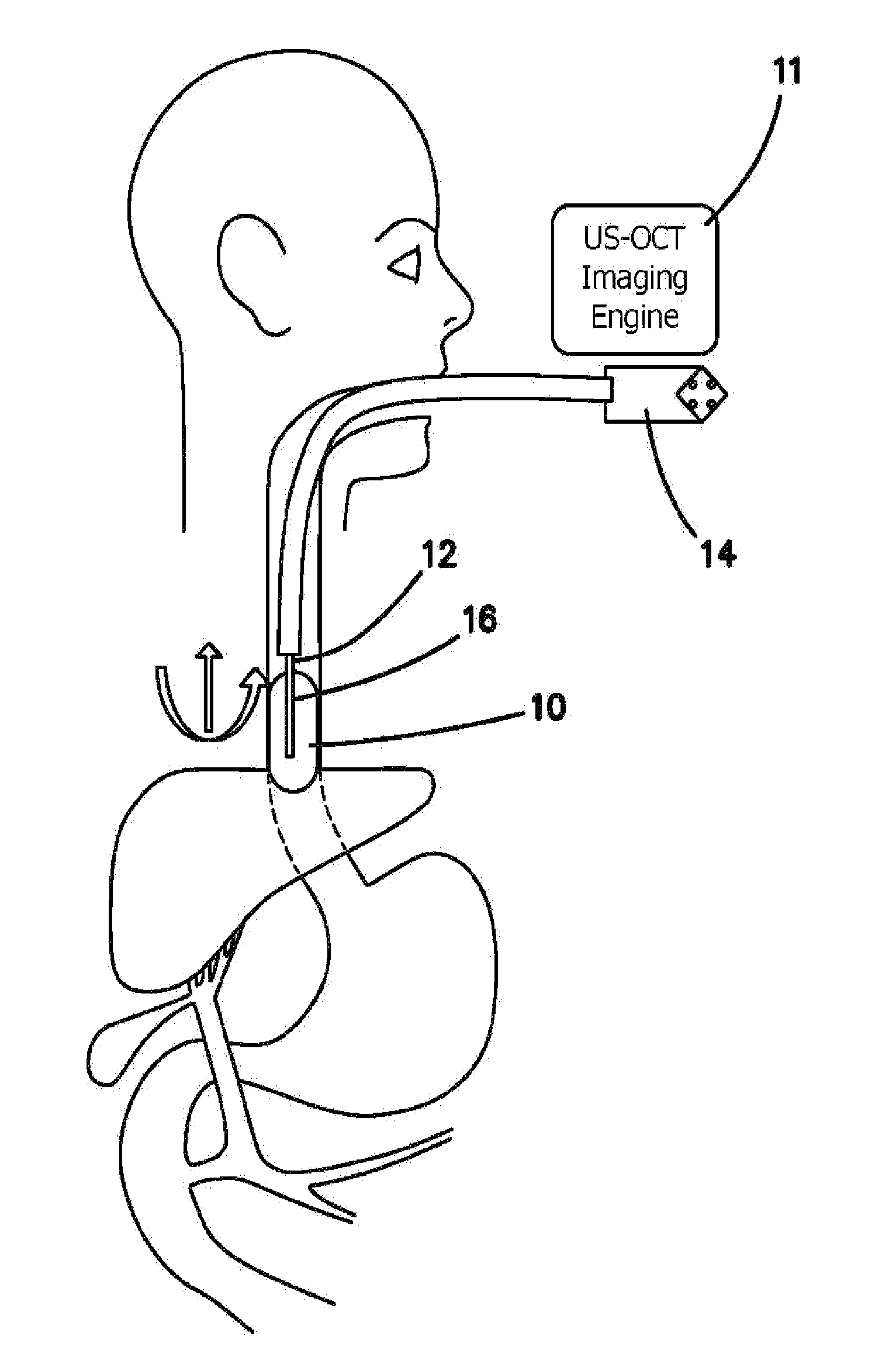
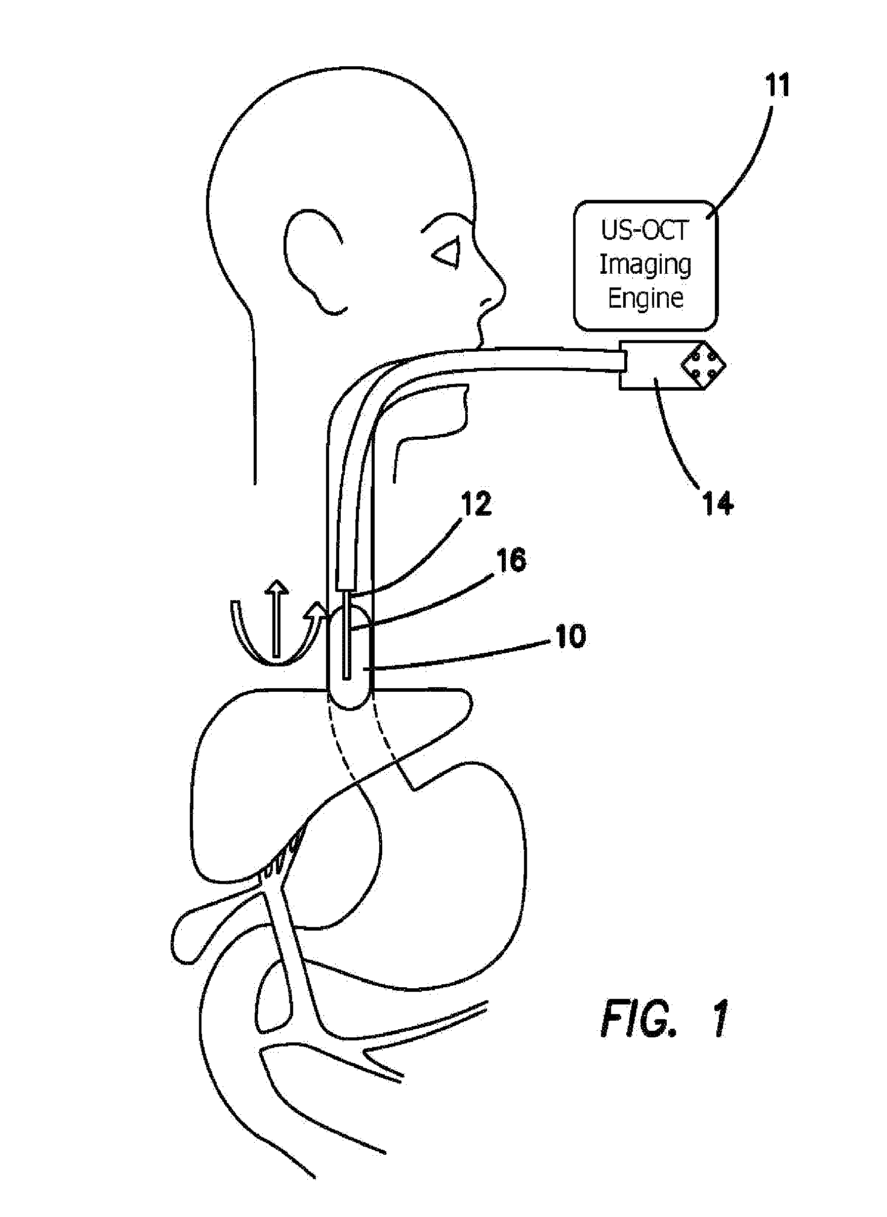
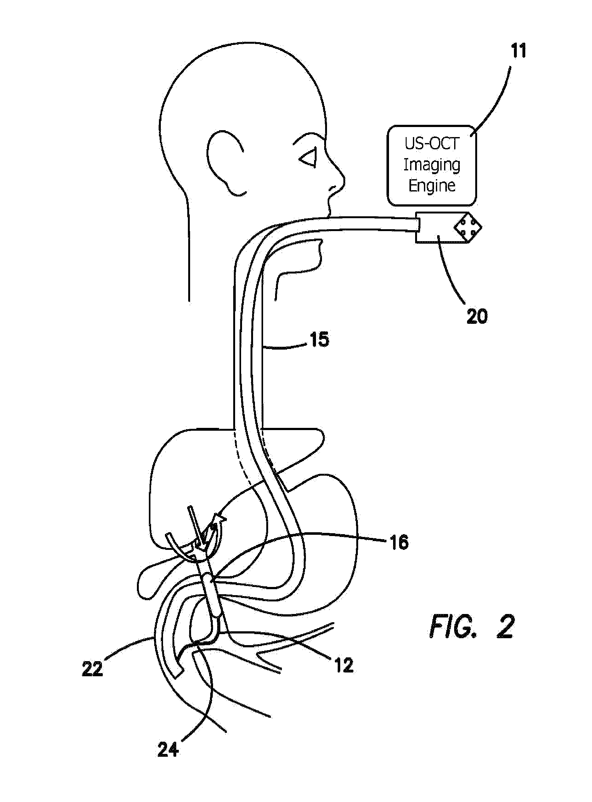
[0046]What is illustrated in the various embodiments of the invention includes a device 10 which is a minimally invasive interventional imaging device 10 with the ability to take a tissue biopsy from a location that is visible on the imaging system. The purpose of device 10 is to enable the physician: to visualize the tissue he or she is about to biopsy with the imaging catheter 12, and simultaneously take a tissue biopsy with the same device 10. The additional imaging information increases the diagnostic accuracy of the tissue biopsy by allowing the physician to first visualize the tissue in the bile duct which he or she deems suspicious and then take a biopsy of that, specific target rather than merely scraping cells off only the inner lining of the bile duct without any indication of where a lesion might be.

[0047]The imaging system of device 10 integrates at least two or more imaging modalities: an ultrasound, an OCT, a PA and / or a floresce...


PUM


Abstract

A multimodality imaging system including ultrasound, optical coherence tomography (OCT), photoacoustic (PA) imaging, florescence imaging and endoscopic catheter for imaging inside the gastrointestinal tract with real-time automatic image co-registration capability, including: an ultrasound subsystem for imaging; an optical coherence tomography (OCT) subsystem for imaging, a PA microscopy or tomography subsystem for imaging and a florescence imaging subsystem for imaging. An invasive interventional imaging device is included with an instrumentality to take a tissue biopsy from a location visible on the ultrasound subsystem for imaging, on the optical coherence tomography (OCT) subsystem for imaging, photoacoustic (PA) subsystem for imaging and florescence subsystem for imaging. The instrumentality takes a tissue biopsy from a visible location simultaneously with the visualization of the tissue about to be biopsied so that the tissue biopsy location is visualized before, during and after the biopsy.

Description

CROSS-REFERENCE TO RELATED APPLICATIONS[0001]This application claims priority to, and the benefit of the earlier filing date of: US provisional patent application entitled INTEGRATED ULTRASOUND AND OPTICAL COHERENCE TOMOGRAPHY (oct) ENDOSCOPE FOR DIAGNOSING CHOLANGIOCARCINOMA AND CYSTIC NEOPLASMS OF THE PANCREAS filed on Oct. 7, 2013, Ser. No. 61 / 887,790, pursuant to 35 USC 119, the contents of which are incorporated herein by reference.GOVERNMENT SUPPORT[0002]This invention was made with government support under contract EB-10090, awarded by the National Institutes of Health. The government has certain rights in the invention.BACKGROUND[0003]1. Field of the Technology[0004]The disclosure relates to the field of endoscopic probes using ultrasound and optical coherent tomography.[0005]2. Description of the Prior Art[0006]Gastrointestinal (gastrointestinal) cancers causes more than 140,000 deaths with more than 290,000 new eases in the United States in 2014. Esophageal cancer is one o...

Claims


Application Information

Patent Timeline
Patent Type & Authority Applications(United States)
IPC IPC(8): A61B8/00A61B10/04A61B10/02A61B8/12
CPCA61B1/05A61B10/0283A61B10/04A61B10/0233A61B5/6853A61B5/6871A61B5/0066A61B5/0071A61B5/0084A61B5/0095A61B5/687A61B1/041A61B1/00082A61B1/0016A61B1/018A61B1/043A61B1/2736A61B8/4416A61B8/445A61B8/4461A61B8/12
Inventor ZHOU, QIFALI, XIANGVAN DAM, JACQUESSHUNG, K. KIRKCUMMINS, THOMASCHEN, ZHONGPING
Owner UNIV OF SOUTHERN CALIFORNIA
Why Eureka
Try Eureka