
Methods and compositions for preserving photoreceptor and retinal pigment epithelial cells

a technology of epithelial cells and photoreceptors, applied in the direction of drug compositions, enzyme inhibitors, peptide/protein ingredients, etc., can solve the problems of z-vad, a pan-caspase inhibitor, failing to prevent photoreceptor loss, etc., to reduce or prevent the loss of photoreceptors and/or retinal pigment epithelial cells, reduce the loss of visual function, and prevent the loss of photoreceptors and/or retinal pigmen

Active Publication Date: 2016-09-01
MASSACHUSETTS EYE & EAR INFARY
View PDF0 Cites 6 Cited by
  • Summary
  • Abstract
  • Description
  • Claims
  • Application Information

AI Technical Summary

Benefits of technology

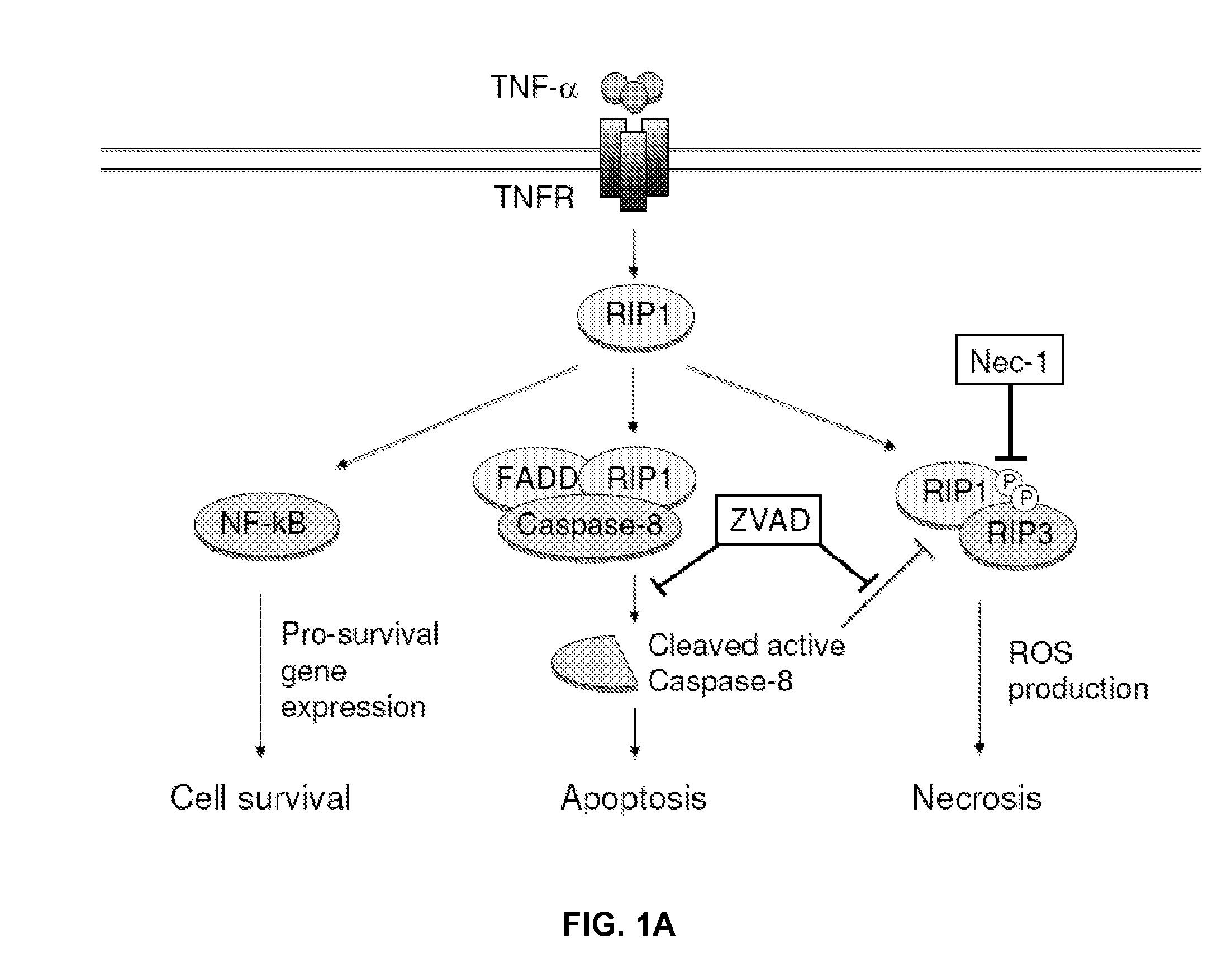
[0011]The invention is based, in part, on the discovery that a necrosis inhibitor, e.g., RIP kinase inhibitor, e.g., a necrostatin, e.g., necrostatin-1, can be used to reduce or prevent the loss of photoreceptor and / or retinal pigment epithelial cell viability, especially when the necrosis inhibitor is combined with an apoptotic inhibitor (e.g., a pan-caspase inhibitor, e.g., Z-VAD and / or IDN-6556). It was previously understood that photoreceptor cell death associated with AMD, RP, and retinal detachment was primarily caused by apoptosis. However, studies have shown that the administration of Z-VAD, a pan-caspase inhibitor, fails to prevent photoreceptor loss in these conditions. The studies described hereinbelow indicate that, in the presence of an apoptosis inhibitor (e.g., a pan-caspase inhibitor), photoreceptors die by necrosis, including necroptosis (or programmed necrosis). These studies show that programmed necrosis is a critical mechanism for ocular conditions wherein a symptom of the condition is the loss of photoreceptor cell viability in the presence of a pan-caspase inhibitor. As a result, it is possible to reduce the loss of visual function associated with an ocular disorder, in particular while the ocular disorder is being treated, by reducing the loss of photoreceptor viabilabilty and / or retinal pigment epithelial cell viability
[0012]In one aspect, the invention provides a method preserving the visual function of an eye of a subject with an ocular condition, wherein a symptom of the ocular condition is the loss of photoreceptor cell viability in the retina of the eye with the condition. The method comprises (a) administering to the eye of the subject an effective amount of a necrostatin and an effective amount of an apoptosis inhibitor thereby preserving the viability of the photoreceptor cells disposed within the retina of the eye, and (b) then measuring the visual function (e.g., visual acuity) of the eye after the administration of the necrosis inhibitor and the apoptosis inhibitor. After administration of the necrosis inhibitor and the apoptosis inhibitor the visual function (e.g., visual acuity) of the eye may be preserved or improved relative to the visual function of the eye prior to administration of the necrosis inhibitor and the apoptosis inhibitor.
[0017]The prevention of photoreceptor death following retinal detachment by co-administration of an apoptotic inhibitor and a necrostatin, e.g., necrostatin-1, at concentrations that exceed 100 μM was surprising because it had been believed that concentrations of necrostatin-1 exceeding 100 μM were toxic and, therefore, could not be administered at such a dosage. Moreover, it was an unexpected finding that the combination of a necrostatin, e.g., necrostatin-1, and a pan-caspase inhibitor, e.g., Z-VAD, achieved a synergistic effect in reducing photoreceptor cell death following retinal detachment compared to either drug alone.
[0020]In another aspect, the invention provides a method of preserving the visual function of an eye of a subject with an ocular condition, wherein a symptom of the ocular condition is the loss of photoreceptor cell viability and / or RPE viability in the retina of the eye. The method comprises (a) reducing the production or activity of a RIP-1 kinase and / or a RIP-3 kinase in the eye thereby to preserve the viability of the photoreceptor cells and / or RPE cells disposed within the retina of the eye; and (b), after treatment, measuring visual function (e.g., visual acuity) of the eye. In certain embodiments, the reduction in the production or activity of the RIP-1 kinase and / or the RIP-3 kinase can be achieved by administering an effective amount of RIPK inhibitor, e.g., a necrostatin. After administration of the RIPK inhibitor the visual function of the eye may be preserved or improved relative to the visual function of the eye prior to administration of the RIPK inhibitor.

Problems solved by technology

However, studies have shown that the administration of Z-VAD, a pan-caspase inhibitor, fails to prevent photoreceptor loss in these conditions.

Method used


Image

  • Methods and compositions for preserving photoreceptor and retinal pigment epithelial cells
  • Methods and compositions for preserving photoreceptor and retinal pigment epithelial cells
  • Methods and compositions for preserving photoreceptor and retinal pigment epithelial cells

Examples


example 1

Increased Expression of RIP-3 and RIP-1 after Retinal Detachment

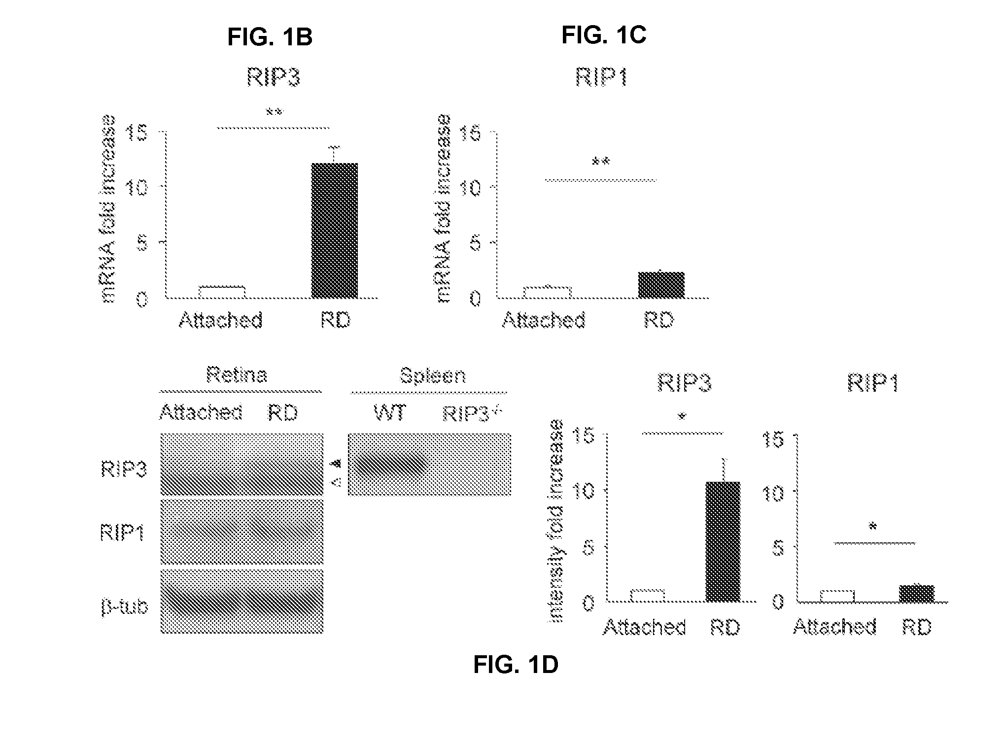
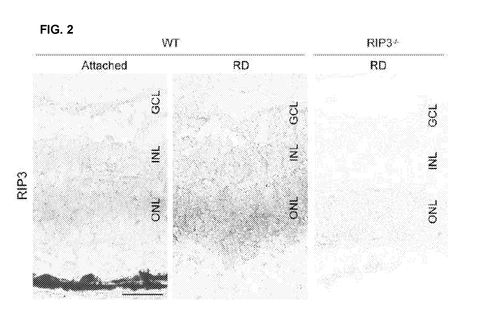
[0380]Photoreceptor death after retinal detachment has been thought to be caused mainly by apoptosis (Cook et al. (1995) INVEST OPHTHALMOL VIS SCI 36:990-996; Arroyo et al. (2005) AM J OPHTHALMOL 139:605-610. Although caspase-dependent pathway is known to be activated after retinal detachment, caspase inhibition by pan-caspase inhibitor fails to prevent photoreceptor death (Zacks et al. (2003) INVEST OPHTHALMOL VIS SCI 44:1262-1267; Hisatomi et al. (2001) AM J OPHTHALMOL 158:1271-1278). In this example, other pathways leading to photoreceptor death were investigated. In particular, the expression levels of RIP-1 and RIP-3 proteins were measured after retinal detachment. RIP-3 is a key regulator of RIP-1 kinase activation (Galluzzi L et al. (2009) J. MOL. CELL BIOL.) and its expression level has been shown to correlate with responsiveness to programmed necrosis (He S et al. (2009) CELL 137:1100-1111). As described below,...

example 2

Necrosis Inhibitor Prevents Retinal Detachment-Induced Photoreceptor Death in Combination with a Caspase Inhibitor

[0389]Necrostatin-1 (Nec-1) is a potent and selective inhibitor of programmed necrosis, which targets RIP-1 kinase activity (Degterev A et al. (2008) NAT. CHEM. BIOL. 4:313-321). The effectiveness of a RIP-1 kinase inhibitor in combination with a capsase inhibitor to prevent retinal detachment induced photoreceptor death was investigated.

[0390]RIP-1 is phosphorylated at several serine residues in its kinase domain during programmed necrosis and Nec-1 inhibits this phosphorylation (Degterev (2008) supra; Cho Y et al. (2009) Cell 137:1100-1111). To investigate the role of RIP-1 kinase in retinal detachment-induced photoreceptor death, the phosphorylation status of RIP-1 was first assessed by immunoprecipitating RIP-1 from the retina (FIG. 3A) and then blotting with an anti-phosphoserine antibody. RIP-1 was immunoprecipitated from retinal lysates. One mg of retinal lysates ...

example 3

Caspase Inhibition Shifts Retinal Detachment-Induced Photoreceptor Death from Apoptosis to Programmed Necrosis

[0398]The morphology of photoreceptors after retinal detachment was examined by transmission electron microscopy (TEM) and propidium iodide (PI) staining and the effect of treatment with Z-VAD and / or a Nec-1 compound of Formula I-C was analyzed.

[0399]TEM was performed as previously described (Hisatomi T et al. (2008). J. CLIN. INVEST. 118:2025-2038). The specimens were observed with Philips CM10 electron microscope. Over 200 photoreceptors each eye were photographed and subjected to quantification of cell death modes in a masked fashion. Photoreceptors showing cellular shrinkage and nuclear condensation were defined as apoptotic cells, while photoreceptors associated with cellular and organelle swelling and discontinuities in nuclear and plasma membrane were defined as necrotic cells. The presence of electron-dense granular materials were reported to occur subsequent to both...


PUM

PropertyMeasurementUnit
diameteraaaaaaaaaa
thickaaaaaaaaaa
volumeaaaaaaaaaa

Abstract

Provided are methods and compositions for maintaining the viability of photoreceptor cells and / or retinal pigment epithelial cells in a subject with an ocular disorder including, for example, age-related macular degeneration (AMD) (e.g., dry or neovascular AMD), retinitis pigmentosa (RP), or a retinal detachment. The viability of the photoreceptor cells and / or the retinal pigment epithelial cells can be preserved by administering a necrosis inhibitor either alone or in combination with an apoptosis inhibitor to a subject having an eye with the ocular condition. The compositions, when administered, maintain the viability of the cells, thereby minimizing the loss of vision or visual function associated with the ocular disorder.

Description

CROSS-REFERENCE TO RELATED APPLICATIONS[0001]This application is a continuation of U.S. patent application Ser. No. 13 / 642,887, filed Feb. 14, 2013, which is the national stage of International (PCT) Patent Application No. PCT / US2011 / 033704, filed Apr. 23, 2011, which claims the benefit of and priority to U.S. Provisional Application No. 61 / 327,476, filed Apr. 23, 2010; U.S. Provisional Application No. 61 / 405,003, filed Oct. 20, 2010; U.S. Provisional Application No. 61 / 409,055, filed Nov. 1, 2010; U.S. Provisional Application No. 61 / 414,862, filed Nov. 17, 2010; U.S. Provisional Application No. 61 / 472,153, filed Apr. 5, 2011; and U.S. Provisional Application No. 61 / 472,144, filed Apr. 5, 2011 the disclosure of each of which is hereby incorporated by reference in its entirety.GOVERNMENT FUNDING[0002]The work described in this application was sponsored, in part, by the National Eye Institute under Grant No. EY14104. The United States Government has certain rights in the invention.FIE...

Claims


Application Information

Patent Timeline
Patent Type & Authority Applications(United States)
IPC IPC(8): A61K31/4178A61K38/00A61K38/06A61K31/40A61K9/00A61K38/05
CPCA61K31/4178A61K9/0048A61K38/005A61K38/06A61K31/40A61K38/05A61K31/12A61K31/195A61K31/4155A61K31/416A61K31/4174A61K31/519A61K45/06A61P27/02A61K2300/00A61B3/028A61B5/4839A61F9/0008
Inventor VAVVAS, DEMETRIOSTRICHONAS, GEORGIOSMILLER, JOAN W.MURAKAMI, YUSUKE
Owner MASSACHUSETTS EYE & EAR INFARY
Learn More