
Mechanically driven ultrasound scanning system and method

a scanning system and mechanical technology, applied in the field of mechanical drive ultrasound scanning system and method, can solve the problems of complex integration, high cost, treatment beam interference,

Inactive Publication Date: 2016-12-08
SONITRACK SYST
View PDF8 Cites 13 Cited by
  • Summary
  • Abstract
  • Description
  • Claims
  • Application Information

AI Technical Summary

Benefits of technology

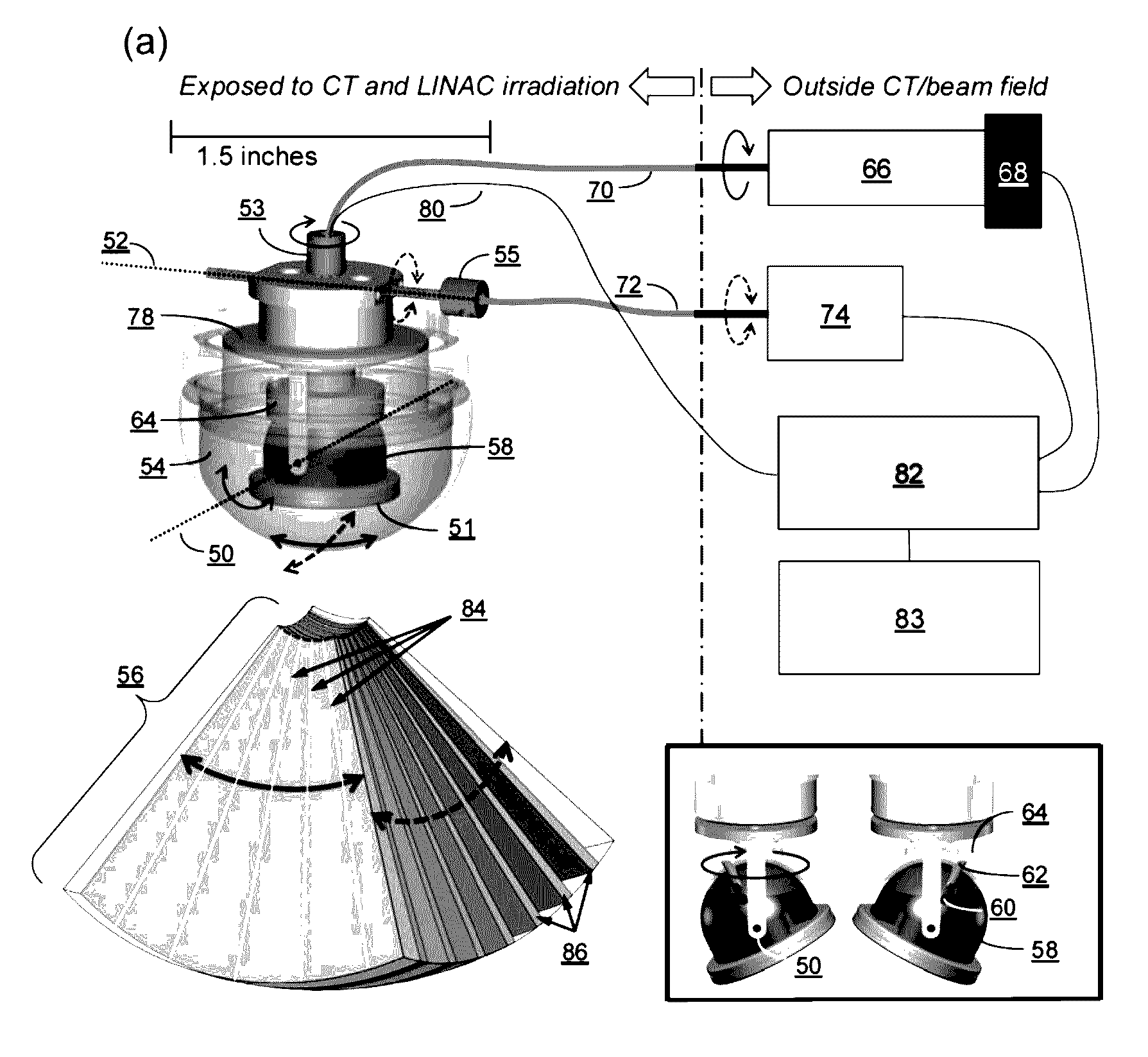
This patent is about a system that uses a single imaging element to capture 3D or 4D imaging information with minimal interference from imaging artifacts and radiation absorption. The system has one or more actuators that can position the imaging element in two degrees of freedom to acquire the desired information. The overall goal is to minimize the impact on the imaged region and improve the accuracy of radiotherapy delivery.

Problems solved by technology

High dose conformality in EBRT is important for delivering curative dose to the target while sparing surrounding healthy structures, but anatomy motion poses a fundamental threat to realizing such conformity.
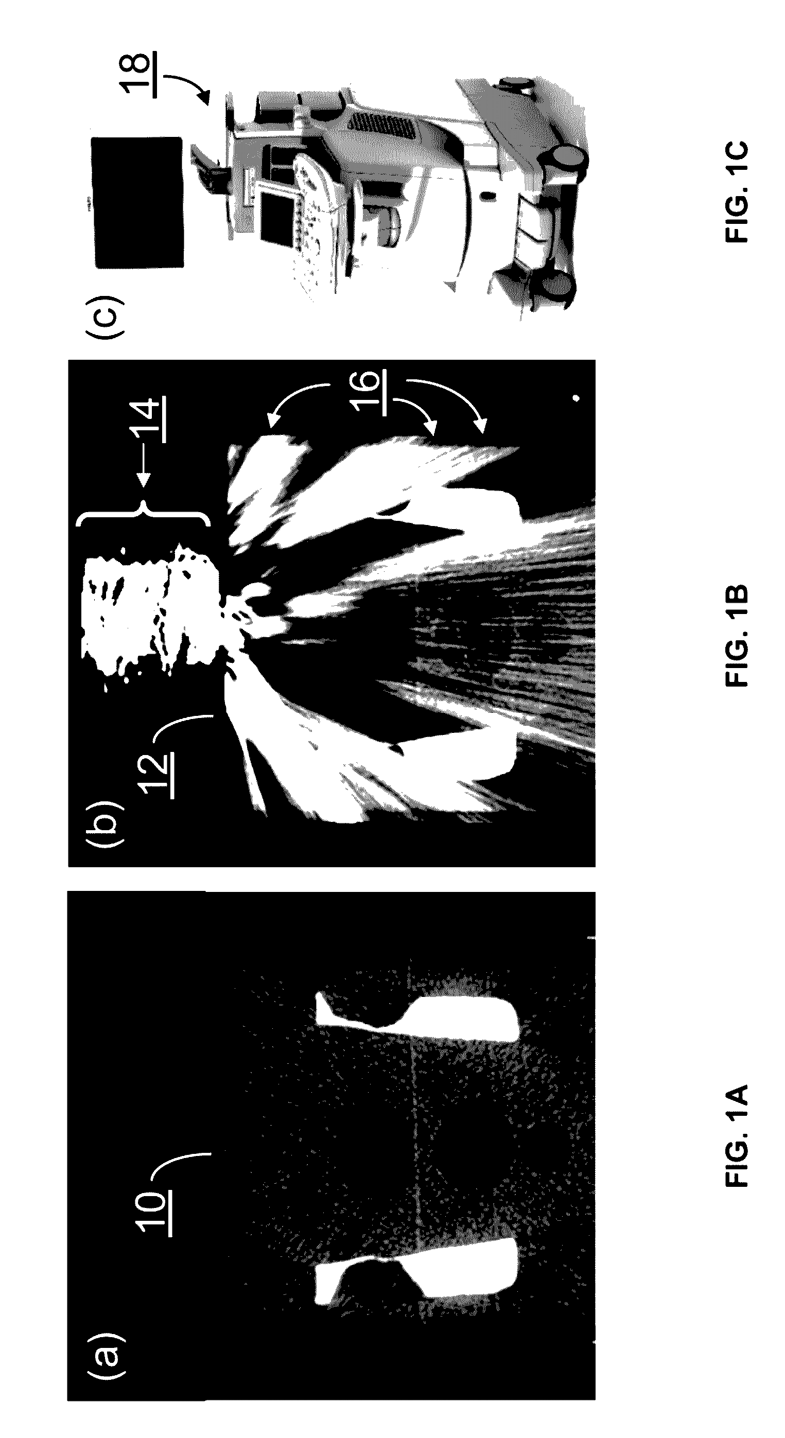
However, integration of a traditional multi-element phased array ultrasound probe with real-time radiotherapy guidance systems has several important challenges including treatment beam interference, computed tomography (CT) imaging interference, workflow obstacles, and high cost.

Method used


Image

  • Mechanically driven ultrasound scanning system and method
  • Mechanically driven ultrasound scanning system and method
  • Mechanically driven ultrasound scanning system and method

Examples


Embodiment Construction

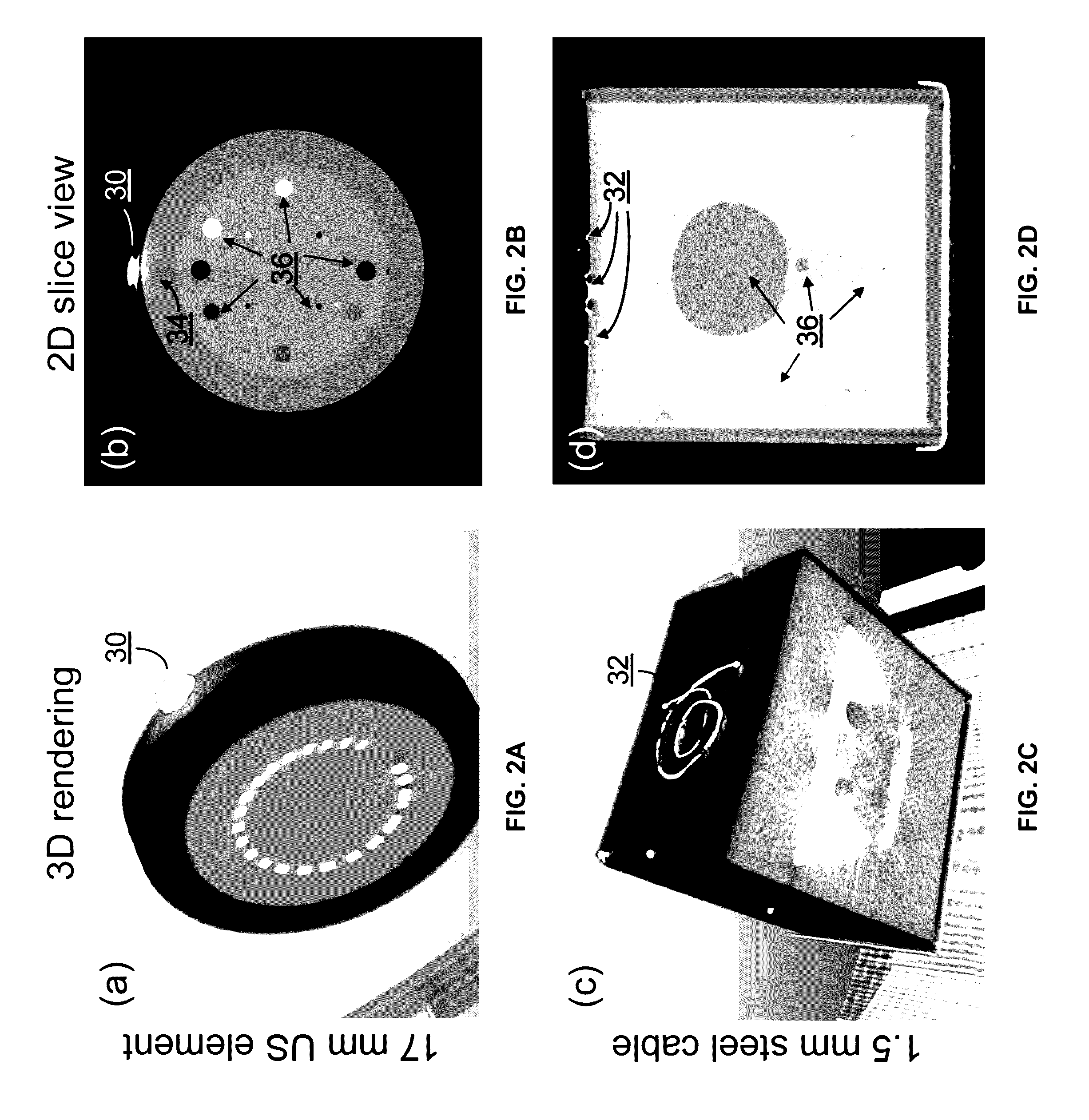
[0024]Use of existing 4D US probes for real-time radiation therapy guidance presents several important technical challenges including treatment beam interference, computed tomography (CT) imaging interference, workflow obstacles, and high cost. The following several paragraphs detail each challenge.

[0025]Treatment beam interference. Presence of a traditional US probe in the treatment field poses a challenge for radiation therapy planning. Radiation beams can pass through the US probe, causing dose deviations that negatively affect patient outcomes. This problem can be addressed using two approaches: (1) plan treatment beam directions to avoid US probe interference; (2) deliver radiation directly through the US probe and incorporate the hardware's perturbing effect into the dose calculation process. However, both approaches have substantial limitations. In the first approach, the US probe can preclude use of certain important beam angles and result in sub-optimal dose sculpting aroun...


PUM


Abstract

Described herein are radiolucent imaging devices and methods capable of acquiring real-time 2D, 3D, or 4D images. By using remote-actuation techniques to mechanically drive an ultrasound transducer element in multiple directions, the majority of dense metallic components typically present in the ultrasound probe itself are eliminated. Therefore the system herein achieves both CT compatibility and radiation beam compatibility.

Description

CROSS-REFERENCE TO RELATED APPLICATIONS[0001]This application is a continuation of International Application No. PCT / US2014 / 068933 filed Dec. 5, 2014, which claims the benefit of priority to U.S. Provisional Patent Application No. 61 / 912,949 filed Dec. 6, 2013, each of which is incorporated herein by reference in its entirety.FIELD OF THE INVENTION[0002]The present invention relates to methods and apparatus for acquiring 3-dimensional (3D) or 4-dimensional (4D) ultrasound (US) images using a mechanically controlled imaging system. The present invention further relates to method and apparatus for acquiring ultrasound (US) images using a radiolucent US probe. More particularly, the present invention relates to the design of an US image system for use during radiation therapy or other medical therapies.BACKGROUND OF THE INVENTION[0003]External Beam Radiation Therapy (EBRT) is used to treat more than half of all cancer patients worldwide. High dose conformality in EBRT is important for ...

Claims


Application Information

Patent Timeline
IPC IPC(8): A61B8/14A61B8/00A61B8/08
CPCA61B8/145A61B8/483A61N5/10A61B8/4405A61B8/4416A61B8/4461
Inventor SCHLOSSER, JEFFREYSOLEK, ROMAN
Owner SONITRACK SYST
Learn More