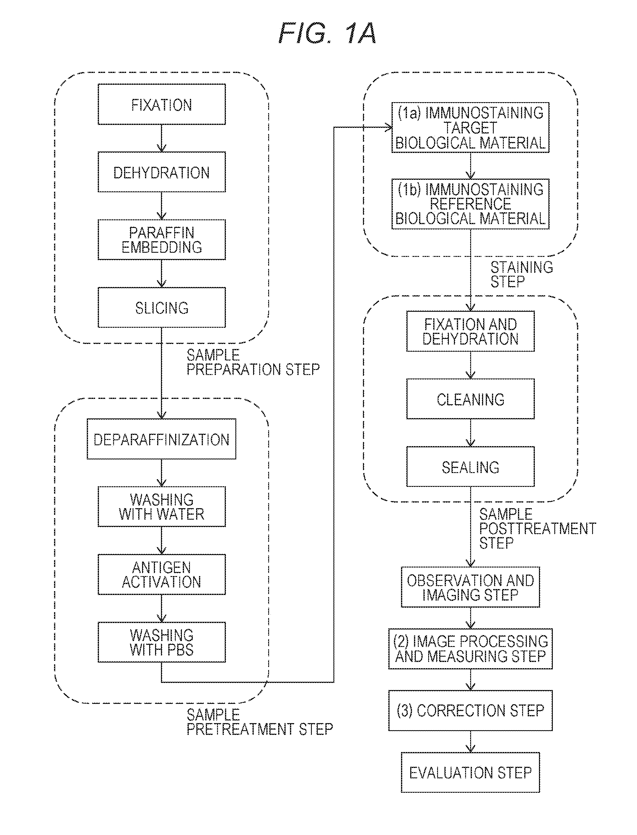
Method for quantifying biological material based on multiple immunostaining
a biological material and immunostaining technology, applied in material analysis, fluorescence/phosphorescence, instruments, etc., can solve the problem that the actual antibody expression level is difficult to estimate from the staining concentration, and achieve the effect of improving the accuracy of pathological diagnosis and increasing the accuracy of extraction and quantification
- Summary
- Abstract
- Description
- Claims
- Application Information
AI Technical Summary
Benefits of technology
Problems solved by technology
Method used
Image

Examples
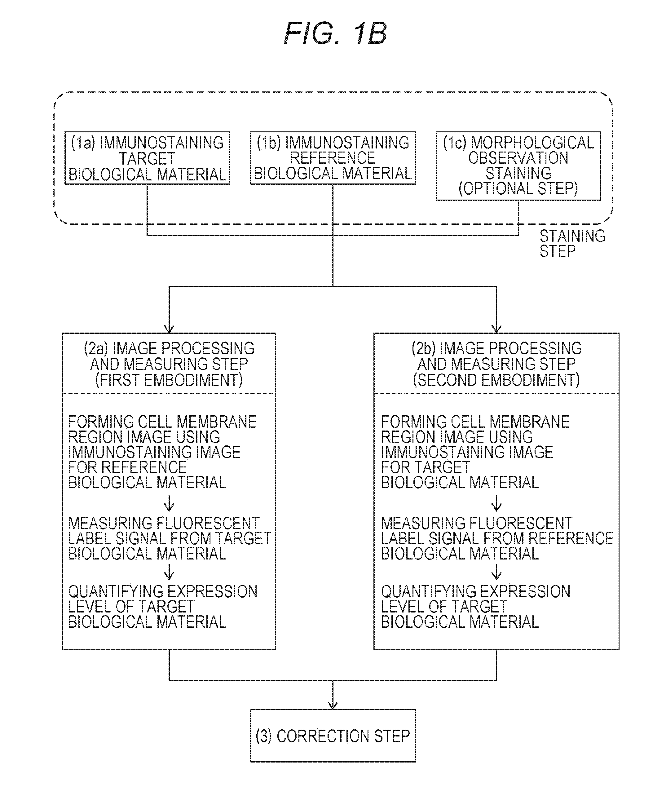
first embodiment
(2a) First Embodiment
[0120]The first embodiment includes the steps of:
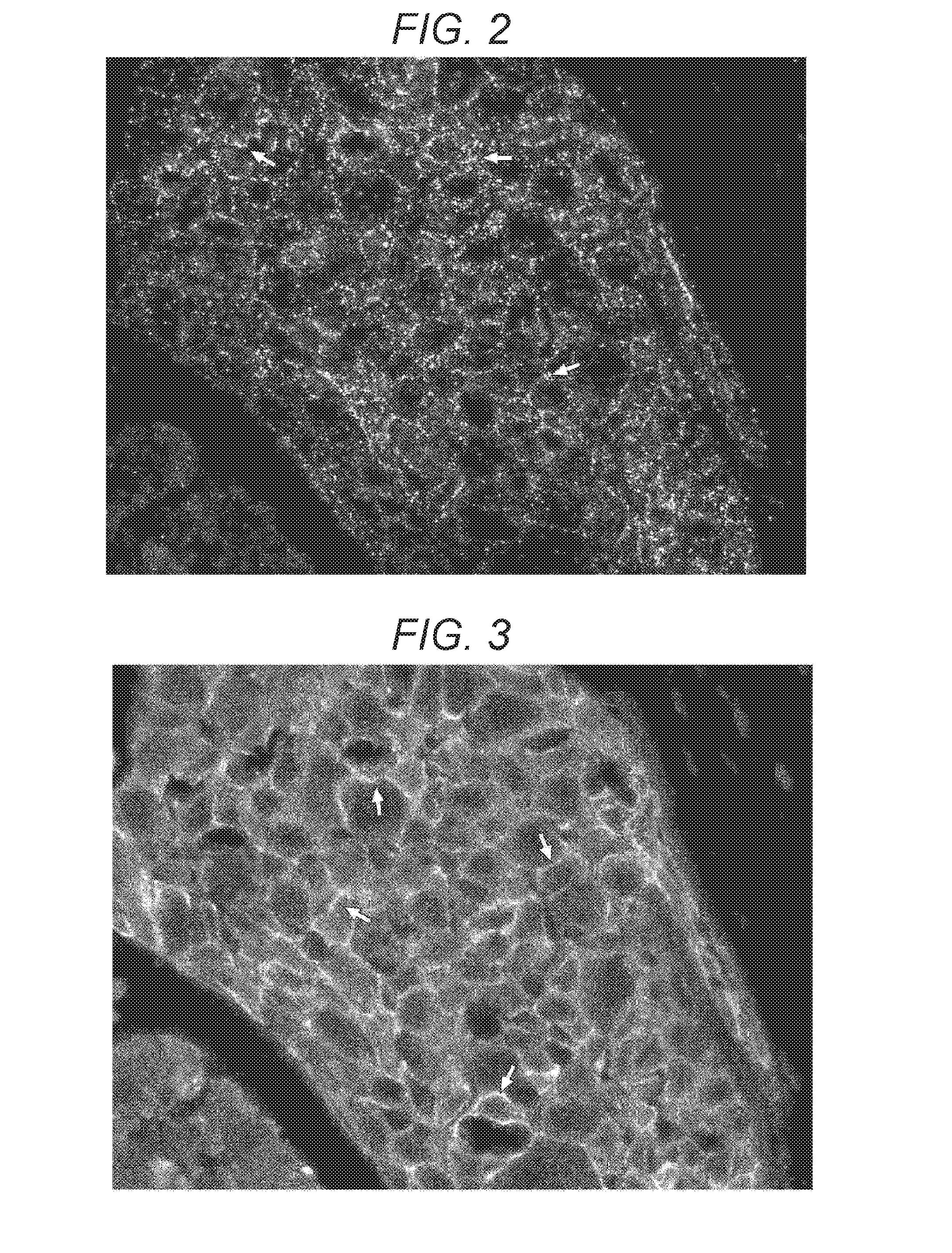
[0121](2a-1) extracting bright spots of the fluorescent dye from the immunostaining image (see FIG. 3) for the reference biological material to form an cell membrane region image and measuring, as the fluorescent label signal from the reference biological material, the sum total of the degrees of brightness of the bright spots in the cell membrane region;
[0122](2a-2) superimposing (see FIG. 4) the immunostaining image (see FIG. 2) for the target biological material on the cell membrane region image and measuring, as the fluorescent label signal from the target biological material, the number of bright spots of the fluorescent dye-containing nanoparticles inside the cell membrane region; and
[0123](2a-3) dividing the number of bright spots in the cell membrane region by the sum total of the degrees of brightness of the fluorescent dye to obtain a corrected measured value as an index value for quantifying the express...
second embodiment
(2b) Second Embodiment
[0125]The second embodiment includes the steps of:
[0126](2b-1) extracting bright spots of the fluorescent dye-containing nanoparticles from the immunostaining image (see FIG. 2) for the target biological material according to a bright spot extraction program to forma cell membrane region image and measuring, as the fluorescent label signal from the target biological material, the number of the bright spots in the cell membrane region;
[0127](2b-2) superimposing the immunostaining image (see FIG. 3) for the reference biological material on the cell membrane region and measuring, as the fluorescent label signal from the reference biological material, the sum total of the degrees of brightness of the fluorescent dye inside the cell membrane region; and
[0128](2b-3) dividing the number of the bright spots in the cell membrane region by the sum total of the degrees of brightness of the fluorescent dye to obtain a corrected measured value as an index value for quantify...
example 1
(E1) Sample Preparation Step and Sample Pretreatment Step
[0150]A cultured breast cancer cell line CRL1500 was fixed with 4% paraformaldehyde and then subjected to dehydration, embedding, and sectioning according to conventional methods, so that samples were obtained. Three samples were prepared for each of a 1 hour-fixation treatment group, a 12 hour-fixation treatment group, and a 96 hour-fixation treatment group.
[0151]The prepared samples were subjected to deparaffinization and then washing for replacement with water. The washed tissue array slides were autoclaved in a 10 mM citric acid buffer solution (pH 6.0) at 121° C. for 15 minutes so that antigen activation was performed. After the activation, the tissue array slides were washed with PBS. The washed tissue array slides were subjected to a blocking treatment with 1% BSA-containing PBS for 1 hour.
(E2) Immunostaining Step
(E2-1) Common Primary Reaction Treatment for First Immunostaining and Second Immunostaining
[0152]A primary r...
PUM
| Property | Measurement | Unit |
|---|---|---|
| wavelength | aaaaa | aaaaa |
| particle size | aaaaa | aaaaa |
| particle size | aaaaa | aaaaa |
Abstract
Description
Claims
Application Information
 - R&D Engineer
- R&D Manager
- IP Professional
- Industry Leading Data Capabilities
- Powerful AI technology
- Patent DNA Extraction
Browse by: Latest US Patents, China's latest patents, Technical Efficacy Thesaurus, Application Domain, Technology Topic.
© 2024 PatSnap. All rights reserved.Legal|Privacy policy|Modern Slavery Act Transparency Statement|Sitemap