Magnetic resonance imaging with motion correction suing pre-pulses and navigators

a technology of magnetic resonance imaging and motion correction, applied in the direction of magnetic measurements, instruments, measurements using nmr, etc., can solve the problems of affecting the operation efficiency of the scanner, affecting the accuracy of the scan, so as to achieve efficient operation

Active Publication Date: 2017-02-09
KONINKLJIJKE PHILIPS NV
View PDF5 Cites 15 Cited by
  • Summary
  • Abstract
  • Description
  • Claims
  • Application Information

AI Technical Summary

Benefits of technology

[0009]The invention may provide a method for magnetic resonance imaging using pre-pulses, a MR imaging system for performing the above method, and a software package for upgrading a MR imaging system to perform the above method, which enable magnetic resonance imaging with high efficiency, high resolution, good image quality and few artifacts for a wide range of subjects of interest, in particular in regions of interest affected by breathing motion of the subject of interest.
[0044]According to another embodiment the step of performing real-time motion correction by adjusting MR scanning parameters for compensation of detected motion of the subject of interest comprises adjusting detection frequency in the readout direction, detection phase in the readout direction, and slice encoding direction. When adjusting the scanning parameters, motion correction can easily be performed for the readout. Motion correction is preferably performed after performing the readout in respect to the readout, for which motion is detected. Alternatively, motion correction can be performed already prior to a readout, i.e. motion can be estimated prior to a readout based e.g. on the previous readout, so that respective motion correction can be applied in advance. Adjusting detection phase enables control of the phase shift applied to the respective k-space lines of the imaging sequence. Adjusting slice encoding direction is particularly suitable when using 3D imaging.

Problems solved by technology

These scan times are not suitable for scanning under breath-hold condition, in particular when considering that the subject of interest, i.e. the patient, is frequently undergoing the MR scan because of serious problems in the upper chest, which may impede maintaining breath-hold condition for the required time of at least an entire scanning sequence.
Therefore, e.g. clinical T1 mapping scans have been largely limited to 2D breath-hold acquisitions with resulting restrictions in the achievable spatial resolution and signal-to-noise ratio.
This however, has not proven reliable and leads to long scan times and low scan efficiencies, which can even be below fifty percent.
The navigator echoes are used to discard data if the heart and / or diagphram are not within a prescribed user position, and is described as being time consuming and technologically challenging to implement.

Method used


Image

  • Magnetic resonance imaging with motion correction suing pre-pulses and navigators
  • Magnetic resonance imaging with motion correction suing pre-pulses and navigators
  • Magnetic resonance imaging with motion correction suing pre-pulses and navigators

Examples


Embodiment Construction

[0058]Like numbered elements in these figures are either equivalent elements or perform the same function. Elements which have been discussed previously will not necessarily be discussed in later figures if the function is equivalent.

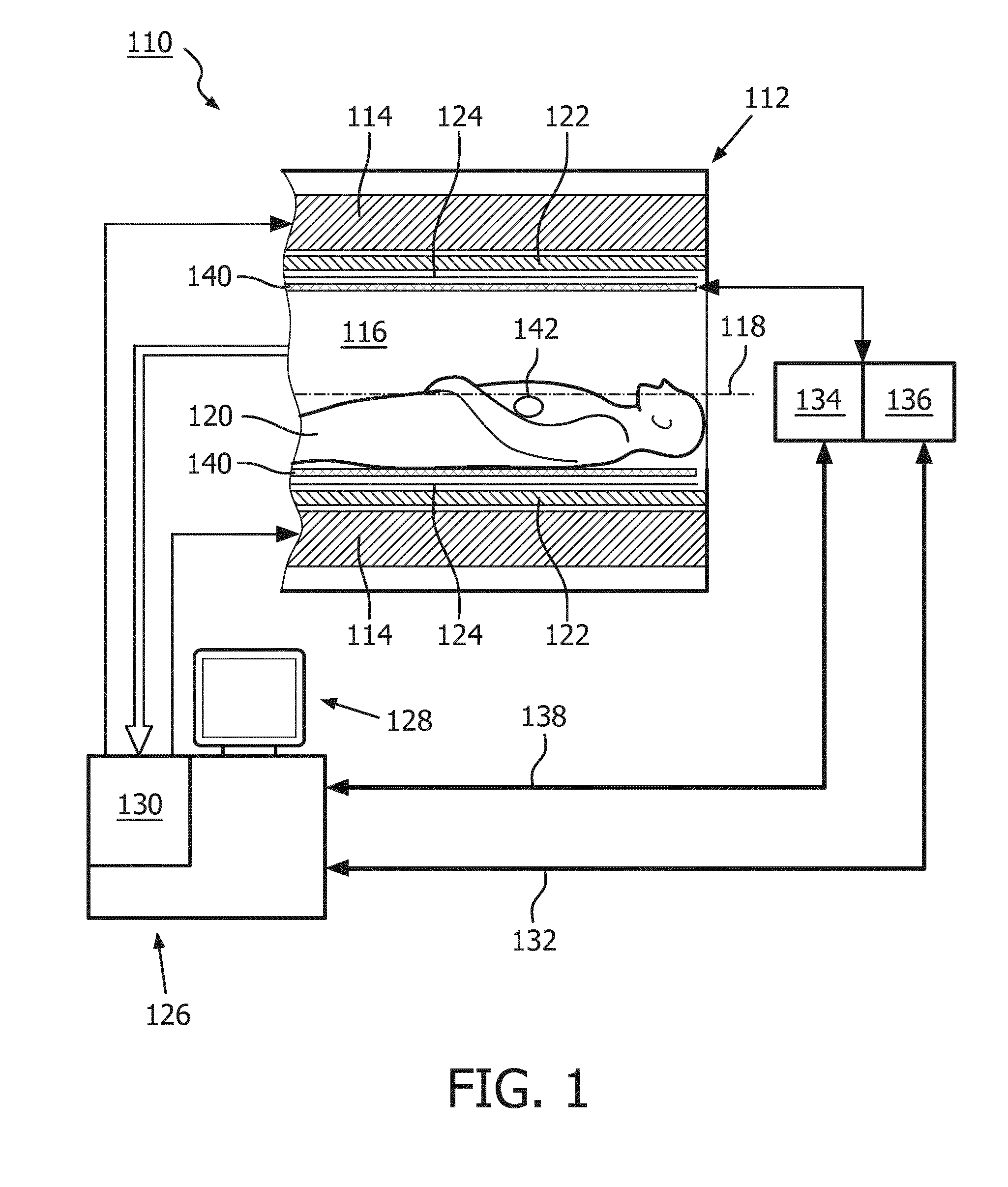
[0059]FIG. 1 shows a schematic illustration of an embodiment of a magnetic resonance (MR) imaging system 110 in accordance with the invention. The MR imaging system 110 can be a MR imaging system known in the Art, which is operated as described later in detail to perform MR imaging, in particular to perform cardiovascular MR imaging.

[0060]The MR imaging system 110 comprises an MR scanner 112. The MR imaging system 110 further includes a main magnet 114 provided for generating a static magnetic field. The main magnet 114 has a central bore that provides an examination space 116 around a center axis 118 for a subject of interest 120, usually a patient, to be positioned within. In this embodiment, the central bore and therefore the static magnetic field of...


PUM


Abstract

The present invention provides a method for magnetic resonance (MR) imaging of a region of interest (142) of a subject of interest (120) under application of a scanning sequence (200) comprising at least one pre-pulse (202, 204) and multiple readouts (206), whereby the multiple readouts (206) are performed after the at least one pre-pulse (202, 204) with different configurations causing different image contrasts, comprising the steps of performing a preparation phase comprising applying at least one scanning sequence (200) to provide a set of reference readouts (206) using the different configurations, and generating a set of navigator images (210) with one navigator image (210) of the region of interest (142) for each configuration of the reference readouts (206), performing an examination phase comprising applying at least one scanning sequence (200), whereby at least one image (212) of the region of interest (142) is generated for each scanning sequence (200), determining motion of the subject of interest (120) by comparing at least one image (212) of the scanning sequence of the examination phase to the navigator image (210) having the same configuration as the compared image (212), performing motion correction of the at least one image (212) based on the determined motion of the subject of interest (120) of the at least one image (212), and providing an MR scan (214) of the region of interest (142) of the subject of interest (120) based on the images (212) after performing motion correction. The invention also provides a MR imaging system (110) adapted to perform the above method and a software package for upgrading a MR imaging system (110), whereby the software package contains instructions for controlling the MR imaging system (110) according to the above method.

Description

FIELD OF THE INVENTION[0001]The invention relates to the field of magnetic resonance imaging using pre-pulses.BACKGROUND OF THE INVENTION[0002]Different techniques for magnetic resonance (MR) imaging using pre-pulses are known in the Art, e.g. in the area of cardiovascular MR imaging. For example longitudinal relaxation time (T1) mapping is becoming an increasingly valuable tool in cardiovascular MR to diagnose a range of cardiomyopathies. T1 mapping is done using pre-pulses to interrogate the recovery behavior of the magnetization and performing readouts after given delays with respect to the pre-pulses. Due to the number of interrogation points required for performing required readouts, typical scanning sequences require at least two pre-pulses. In other configurations, different flip angles can be used for the readout, also resulting in images having different image contrasts. In other setups, different b-values can be used, e.g. in diffusion weighted imaging, also resulting in i...

Claims


Application Information

Patent Timeline
Patent Type & Authority Applications(United States)
IPC IPC(8): G01R33/565G01R33/561G01R33/483G01R33/567
CPCG01R33/56509G01R33/4838G01R33/5619G01R33/5673G01R33/50
Inventor VOIGT, TOBIAS RATKOHENNINGSSON, MARKUS OLAF SAMUEL
Owner KONINKLJIJKE PHILIPS NV
Why Eureka
Try Eureka