Ultrashield devices and methods for use in ultrasonic procedures

a technology of ultrasonic procedures and ultrasonic shields, applied in the field of ultrasonic shield devices and methods for use in ultrasonic procedures, can solve the problems of difficult to remove from the patient, fluid or gel is difficult to contain on the patient, and cannot be readily achieved

Active Publication Date: 2017-05-11
CAL TENN INNOVATION INC
View PDF16 Cites 31 Cited by
  • Summary
  • Abstract
  • Description
  • Claims
  • Application Information

AI Technical Summary

Benefits of technology

[0024]In some embodiments, the ultrashield further comprises a probe contact layer configured to mate with the couplant layer and adhere to the probe cover. In other embodiments, the ultrashield is integral with the probe cover. Optionally, the probe cover may further comprise a removable protective pouch extending over the ultrashield, wherein the pouch resists loss of couplant from the couplant layer.

Problems solved by technology

Clearly, in the case of ultrasound waves passing from the probe to the tissues, this cannot be readily achieved.
To begin, patients often find the fluid or gel to be cold, sticky and messy.
The fluids or gels are difficult to contain on, and remove from, the patient during and after the ultrasound procedure.
Further, commercially available oils and water based gels often introduce problems to the electronics by their chemically degrading nature.
They may react with the adhesives, elastomers, and epoxies used in the construction of medical ultrasound transducers, thus appreciably degrading performance and shortening their service life.
It has also resulted in closing of a company.
Such chemicals are not approved or suitable for use in applications where they may be carried into the body, such as during biopsy, intra-operative procedures, or when the transducer is placed inside a body orifice.
In instances where sterile latex rubber or synthetic covers containing thickened ultrasound coupling gels are used in surgery, tearing, cutting, or rupture of the cover results in the tissue incompatible ultrasound coupling gel spilling into the body cavity.
In addition, many practitioners also have difficulty with consistency of application.
It is difficult for many practitioners to apply enough gel within a probe cover to prevent air pockets, to remain thick enough and evenly applied between the probe and cover throughout the examination without ‘spilling around’ edges of the probe, and without causing uneven ‘wrinkles’ or curvatures of the cover which causes air pockets outside the probe cover.
This increases procedure time and results in suboptimal visualization of underlying structures and interferes with the quality of examination or procedure.

Method used


Image

  • Ultrashield devices and methods for use in ultrasonic procedures
  • Ultrashield devices and methods for use in ultrasonic procedures
  • Ultrashield devices and methods for use in ultrasonic procedures

Examples


Embodiment Construction

[0055]Specific embodiments of the disclosed devices and methods will now be described with reference to the drawings.

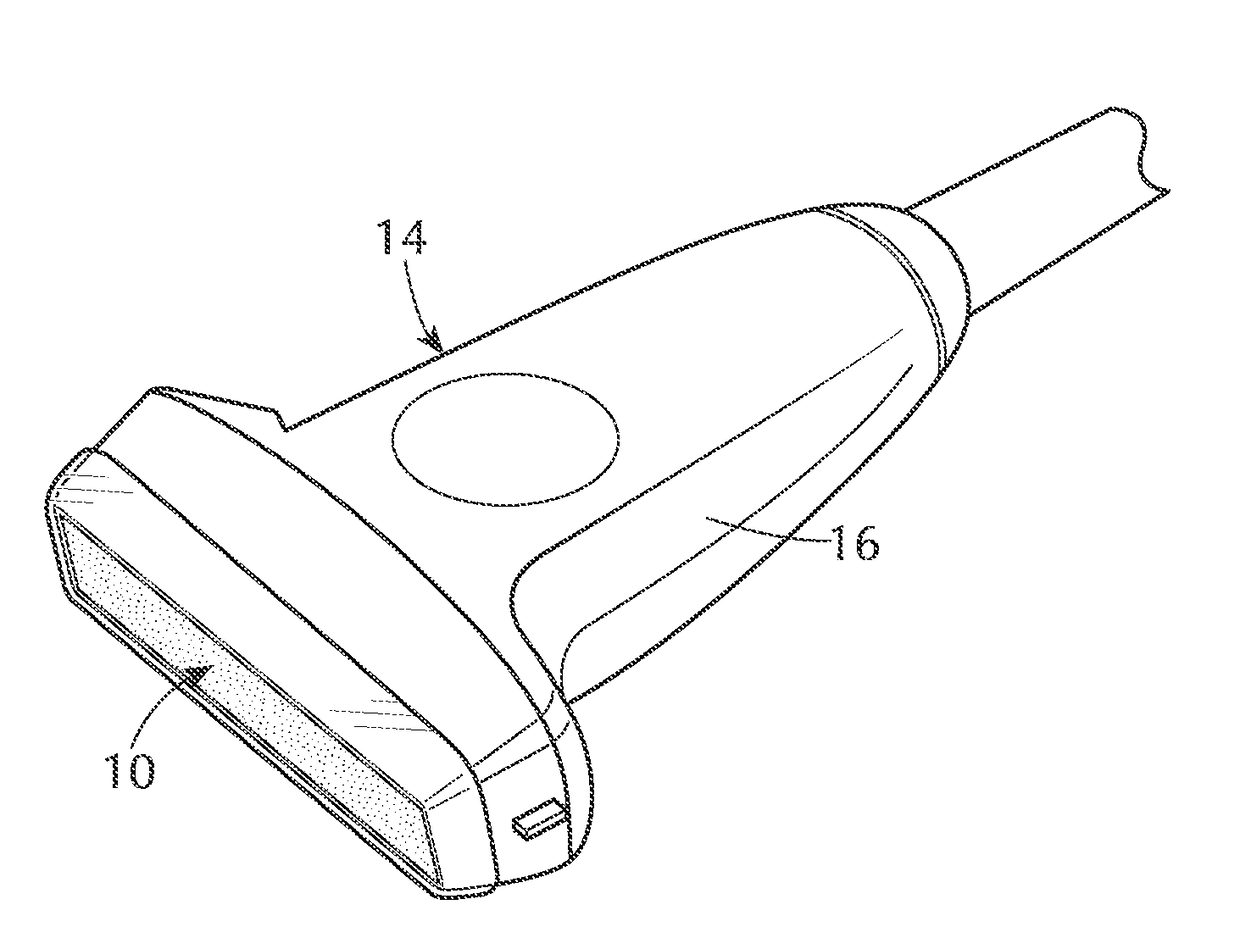
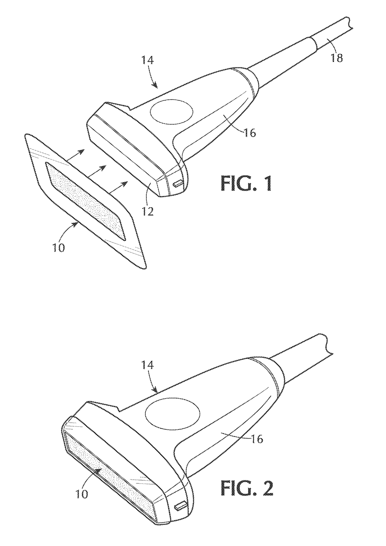
[0056]Devices and methods are provided for specific use with ultrasound machines to enable a clinician or a technician to use a conventional ultrasound probe, such as to generate an ultrasonic image, without the need for external ultrasonic gel or similar couplant. In particular, an ultrashield is provided for use with a conventional ultrasound probe that eliminates the need for additional ultrasonic couplants, such as gels. The ultrashield is a cover or shield which is positioned over the faceplate of an ultrasound probe, either alone or in conjunction with a probe cover. FIG. 1 illustrates an embodiment of an ultrashield 10 which is sized and configured to affix to the faceplate 12 of an ultrasound probe 14, as indicated by arrows. Typically, the ultrashield 10 is sized and configured so that portions of its exterior edge wrap around the probe 14, beyond the facepla...


PUM


Abstract

Devices and method are provided for ultrasound transmission without the need for external couplants, such as gels, which are typically used in conventional ultrasound procedures. In particular, ultrashields are provided for use with ultrasound probes, wherein the ultrashields have specialized layers to provide an uninterrupted pathway of acoustic conductance from the probe to the surface of the body throughout the procedure while introducing minimal to no attenuation of ultrasound wave transmission. In addition, combinations of ultrashields and probe covers are provided to provide additional features such as a microbial barrier.

Description

CROSS-REFERENCE[0001]This application claims priority to U.S. Provisional Patent Application No. 62 / 285,758 entitled An Ultrasonic Couplant Design and Probe Cover Design to Replace Use of Ultrasonic Gel During Ultrasound Imaging, filed on Nov. 9, 2015, incorporated herein by reference for all purposes.BACKGROUND OF THE INVENTION[0002]The term “ultrasound” typically applies to acoustic energy with a frequency above human hearing (20,000 hertz or 20 kilohertz). When used in medical applications, ultrasound is typically between 1 and 30 MHz for imaging and flow measurements and between 0.05 and 1.00 MHz for therapy. The application of ultrasound in medicine began in the 1950s. It was first introduced in the field of obstetrics. Obstetric sonography is the use of medical ultrasonography in pregnancy, in which sound waves are used to create real-time visual images of the developing embryo or fetus in its mother's uterus. The procedure is a standard part of prenatal care in many countries...

Claims


Application Information

Patent Timeline
Patent Type & Authority Applications(United States)
IPC IPC(8): A61B8/00
CPCA61B8/4281A61B8/4422A61B8/4444A61B8/4455
Inventor DESAI, SIDDHARTHJONES, JERRY
Owner CAL TENN INNOVATION INC
Why Eureka
Try Eureka