Method for segmenting and predicting tissue regions in patients with acute cerebral ischemia

a technology of cerebral ischemia and tissue regions, applied in the field of multi-dimensional imaging, can solve the problems of limited accuracy of classification models, insufficient reliability or accuracy of outputs, and insufficient reliability of classification models

Inactive Publication Date: 2017-05-18
UNIVERSITY OF BERN
View PDF5 Cites 17 Cited by
  • Summary
  • Abstract
  • Description
  • Claims
  • Application Information

AI Technical Summary

Benefits of technology

[0010]FIG. 1 shows a simplified flow diagram of an example segmentation method for use in a segmentation/prediction method according to the invention.
[0011]FIG. 2 shows a simplified flow diagram of an

Problems solved by technology

Approaches have been proposed which consider both regions simultaneously, but these have used relatively simplistic classification models and have limited accuracy.
The prior art methods have the

Method used


Image

  • Method for segmenting and predicting tissue regions in patients with acute cerebral ischemia
  • Method for segmenting and predicting tissue regions in patients with acute cerebral ischemia
  • Method for segmenting and predicting tissue regions in patients with acute cerebral ischemia

Examples


first embodiment

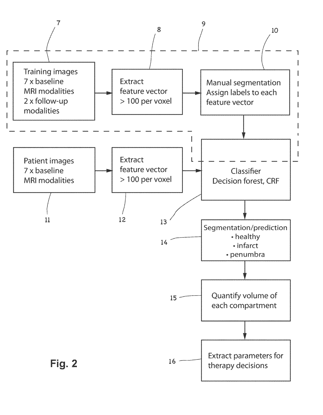
[0079]As will be described in relation to the invention, the training data may comprise image datasets, 7, whose modalities and feature vectors, 8, correspond to the image dataset(s), 11, and feature vector(s), 12, of patients. The training data comprises pre-treatment images comprising hypoxic regions of previous stroke patients, and the voxels may be manually segmented, 10, for example by an experienced neuroradiologist, in order to generate training data for training the classifier, 13.

second embodiment

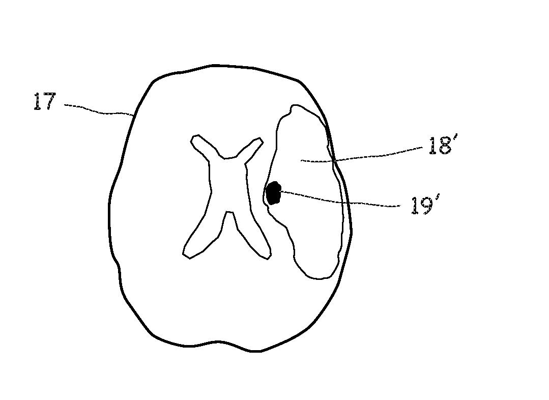
[0080]As will described in relation to the invention, and as illustrated in FIG. 2, the training data 7 may additionally comprise follow-up image datasets, for example post-treatment image datasets corresponding to (i.e. relating to the same patients as) at least some of the pre-treatment MRI images of the hypoxic regions of the previous stroke patients mentioned above. In the example illustrated in FIG. 2, the follow-up MRI image datasets may comprise only structural modalities (e.g. T1contrast and T2) This allows the learning process to benefit from the outcome information present in the structural modality information. Advantageously, the training data 7 may optionally include information about the treatment which was carried out on the patients whose follow-up MRI image data is included. Such treatment parameter information (for example the type of treatment, or the frequency, dosage, drug details, therapy duration, surgical interventions etc) may also be included in the trainin...


PUM


Abstract

A segmentation/prediction method is described for differentiating between infarct, penumbra and healthy regions in a tomographic (e.g. MRI or CT) image dataset of the brain of a stroke patient under examination. The method comprises deriving (7, 11) a multidimensional set of feature vectors from a plurality of baseline modalities, where the modalities comprising both structural and functional modalities. For each volume element of image dataset, an n-dimensional feature vector is extracted (8, 12), such that it represents both structural and functional modalities of the volume element. A classification (13) is performed on the volume element and the classification is used to inform the segmentation (14) in order to label the volume element as belonging to healthy tissue, penumbra tissue, or infarct tissue. The classification operation (13) uses a learning-based classifier, trained using pre-treatment image datasets comprising a plurality of second hypoxic regions, the second hypoxic regions being of the brains of previous stroke patients. In a second embodiment, follow-up (post-treatment) image datasets are used for training the classifier.

Description

TECHNICAL FIELD OF THE INVENTION[0001]The present invention relates to the field of multi-dimensional imaging and, in particular, to the field of classifying volumetric elements of affected regions of the brains of acute ischemic stroke patients in order to differentiate between salvageable and non-salvageable brain tissue.BACKGROUND OF THE INVENTION[0002]Acute ischemic stroke, or cerebral ischemia, is a neurological emergency which may be reversible if treated rapidly. Outcomes for stroke patients are strongly influenced by the speed and accuracy with which the ischemia can be identified and treated. Effective reperfusion and revascularization therapies are available for salvaging regions of brain tissue which are characterized by reversible hypoxia, and these regions must be identified and distinguished from tissue which is destined to infarct. Volumetric imaging of the brain tissue, using computer tomography (CT) or magnetic resonance imaging (MRI) may be used to generate 4D (spa...

Claims


Application Information

Patent Timeline
IPC IPC(8): G06T7/62G06K9/66G06T7/174G06K9/62G06T7/00G06T7/11
CPCG06T7/62G06T7/0012G06T7/11G06T7/174G06K9/6256G06T2207/30096G06K9/6267G06T2207/20081G06T2207/10088G06T2207/30016G06T2207/10081G06K9/66G06F18/41G06F18/24G06F18/214
Inventor BAUER, STEFANREYES, MAURICIOWIEST, ROLAND
Owner UNIVERSITY OF BERN
Why Eureka
Try Eureka