
Method and apparatus for actuation of a magnetic resonance scanner for the simultaneous acquisition of multiple partial volumes

Active Publication Date: 2017-05-25
SIEMENS HEALTHCARE GMBH
View PDF2 Cites 12 Cited by
  • Summary
  • Abstract
  • Description
  • Claims
  • Application Information

AI Technical Summary

Benefits of technology

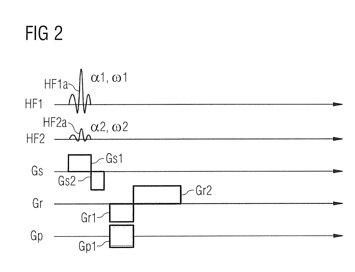
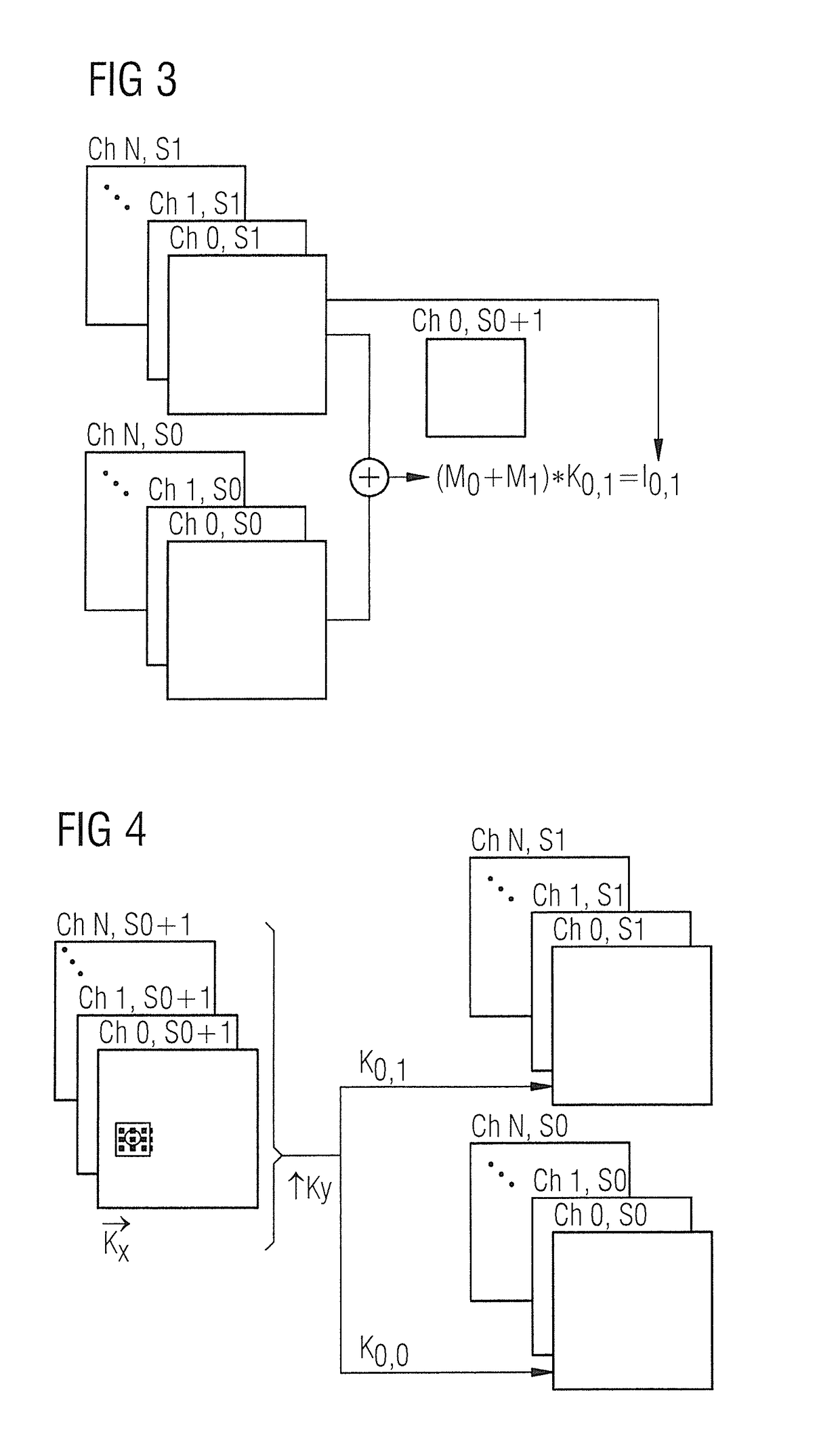
The present invention provides a magnetic resonance imaging (MRI) method that allows for faster MRI with lower SAR stress and higher precision in recording images with different contrasts. The method involves simultaneously actuating a MRI data acquisition scanner to acquire magnetic resonance raw data of an object using different transverse magnetizations. This results in images with different contrasts that can be recorded simultaneously, reducing the time required for the MRI process and avoiding cumbersome post-editing of images with different contrasts.

Problems solved by technology

However, due to the long relaxation times of the spins, the imaging times are very long and this, impairs the comfort of a patient to be examined.
In addition, during the data recording, patients are not able to leave the magnetic resonance tomography system briefly or even to change their position because a change in position of the patient while data are being acquired would render the imaging process useless, and the entire process would have to be restarted from the beginning.
Advantageously, these methods reduce the measuring time and / or increase the temporal sampling rate.
Depending upon the pulse sequence of the sequence to be accelerated, it is not automatically possible to apply all pulses to a number of slices simultaneously without, for example, exceeding SAR thresholds (SAR=specific absorption rate=measure for the absorption of electromagnetic fields in a material) or the available peak power of the RF amplifier.
If, for example, imaging is initially performed with simultaneous excitation of a number of slices with a first flip angle α1 for all slices, and then further simultaneous excitation is performed with a second flip angle α2 with another value for all slices, this has the drawback that for the MR imaging with the higher flip angle, both the SAR stress and the required RF peak power would be higher by the factor N of the number of simultaneously excited slices.
This steep increase means stress and power thresholds are easily exceeded in the case of SMS measurements.
A further problem with SMS imaging relates to the chronological consistency of image data with different contrasts.
Hence, the assignment of image information between the different contrasts requires complex and error-prone registration methods.
A similar problem also occurs if different sequence types are to be applied for different slices.
However, in many scenarios, it is no longer possible to achieve acceleration factors of 2.
However, the latter instance represents a restriction of the method since both slices initially undergo slightly different dephasing, which has to be taken into account by suitable compensation measures (for example considered during the application of a refocusing pulse).
One drawback of this technique is that the recurring excitation pattern continues ad infinitum and hence, depending upon the positioning of the image volume, (unwanted) more remote anatomical structures are also excited.

Method used


Image

  • Method and apparatus for actuation of a magnetic resonance scanner for the simultaneous acquisition of multiple partial volumes
  • Method and apparatus for actuation of a magnetic resonance scanner for the simultaneous acquisition of multiple partial volumes
  • Method and apparatus for actuation of a magnetic resonance scanner for the simultaneous acquisition of multiple partial volumes

Examples


Embodiment Construction

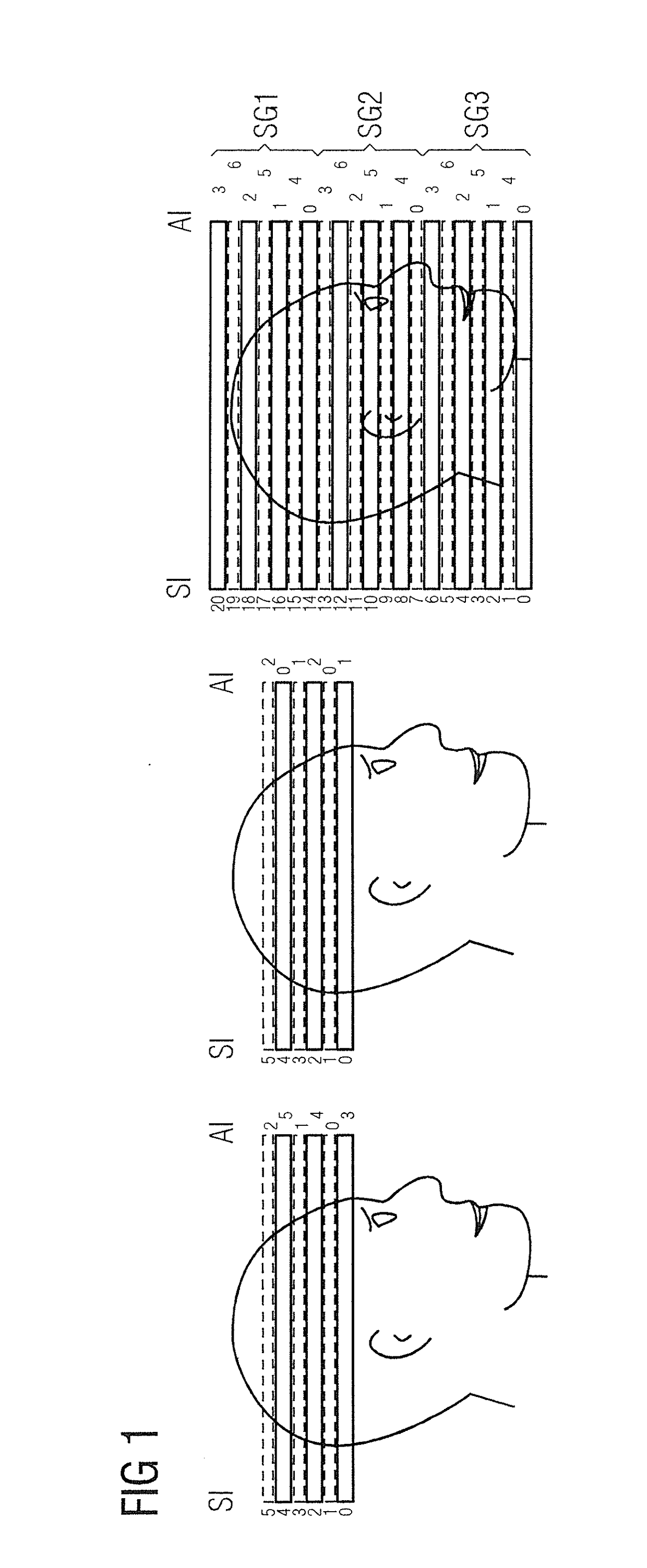
[0066]In FIG. 1 shows three diagrams illustrating an acquisition scheme for simultaneous multi-slice imaging (SMS). The left diagram shows an acquisition scheme for a MR-magnetic resonance imaging method with single-slice recording. This depicts a non-accelerated measurement with 6 interleaved recordings of slices. In the case of echo-planar imaging this requires 6 slice excitations and 6 readout cycles. The left side of the left diagram is a slice index SI of a respective slice. In this case, the slice index SI goes from 0 to 5. The right side of the left diagram shows acquisition indices AI. These indicate the sequence in which each slice is excited and read-out. The left diagram shows an interleaved excitation of single slices, i.e., the slices are excited and read-out in the sequence 1, 3, 5, 0, 2, 4.

[0067]In the middle diagram, once again 6 slices are recorded but with an accelerated SMS imaging procedure with the acceleration factor 2. I.e., 2 slices are always excited and als...


PUM


Abstract

In a method and a magnetic resonance imaging system to generate magnetic resonance image data of an object, during the acquisition of magnetic resonance raw data, different transverse magnetizations are excited in multiple sub-volumes to be depicted and used for imaging. These different transverse magnetizations are simultaneously present in at least one time interval of the measurement. Image data are reconstructed from the acquired raw data.

Description

BACKGROUND OF THE INVENTION[0001]Field of the Invention[0002]The invention concerns a method for the actuation of a magnetic resonance imaging scanner for the generation of magnetic resonance image data of an object under examination from whom magnetic resonance raw data are acquired. The invention also relates to an actuation-sequence-determining system. The invention also relates to a magnetic resonance imaging system.[0003]Description of the Prior Art[0004]Modern imaging methods are frequently used to generate two-dimensional or three-dimensional image data, or a time series of image data, which can be used for the visualization of an object under examination. Imaging systems based on magnetic resonance imaging, called magnetic resonance tomography systems, have been successfully established and proven in a multitude of applications. With this type of imaging, generally a static basic magnetic field B0, which is used for initial alignment and homogenization of magnetic dipoles (n...

Claims


Application Information

Patent Timeline
IPC IPC(8): G01R33/483G01R33/48G01R33/54
CPCG01R33/4835G01R33/4828G01R33/543
Inventor BECK, THOMASFEIWEIER, THORSTEN
Owner SIEMENS HEALTHCARE GMBH
Learn More