Differentiation of macrophages from pluripotent stem cells

Active Publication Date: 2018-01-11
AGENCY FOR SCI TECH & RES
View PDF2 Cites 2 Cited by
  • Summary
  • Abstract
  • Description
  • Claims
  • Application Information

AI Technical Summary

Problems solved by technology

However, in depth insights into the differentiation of primitive macrophages from human pluripotent stem cells, their homeostasis, as well as their defined activities in a tissue-specific context are currently lacking.
However, while monocytes clearly give rise to macrophages in pathological settings and inflammation, recent evidence has shown that (1

Method used


Image

  • Differentiation of macrophages from pluripotent stem cells
  • Differentiation of macrophages from pluripotent stem cells
  • Differentiation of macrophages from pluripotent stem cells

Examples


example 1

[0064]Differentiation of Mouse Pluripotent Stem Cells into Primitive Macrophages

[0065]If mESC / miPSC are expanded on MEF, deplete MEF by 1-2× passage on gelatin-coated 6-well plates before commencing with differentiation.

[0066]On day 0, colonies are dissociated and resuspended at 175,000-200,000 cells / ml in serum-free differentiation medium (recipe below) supplemented with 5 ng / ml hFGF2 and 5 ng / ml hBMP4. These are dispensed into non-adherent plates to allow for embryoid body (EB) formation.

[0067]After 48 h, EBs are pooled and dissociated with TrypLE (preferred) or Trypsin.

[0068]EBs are then reaggregated at 175,000 cells / ml in serum-free differentiation medium supplemented with 5 ng / ml hFGF2, 2 ng / ml hBMP4, 2 ng / ml human Activin A, 5 ng / ml hVEGF.

[0069]After a further 48 h, EBs are dissociated and sorted for Flk-1+ cells.

[0070]Sorted cells are then reaggregated at 500,000 cells / ml in serum-free differentiation medium supplemented with 5 ng / ml VEGF, 300 ng / ml DKK1, 100 ng / ml M-CSF. The...

example 2

[0079]Differentiation of Human Pluripotent Stem Cells into Primitive Macrophages

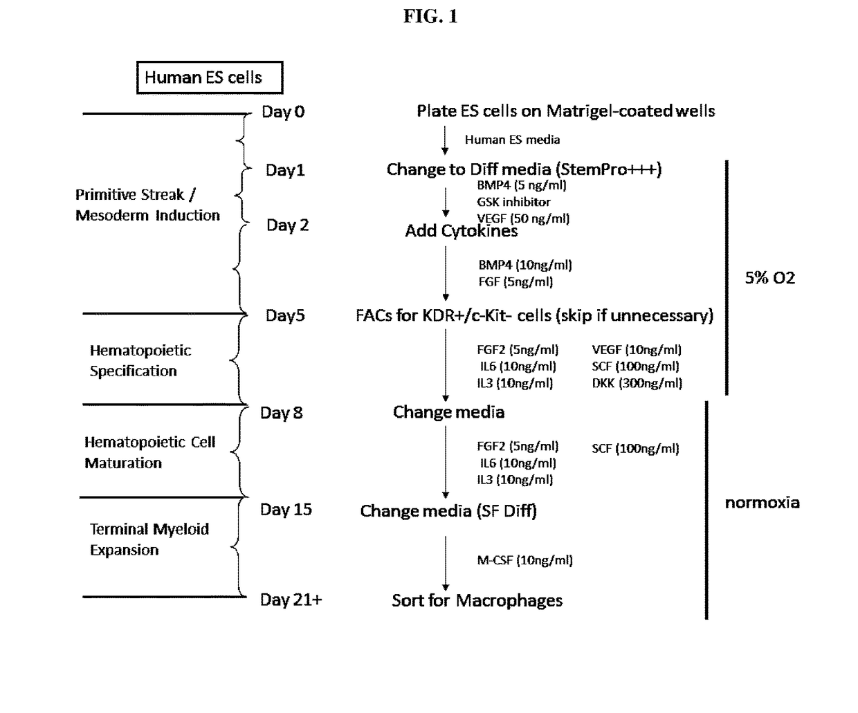
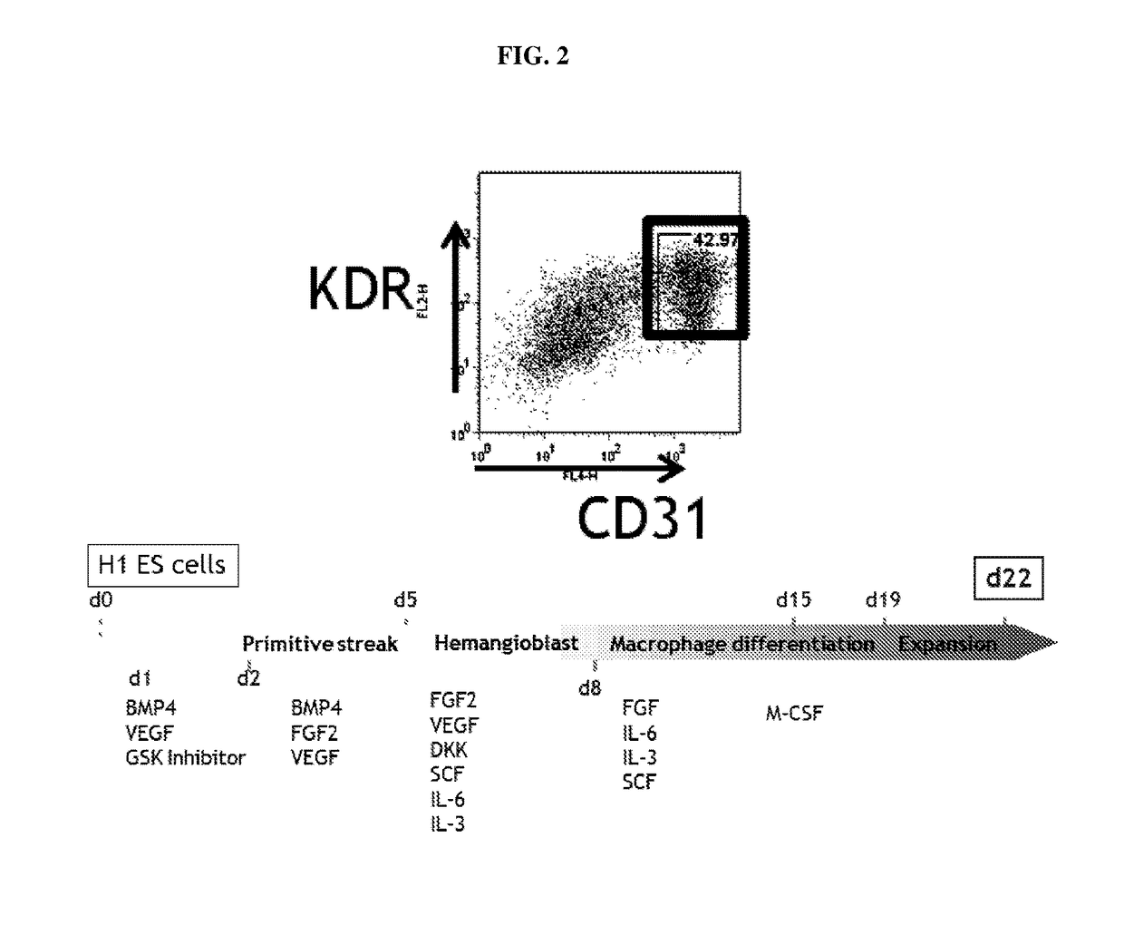
[0080]The differentiation strategy can be divided into four stages: primitive streak / mesoderm induction, hematopoietic specification, hematopoietic cell maturation, and finally terminal myeloid expansion. The experimental details of each stage are illustrated in FIGS. 1 and 2, and described below:

[0081]Stage 0: ES or iPS cells are maintained on Matrigel-coated dishes in CF1 MEF-conditioned media. The media recipe consists of 80% DMEM / F12, 20% knockout serum replacement, L-glutamine, non-essential amino acids, beta-mercaptoethanol, and 6 ng / ml FGF-2 and media is replaced daily. Cells are passaged every 5-6 days using collagenase IV treatment and gentle detachment with a cell scraper. 24 hours prior to initiation of differentiation, cells are plated at a very low density (2 on Matrigel-coated 6-well plates and allowed to attach overnight.

[0082]Stage 1 (primitive streak / mesoderm induction): In this initial ...

example 3

[0096]Differentiation of Human iPSC-Derived Primitive Macrophages

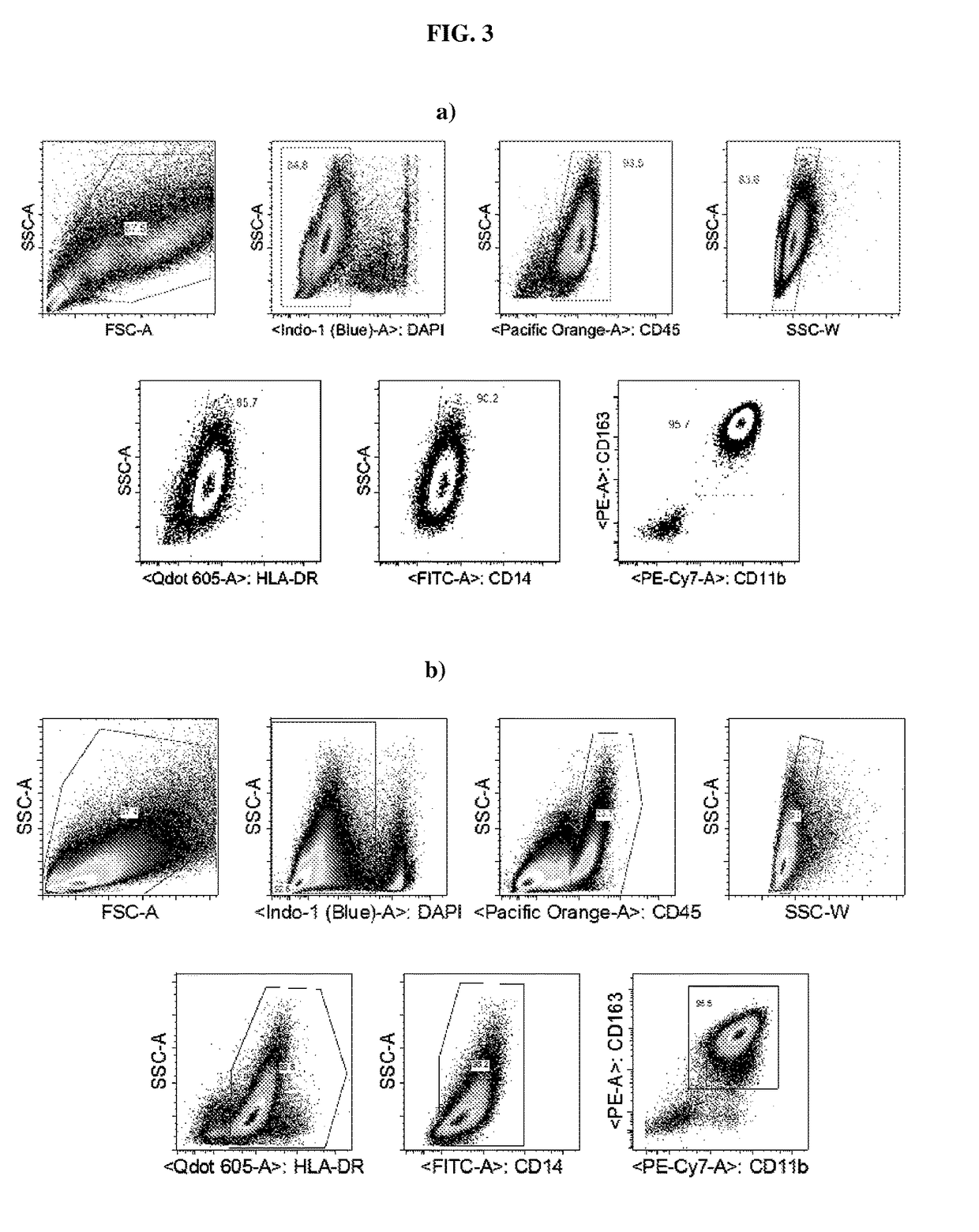
[0097]Phagocytosis assays were performed with day 12-sorted iPSCs-derived primitive macrophages (CD163+CD11b+HLA-DR+IBA-1+CX3 CR1+CD14+), wherein the macrophages have been differentiated from patient derived WT iPSCs and the expression profile is CD45+ CD11b+, and CD163+. Specifically, the day 12-sorted iPSCs-derived primitive macrophages were exposed to fluorescently labeled latex beads or amyloid Aβ peptides in order to assess their capacity to phagocyte as macrophages and microglia.

[0098]As shown in FIG. 4, the capacity of said iPSC-derived primitive macrophages to phagocyte as macrophages and microglia is supported. This is of importance since amyloid beta (Aβ or Abeta) peptides are crucially involved in Alzheimer's disease as the main component of the amyloid plaques found in the brains of Alzheimer patients.

[0099]Procedure

[0100]The method for the analysis of macrophage phagocytic ability used a control line of Hu...


PUM


Abstract

The present invention relates to a method of culturing primitive-like macrophages from stem cells, a kit when used in the method thereof and uses of the primitive like macrophage for in-vitro disease models and for screening compounds for therapy. One embodied culture method comprises contacting and incubating embryonic stem cells or induced pluripotent stem cells with a serum-free culture media comprising a GSK3 inhibitor to differentiate stem cells into cells of the mesoderm lineage, followed by incubation with a culture media comprising Dickkopf-related protein 1 (DKK1) to differentiate the mesoderm into cells of hematopoietic lineage, maturing hematopoietic cells and incubating these cells with a culture media comprising M-CSF to drive differentiation into primitive-like macrophages. Another embodiment comprises incubating the stem cells with serum-free culture media comprising FGF2 and BMP4 to induce differentiation into cells of the mesoderm lineage, followed by incubating the cells with a culture media comprising FGF2, BMP4, Activin A and VEGF to differentiate the cells of the mesoderm lineage into cells of the hematopoietic cell lineage, maturing the cells of the hematopoietic cell lineage and lastly, incubating the matured hematopoietic cells with culture media comprising M-CSF to drive the differentiation of hematopoietic cells into primitive-like macrophages.

Description

CROSS-REFERENCE TO RELATED APPLICATIONS[0001]This application claims the benefit of priority of Singapore application number 10201500366V, filed Jan. 16, 2015, the contents of it being hereby incorporated by reference in its entirety for all purposes.FIELD OF THE INVENTION[0002]The invention relates to a differentiation of macrophages from stem cells. Specifically, the invention relates to a serum-free, feeder-free and embryoid body-free method for differentiation of primitive macrophages from stem cells.BACKGROUND OF THE INVENTION[0003]Macrophages are mononuclear phagocytes that play a crucial role in tissue homeostasis and immunity, but also contribute to a broad spectrum of pathologies and thus represent key therapeutic targets. However, in depth insights into the differentiation of primitive macrophages from human pluripotent stem cells, their homeostasis, as well as their defined activities in a tissue-specific context are currently lacking.[0004]According to the mononuclear ph...

Claims


Application Information

Patent Timeline
IPC IPC(8): C12N5/0786G01N33/50
CPCC12N5/0645G01N33/5055A61K35/15C12N2500/90C12N2501/415C12N2501/22C12N2506/02C12N2506/45C12N2501/727C12N2501/165C12N2501/115C12N2501/125C12N2501/2306C12N2501/2303G01N33/5058G01N33/5073C12N2533/90A61P17/02
Inventor LIM, HWEI-IN SHAWNHUBER, TARAGINHOUX, FLORENT
Owner AGENCY FOR SCI TECH & RES
Why Eureka
Try Eureka