
Methods for quantitative assessment of mononuclear cells in muscle tissue sections

a mononuclear cell and muscle tissue technology, applied in image enhancement, instruments, editing/combining figures or texts, etc., can solve problems such as impairment of quality of life, failure of additional organ systems during disease progression, and compromise of muscle fiber structural integrity

Inactive Publication Date: 2018-02-08
FLAGSHIP BIOSCI
View PDF23 Cites 4 Cited by
  • Summary
  • Abstract
  • Description
  • Claims
  • Application Information

AI Technical Summary

Benefits of technology

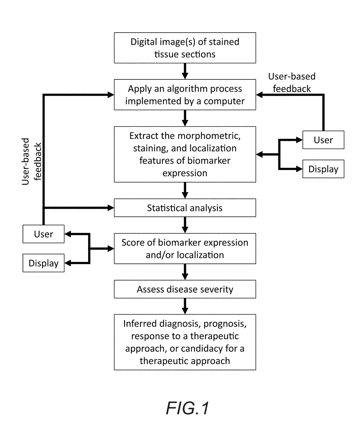
The patent describes a way to evaluate muscle tissue using digital analysis of images. It explains how to extract features from stained tissue sections and analyze them to identify different cells and measure their size. These features are then used to create a score that can help doctors diagnose and treat diseases. The method can help with various stages of diseases, such as determining if it is present, how severe it is, and how well treatment is going.

Problems solved by technology

Generally, myopathies include progressive weakening and wasting of skeletal muscle and may include failure of additional organ systems during disease progression.
Muscular dystrophies represent a group of myopathies that are incurable, significantly impair quality of life and are frequently fatal.
In DMD and BMD, muscle fiber structural integrity is compromised due to a failure to produce functional dystrophin.
Muscle fiber damage triggers the cellular repair process, resulting in an influx of immune and inflammatory cells.
In myopathies such as DMD and BMD, this process is disrupted, resulting in inappropriate accumulation of certain inflammatory cells (e.g., M1 macrophages) and inadequate presence of other inflammatory cells (e.g., M2 macrophages) [Madaro, L., & Bouché, M. BioMed Research International, 2014; 438675].
Dysregulation of this immune cycle contributes to the failure of muscle regeneration.

Method used


Image

  • Methods for quantitative assessment of mononuclear cells in muscle tissue sections
  • Methods for quantitative assessment of mononuclear cells in muscle tissue sections
  • Methods for quantitative assessment of mononuclear cells in muscle tissue sections

Examples


Embodiment Construction

[0019]In the following description, for purposes of explanation and not limitation, details and descriptions are set forth in order to provide a thorough understanding of the present invention. However, it will be apparent to those skilled in the art that the present invention may be practiced in other embodiments that depart from these details and descriptions without departing from the spirit and scope of the invention.

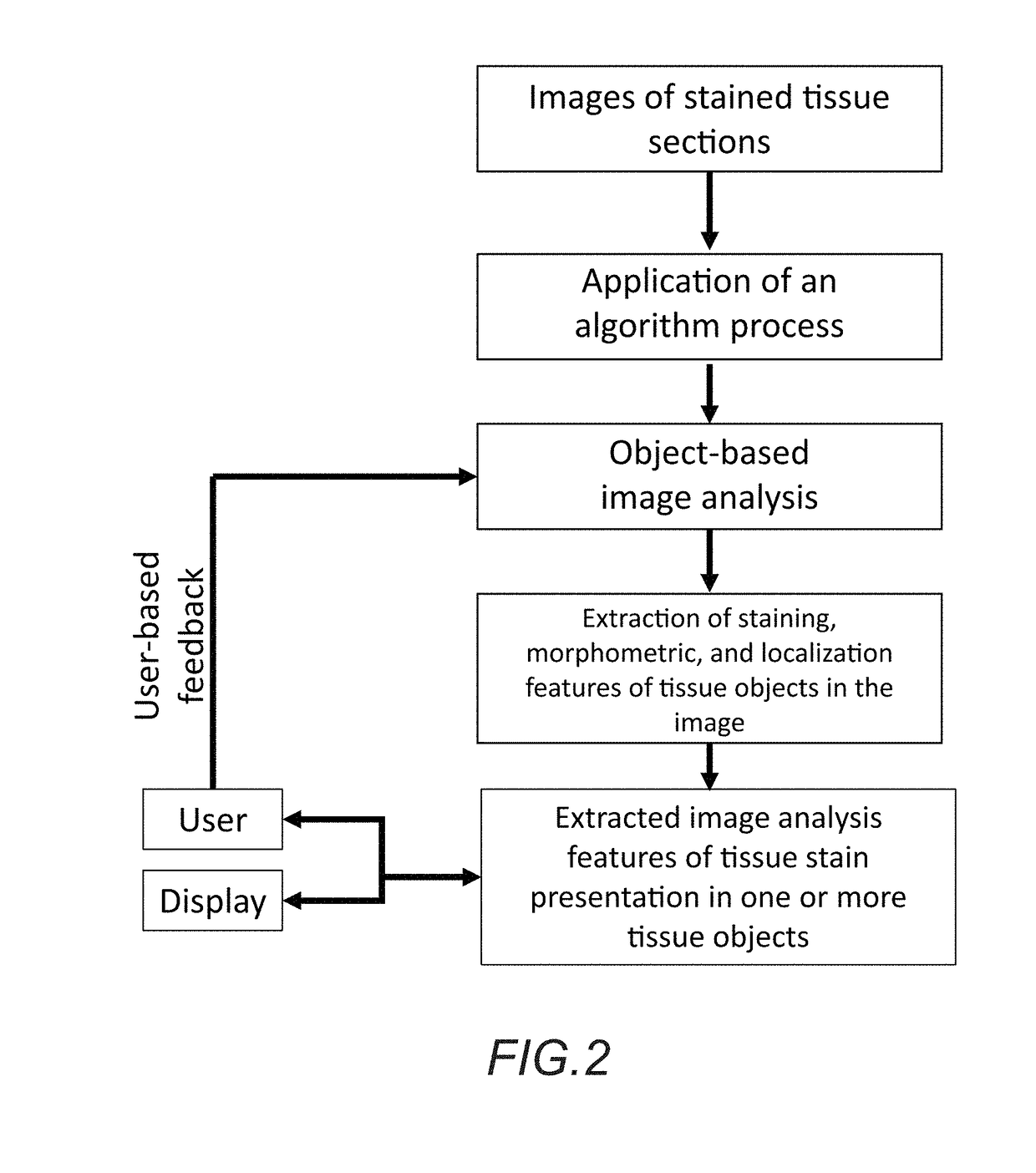
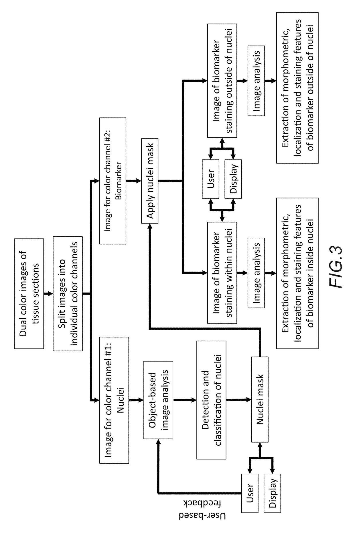
[0020]In an illustrative embodiment, the method for assessment of biomarker protein and / or transcript expression in nucleated and non-nucleated cells within muscle tissue using digital image analysis may generally comprise nine consecutive steps, including: 1) obtaining muscle tissue embedded in a tissue block from patients submitted for evaluation; 2) processing said tissue block using standard histologic procedures to generate one or more tissue sections attached to a glass histology slide; 3) contacting said tissue sections with one or more antibodies, nucleotide...


PUM

PropertyMeasurementUnit
immunofluorescent fluorescentaaaaaaaaaa
fluorescenceaaaaaaaaaa
mass spectrometryaaaaaaaaaa

Abstract

In accordance with the embodiment described herein, we describe a method for evaluating muscle fiber nuclei and non-muscle fiber mononuclear cells, and biomarkers expressed within these and muscle fibers, within the context of muscle tissue using digital tissue image analysis. An algorithm process is applied to histologically stained tissue sections to extract the morphometric, staining, and localization features of a plurality of tissue objects. These features can be further analyzed to describe relationships between tissue objects or tissue object image analysis features. One or more of these image analysis features or relationships between objects and features are summarized to derive a patient-specific score. Patient stratification criteria are applied to the patient-specific score and patient strata membership is evaluated to infer presence of disease, natural course of disease, disease severity, treatment efficacy, or response to a therapy and eligibility for said therapy.

Description

CROSS-REFERENCE TO RELATED APPLICATIONS[0001]This application is a continuation-in-part (CIP) of commonly owned U.S. Ser. No. 14 / 983,296, filed Dec. 29, 2015, titled “METHODS FOR QUANTITATIVE ASSESSMENT OF MUSCULAR FIBERS IN MUSCULAR DYSTROPHY”;[0002]which claims benefit of priority with U.S. Provisional Ser. No. 62 / 097,543, filed Dec. 29, 2014, titled “METHODS FOR QUANTITATIVE ASSESSMENT OF MUSCULAR FIBERS IN MUSCULAR DYSTROPHY”; the contents of each of which are hereby incorporated by reference.BACKGROUNDField of the Invention[0003]This invention relates to methods for assessing immunohistochemistry or immunofluorescence stained muscle tissue with digital image analysis for the purpose of evaluating myopathy disease status; specifically for digital image analysis-bases scoring of nucleated cells relative to muscle fibers in muscle tissues obtained from myopathy patients.Description of the Related Art[0004]Myopathies categorize a group of genetic disorders which result in muscle dy...

Claims


Application Information

Patent Timeline
Patent Type & Authority Applications(United States)
IPC IPC(8): G06T7/00G06K9/46G06T11/60G01N1/30
CPCG06T7/0012G01N1/30G06K9/4604G06T2207/10056G06T2207/30204G06T2207/30024G06T11/60G01N33/6887G01N2333/4712G01N2800/10G06T2207/10024G06V20/695G06V20/698
Inventor FAELAN, CRYSTAL DAWNMILICI, ANTHONY J.BLACK, JOSHUA C.MARTIN, NATHAN T.
Owner FLAGSHIP BIOSCI
Learn More