
Process for printing 3D tissue culture models

Active Publication Date: 2018-08-16
INVENTIA LIFE SCI PTY LTD
View PDF1 Cites 17 Cited by
  • Summary
  • Abstract
  • Description
  • Claims
  • Application Information

AI Technical Summary

Benefits of technology

The patent is about a process using a 3D bio-printer to create tissue models with high concentrations of cells that are alive and functional after printing. The bio-printer is capable of printing drops containing cells and activators using a process that ensures cell integrity and viability. The process is affordable, repeatable, and scalable. The results can be printed onto various substrates and the printing platform can be heated to aid cell proliferation. Overall, this technology allows for the creation of precise and functional tissue models for various research applications.

Problems solved by technology

This leads to complex intercellular interactions that cannot be mimicked in two-dimensional monolayers of cell cultures (Pampaloni, Ansari and Stelzer, 2013).
It has been demonstrated that cells behave more natively when cultured in three-dimensional environments but the formation of three-dimensional cell culture is difficult, expensive and laborious to generate.
While many previous studies describe printing of cells using drop-on-demand technology, there are no reports of printing a high cell density (greater than 107 cells / ml) in bio-ink via the drop on demand method.
Generating cellular spheroids using a drop-on-demand device presents difficulties due to the high viscosity associated with high cell densities and the small feature size in a drop-on-demand device.

Method used


Image

  • Process for printing 3D tissue culture models
  • Process for printing 3D tissue culture models
  • Process for printing 3D tissue culture models

Examples


example 1

Materials and Methods

[0167]Alginate (high >55% guluronic acid content, 50,000-80,000 g / mol, FMC Biopolymer), calcium chloride (BioReagent, Sigma Aldrich), phosphate buffered solution without calcium chloride (PBS, Gibco), Dulbecco's modified eagle medium (DMEM, Gibco), calcium free DMEM (Gibco), sodium pyruvate (Gibco), L-glutamine (Gibco), Gellan gum (Sigma Aldrich), trypsin (Sigma Aldrich), 0.22 μm syringe filter (polyethersulfone membrane, Merck Millipore), T150 flask (Corning), 15 ml centrifuge tube (Corning), ethanol (Sigma Aldrich), were used as received. Tissue culture media was made by mixing DMEM with FCS at 10 v / v %.

Cell Type

[0168]SK-N-BE(2) neuroblastoma cells were used.

Cell Culture

[0169]Neuroblastoma at 80% confluency in a T150 flask was washed with 3 ml PBS. After aspiration of excess PBS, 3 ml of trypsin was added and the flask was incubated at 37° C. to dissociate cells from flask surface for 5 minutes. Subsequently, 7 ml of tissue culture media was added and the diss...

example 2

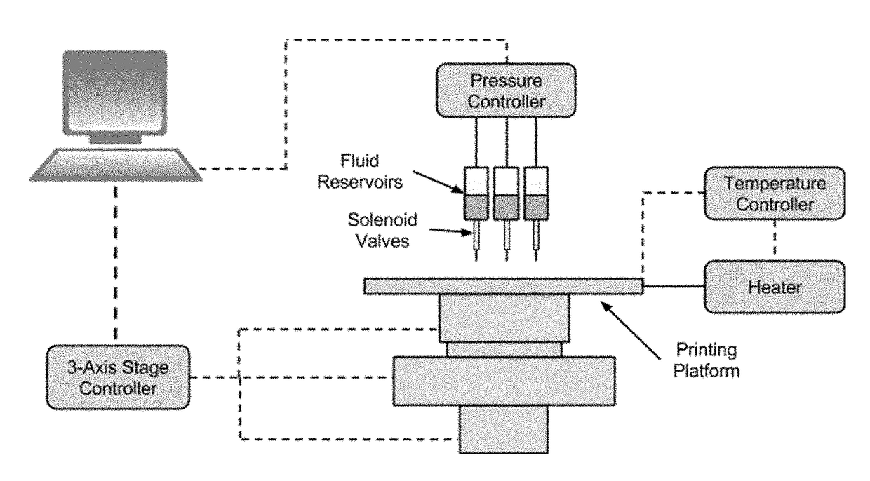
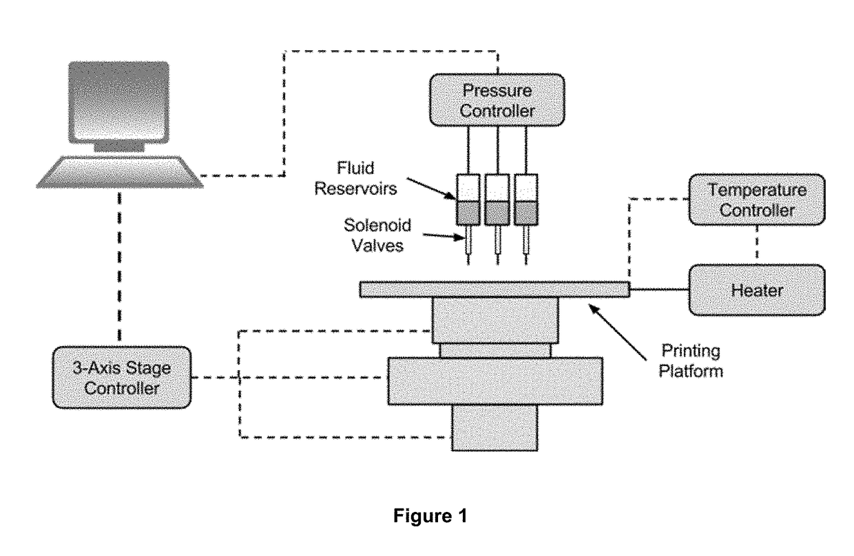
[0181]Hydrogel mold or cup was printed using bio-ink, cell-ink and activator, as detailed in Example 1, at a supply pressure of 40 psi, 12 psi and 5 psi respectively. SK-N-BE(2) cells in cell-ink were printed into centre of hydrogel cup at a concentration of 100 million cells / ml. Microscope images of the printed 3D cell culture model showed cell spheroid formation after incubation for 24, 48 and 120 hours.

example 3

[0182]Hydrogel mold or cup was printed using bio-ink and activator, as detailed in Example 1, at a supply pressure of 40 psi and 5 psi respectively. The cell-ink was Ficoll™ (Sigma Aldrich, 1.65 g) dissolved in PBS (10 ml) to give 16.5 w / v %. SK-N-BE(2) cells in cell-ink were printed into centre of hydrogel cup at a concentration of 250 million cells / ml, at a supply pressure of 20 psi. Microscope images of the printed 3D cell culture model showed cell spheroid formation after incubation for 24, 48 and 72 hours.


PUM

PropertyMeasurementUnit
Cell viabilityaaaaaaaaaa
Temperatureaaaaaaaaaa
Concentrationaaaaaaaaaa

Abstract

A process for producing a 3D tissue culture model by (a) printing a drop of bio-ink to a substrate; (b) printing a drop en.) of activator to the drop of bio-ink to form a hydrogel droplet; (c) repeating steps (a) and (b) in any order to form a hydrogel mold adapted to receive a drop containing cells; (d) printing a drop containing cells to the hydrogel mold; and (e) repeating steps (a) and (b) in any order to form a 3D tissue culture model comprising the cells encapsulated in the hydrogel mold.

Description

RELATED APPLICATION[0001]This application claims the benefit of Australian Provisional Patent Application No 2015902917, filed on 22 Jul. 2015, the disclosures of all of which are hereby expressly incorporated by reference in their entireties.TECHNICAL FIELD[0002]The technology relates to processes for forming three-dimensional tissue cultures such as 3D tissue culture models and uses in 3D tissue culture models.BACKGROUND[0003]Most cell structures in the human and animal body are organised in three-dimensions. This leads to complex intercellular interactions that cannot be mimicked in two-dimensional monolayers of cell cultures (Pampaloni, Ansari and Stelzer, 2013). However, two-dimensional cell culture has been the paradigm for typical in vitro cell culture. It has been demonstrated that cells behave more natively when cultured in three-dimensional environments but the formation of three-dimensional cell culture is difficult, expensive and laborious to generate.[0004]Cellular sphe...

Claims


Application Information

Patent Timeline
IPC IPC(8): C12N5/00G01N33/50B29C64/112B29C64/40B29C64/188B33Y10/00B33Y40/00B33Y70/00
CPCC12N5/0062C12N5/0012G01N33/5082B29C64/112B29C64/40B29C64/188B33Y10/00B33Y40/00B33Y70/00C12N2513/00C12N2503/02B29K2005/00B29L2031/40A61L27/52A61L27/20A61L2300/62A61L2300/64C12M21/08C12M33/00C08L5/04B33Y80/00
Inventor O'MAHONY, AIDAN PATRICKUTAMA, ROBERT HADINOTOFIFE, CHRISTOPHER MICHAELATAPATTU, LAKMALIRIBEIRO, JULIO CESAR CALDEIRAKAVALLARIS, MARIAGOODING, JOHN JUSTIN
Owner INVENTIA LIFE SCI PTY LTD
Features
  • Generate Ideas
  • Intellectual Property
  • Life Sciences
  • Materials
  • Tech Scout
  • Unparalleled Data Quality
  • Higher Quality Content
  • 60% Fewer Hallucinations
Learn More