Nf-kb signaling pathway-manipulated dendritic cells
a signaling pathway and dendrite cell technology, applied in the field of dendrite cells, can solve the problems of unable to meet the needs of patients, the options of genetically modifying the most widely used human dcs in medicine (monocyte-derived dcs) are very limited, and the clinical outcomes remain below expectations
- Summary
- Abstract
- Description
- Claims
- Application Information
AI Technical Summary
Benefits of technology
Problems solved by technology
Method used
Image
Examples
example 1
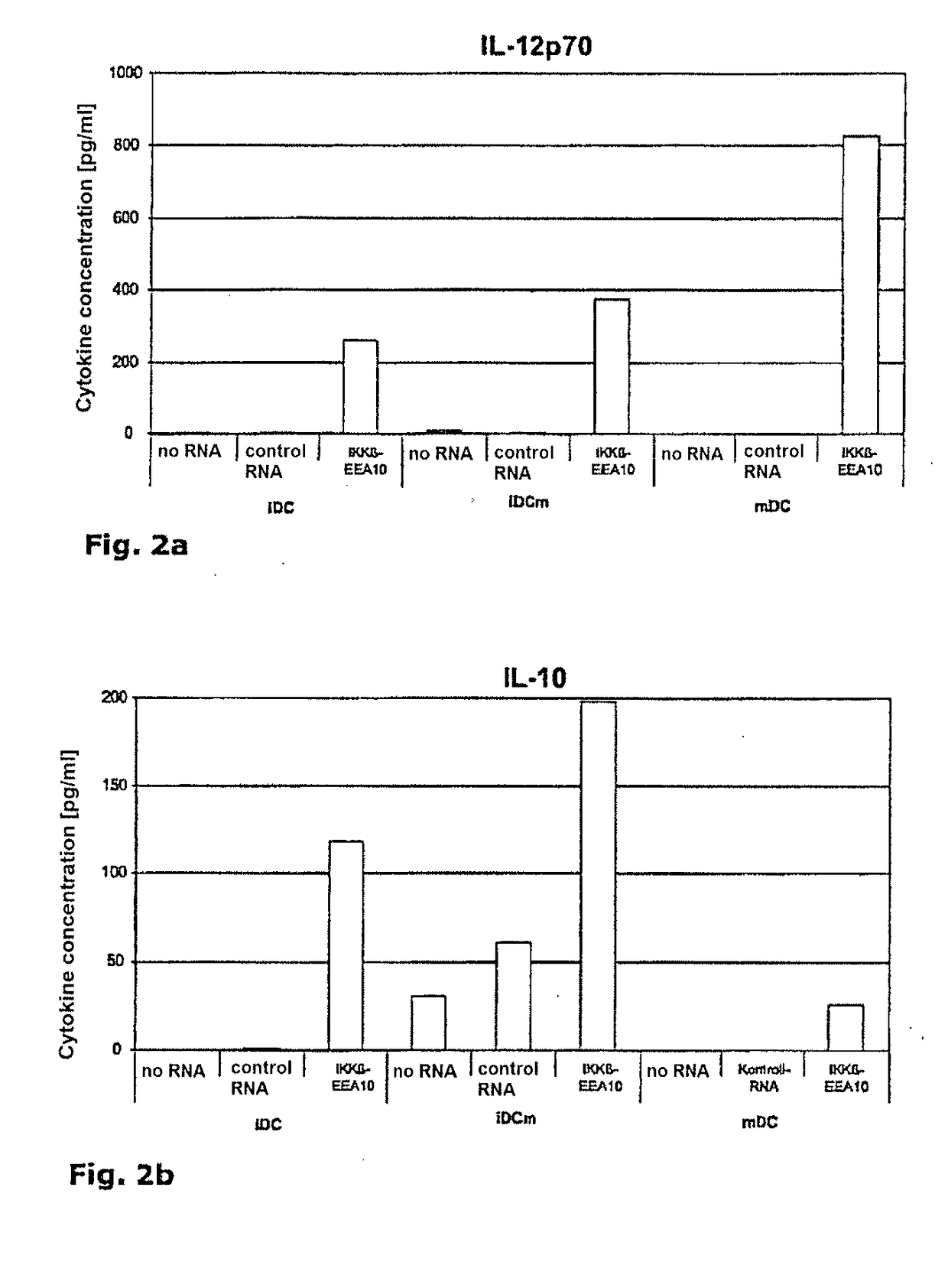
of IL-12p70 and IL-10 by IKKβ-EEA10-RNA-Electroporated Dendritic Cells
[0086]Dendritic cells, immature (iDC) or mature (mDC) without RNA, were electroporated with a control RNA or IKKβ-EEA10-RNA (SEQ ID NO:6). Immediately after electroporation, half of the immaturely electroporated cells were matured (iDCm). Twenty-four hours after electroporation, the cytokine concentrations (IL-12p70 and IL-10) in the supernatants were determined in a cytometric bead array (CBA). FIGS. 2(a) and (b), respectively, show data of one representative of four independent experiments.
example 2
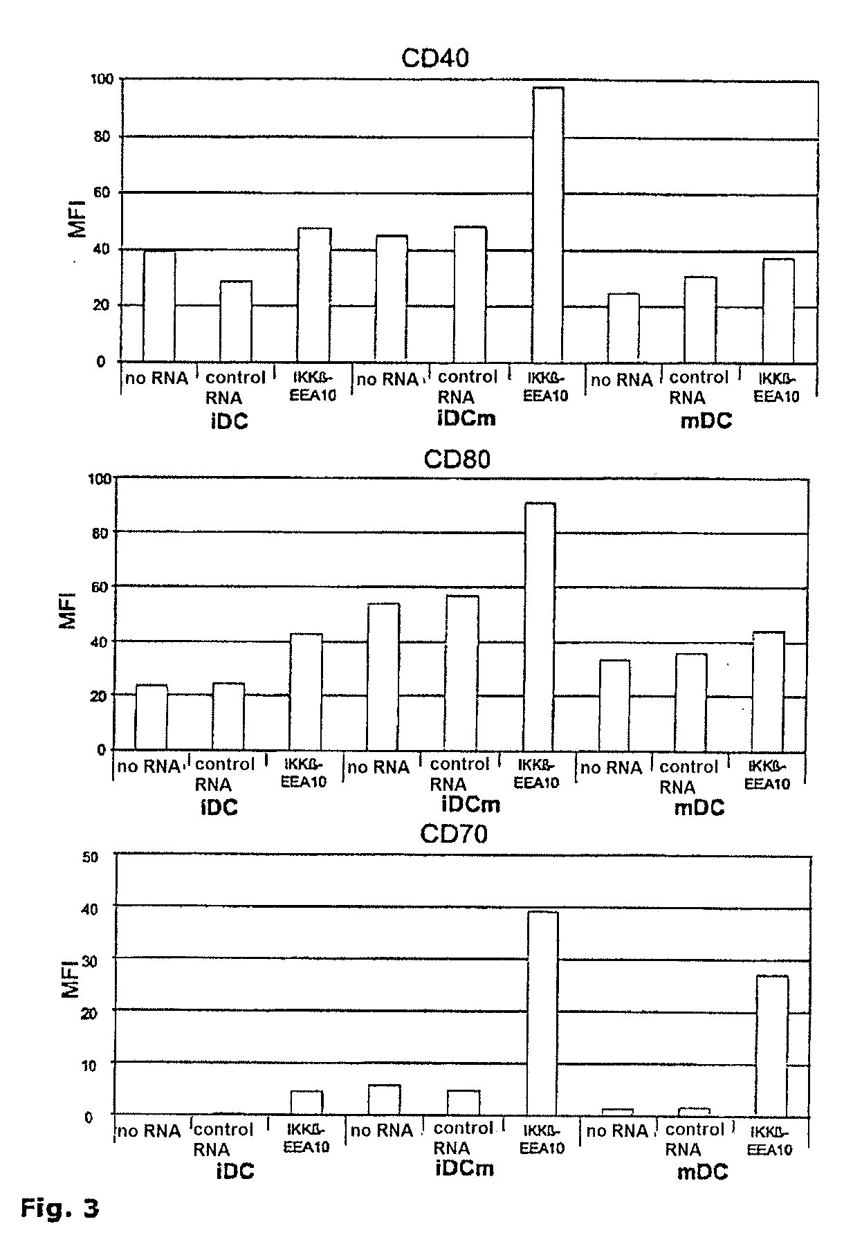
n of Surface Markers on Dendritic Cells Transfected with the NFκB Signaling Pathway Component IKKβ-EEA10
[0087]Immature (iDC) and mature (mDC) dendritic cells were electroporated with RNA encoding IKKβ-EEA10 (SEQ ID NO:6). After electroporation, half of the immaturely electroporated cells were treated with maturation cocktail (iDCm). As control conditions, DCs were electroporated without RNA or with irrelevant RNA (control RNA). After electroporation, the DCs were cultured in DC medium for 24 h, harvested and stained with a PE-labeled antibody against CD40, CD80 and CD70. The PE label identifies the coupling of the pigment phycoerythrin and an antibody. The mean fluorescence intensity (MFI) of the electroporated dendritic cells was determined by flow cytometry. The values given in FIG. 3 show the specific MFI, which was calculated from the measured relative fluorescence minus the measured fluorescence of the isotype antibody. The data represent one representative of four independent ...
example 3
Staining of the Stimulation of Autologous T Cells with Dendritic Cells Electroporated with RNA of NFκB Signaling Pathway Components
[0088]Mature dendritic cells were electroporated with control RNA, IKKβ-EEA10-RNA (SEQ ID NO:6) and IKKα-EE-RNA, or with a combination of IKKβ-EEA10 and IKKα-EE-RNA. A portion of the cells was co-electroporated with RNA encoding the tumor marker MelanA, (+ MelanA RNA). Three hours after electroporation, one half of the condition series without MelanA was loaded with MelanA / A2-peptide for 1 h (+ peptide loading). Four hours after electroporation, autologous CD8+ T cells were stimulated with said dendritic cells in the ratio 10:1. After one week, the number of antigen-specific T cells was analyzed and their phenotype determined by CCR7 and CD45RA staining. Said T cells were analyzed following a priming FIG. 4(a) and after a restimulation FIG. 4(b).
[0089]The figures show data from one donor.
PUM
 Abstract
Description
Claims
Application Information
 - R&D
- Intellectual Property
- Life Sciences
- Materials
- Tech Scout
- Unparalleled Data Quality
- Higher Quality Content
- 60% Fewer Hallucinations
Browse by: Latest US Patents, China's latest patents, Technical Efficacy Thesaurus, Application Domain, Technology Topic, Popular Technical Reports.
© 2025 PatSnap. All rights reserved.Legal|Privacy policy|Modern Slavery Act Transparency Statement|Sitemap|About US| Contact US: help@patsnap.com


