Methods, Systems, and Devices for Optical Sectioning

Active Publication Date: 2018-08-23
NORTHEASTERN UNIV
View PDF3 Cites 8 Cited by
  • Summary
  • Abstract
  • Description
  • Claims
  • Application Information

AI Technical Summary

Benefits of technology

[0005]The existing methods for optical sectioning are inadequate, in part because they require multiple image samples to be acquired over time, making in-vivo imaging difficult and otherwise limiting optical sectioning to cases in which a subject specimen to be imaged (target) remains very stationary with respect to an imaging device. While confocal microscopy provides sectioning, it rejects a disproportionately large amount of light, requiring a high-power source of light in order to function properly. Additionally, because the pinhole only allows a single pixel or limited group of pixels to be imaged at a time, raster scanning is required to build a full two-di

Problems solved by technology

The existing methods for optical sectioning are inadequate, in part because they require multiple image samples to be acquired over time, making in-vivo imaging difficult and otherwise limiting optical sectioning to cases in which a subject specimen to be imaged (target) remains very stationary with respect to an imaging device.
Additionally, because the pinhole only allows a single pixel or limited group of pixels to be imaged at a time, raster scanning is required to build a full two-dimensional image.
This raster scanning takes time, and any movement of a patient or other target object to be imaged can significantly impair the ability of confocal microscopy to produce a proper image of high resolution.
While structured illumination microscopy (SIM) does not require the use of a pinhole, nor raster scanning, the need to acquire at least three images in order to decouple in-focus lig

Method used


Image

  • Methods, Systems, and Devices for Optical Sectioning
  • Methods, Systems, and Devices for Optical Sectioning
  • Methods, Systems, and Devices for Optical Sectioning

Examples


Embodiment Construction

[0045]A description of example embodiments follows.

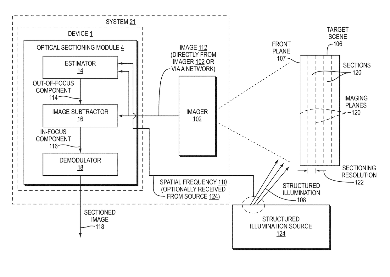
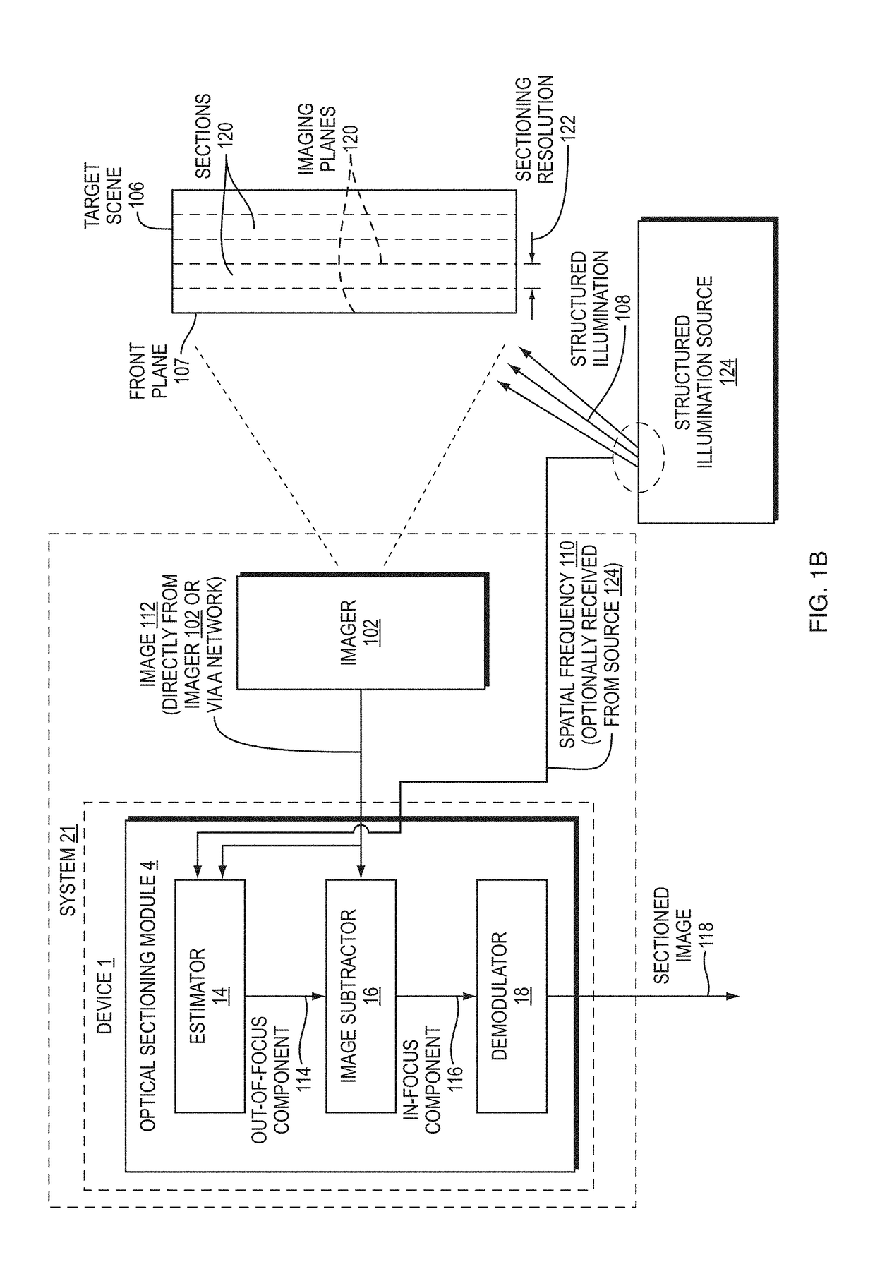
[0046]Disclosed herein are embodiment methods, systems, and devices that enable confocal-like optical sectioning in real time. A single N×M pixel sample may be used to produce an optical sectioned image, and imaging may be time-limited only by a frame rate of an imager, such as a CCD camera. Consistent with embodiments, target samples can be sectionally imaged at physiologically relevant timescales, for example. This is in contrast to traditional confocal microscopy and traditional structured illumination, which typically require either sequential scanning of N×M pixels or at least three frames of data, respectively. Disclosed embodiments can also produce more robust sectioning within a turbid medium than a traditional structured illumination.

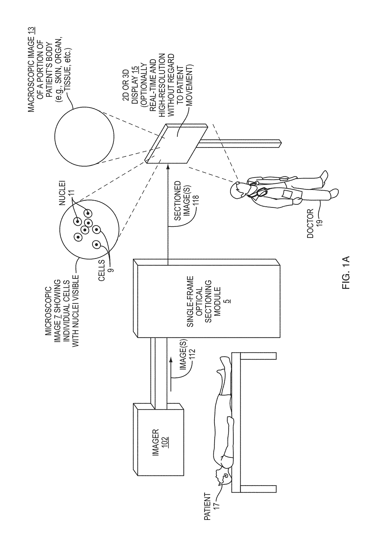
[0047]Optical sectioning has provided pathologists and clinicians the ability to image biological samples noninvasively at or below a surface of the sample. In example cases such as skin cancer...


PUM


Abstract

Methods, systems, and devices described herein enable single-image optical sectioning, even at depth within turbid media, such as human skin or other tissue. Embodiments can eliminate the need for multiple image samples or raster scans, making in-vivo or live biological imaging easier and faster than multi-image sectioning techniques. Better contrast and resolution than traditional three-phase structured illumination microscopy (SIM) is possible in turbid media. Embodiments enable imaging of cell nuclei. Resolution and contrast resulting from disclosed embodiments are less sensitive to motion of or within patients or other targets than confocal microscopy and three-phase SIM techniques. Three-dimensional images of target specimens can be provided based on a group of single-image optical sections. Real-time imaging can also be provided.

Description

RELATED APPLICATION(S)[0001]This application claims the benefit of U.S. Provisional Application No. 62 / 454,677, filed on Feb. 3, 2017. The entire teachings of the above application are incorporated herein by reference.GOVERNMENT SUPPORT[0002]This invention was made with government support under CBET-1510281 from National Science Foundation. The government has certain rights in the invention.BACKGROUND[0003]Various optical sectioning techniques have provided pathologists and clinicians with some ability to image biological samples noninvasively at or below a surface of a target specimen, such as the skin of a person. Existing methods for optical sectioning include confocal microscopy, which is the most common method. Confocal microscopy works by passing received light through a pinhole, which rejects out-of-focus light from reaching a detector. Another optical sectioning method, known as structured illumination microscopy (SIM), does not require the use of a pinhole. SIM has an advan...

Claims


Application Information

Patent Timeline
IPC IPC(8): G06K9/00G06K9/34G02B21/36
CPCG06K9/0014G06K9/346G02B21/365G02B21/06G02B21/367G02B27/58G06V20/695G06V10/273
Inventor HOFFMAN, ZACHARY R.DIMARZIO, CHARLES A.
Owner NORTHEASTERN UNIV
Why Eureka
Try Eureka