Methods for using artificial neural network analysis on flow cytometry data for cancer diagnosis

a flow cytometry and artificial neural network technology, applied in the field of cancer diagnosis by using artificial neural network analysis on flow cytometry data, can solve the problems of generating numerous false positives and negatives, unable to detect early, and unable to accurately predict the effect of cancer treatment progress

Active Publication Date: 2018-08-30
ANIXA DIAGNOSTICS CORP
View PDF3 Cites 45 Cited by
  • Summary
  • Abstract
  • Description
  • Claims
  • Application Information

AI Technical Summary

Benefits of technology

[0006]Described herein are methods relating to flow cytometry data analysis that use flow cytometer output data to classify characteristics of cell populations. Described herein are methods relating to flow cytometry that eliminate the need for most manual or automatic gating of cell populations. Described herein are methods relating to flow cytometry data analysis for diagnosing cancer by analyzing target cell populations, such as MDSCs, in concert with other cell populations including myeloid cells and lymphoid cells. A diagnosis of cancer as used hereinafter means a diagnosis of there being a malignant tumor present. Described herein are methods of flow cytometry data analysis that include a computationally efficient representation of cell populations in a multidimensional data space, wherein the axes of the data space are the measurement channels of a flow cytometer or a transformation thereof. The methods may further comprise a neural network analysis that classifies samples based on the learned characteristics of the distributions of target cells in a multidimensional data space. Described herein are methods relating to flow cytometry data analysis that can be performed on an entirety of a target cell distribution and therefore detect distinctions between samples that are undetectable by conventional flow cytometry data gating. Data analysis methods as described herein can be used for malignant tumor detection by detecting cell population and subpopulation distribution differences, including differentiation of MDSC subpopulations present, for example, in peripheral blood samples taken from non-tumor-bearing subjects and subjects having malignant tumors (tumor-bearing subjects). By providing sensitive and specific methods and devices for differentiating various cell types and detecting cell population differences, cancer diagnostic devices and assays can be developed for early cancer detection in subjects or various patients.

Problems solved by technology

Moreover, the financial costs of cancer treatment at an advanced stage can be an additional burden.
However, methods for early detection often lack sensitivity and generate numerous false positives and negatives.
False negatives lead to missed opportunities to intervene early, and false positives lead to additional unnecessary testing, which can include biopsies and other painful, stressful and expensive procedures.
In addition, conventional screening tests, such as colonoscopies, are often invasive; hence, individuals are often resistant to undertake them.
When considering a confirmatory test, it is well known that up to 95% of biopsies return negative results (meaning the early indication of cancer was not verified by the painful, expensive procedure).
However, many of these liquid biopsy methodologies rely on expensive analytical techniques such as DNA or RNA sequencing.
These tests often require extensive levels of multiplexing that make them expensive, difficult to operate, and also difficult to interpret.
However, conventional methods of flow cytometry data analysis as applied to MDSCs in blood samples are not sufficiently accurate to allow MDSCs to be used as a sole biomarker for a cancer detection screening test for the general public.
While this method is suitable for research studies and some diagnostic applications, for cancer detection, this method of analysis can be labor intensive, subjective, and results in a very coarse representation of the flow cytometer output data, often obscuring or ignoring a great deal of information available regarding the relative distributions of all the cell populations and the shape of the distributions of the isolated cell populations.

Method used


Image

  • Methods for using artificial neural network analysis on flow cytometry data for cancer diagnosis
  • Methods for using artificial neural network analysis on flow cytometry data for cancer diagnosis
  • Methods for using artificial neural network analysis on flow cytometry data for cancer diagnosis

Examples


example 1

MDSC Detection

[0317]Populations of MDSCs are identified in patients with cancer by flow cytometry and convoluted neural networks.

[0318]A peripheral blood sample from a patient with cancer is taken. The blood sample is centrifuged to pellet the cells. The cells are resuspended to a concentration of 107 cells / mL (cells per mL) in Phosphate Buffer Solution (PBS).

[0319]Cells are then labeled with anti-human monoclonal antibodies. Antibodies that are used include anti-lineage-FITC (fluorescein isothiocyanate), including anti-CD3, -CD14, -CD16, -CD19, -CD20 and -CD56, anti-CD33-PE, anti-HLA-DR-ECD, anti-CD11b-PE-Cy5, anti-CD14-PE, anti-CD15-PE-Cy5, anti-CD33-PE-Cy7. Cells are then analyzed by flow cytometry with at least 4×10⁴ events acquired for analysis.

[0320]Following the initial FSC / SSC discrimination, the gate is set on DR− / LIN− cells. Subpopulations are then gated to identify MDSCs, including CD14−, CD11b+, CD15+, CD66+, CD14+, CD15−, and their combinations.

[0321]The Matlab 2016b ma...

example 2

MDSC Detection with Recombinant Antibodies

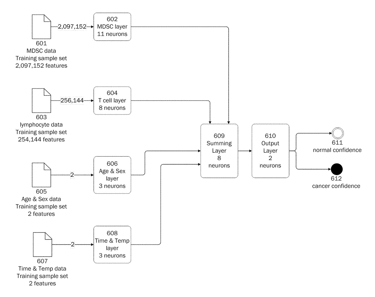
[0327]Blood samples were collected from tumor bearing and non-tumor bearing individuals. Each blood sample was processed and prepared for flow cytometry analysis; this includes being stained with recombinant monoclonal antibodies conjugated to different fluorophores. Four panels were used, and the panels included:

[0328]Panel 1: Peripheral Blood Mononuclear Cell Staining for MDSC

[0329]Panel 2: Peripheral Blood Mononuclear Cell Staining for Lymphocytes

[0330]Panel 3: Whole Blood Staining for MDSC for MDSC

[0331]Panel 4: Whole Blood Staining for MDSC for Lymphocytes

[0332]Prior to analyses by the flow cytometer, the four panels were processed for staining with antibodies. The recombinant monoclonal antibodies used in the staining process for Panel 1 and Panel 3 include: recombinant PE anti-human CD3, recombinant APC-Vio770 anti-human CD14, recombinant PE-Vio770 anti-human CD15 (SSEA-1), recombinant PE-Vio615 anti-human CD16, recombinant VioBright ...


PUM


Abstract

The present disclosure provides methods for applying artificial neural networks to flow cytometry data generated from biological samples to diagnose and characterize cancer in a subject. The disclosure also provides methods of training, testing, and validating artificial neural networks.

Description

CROSS REFERENCE[0001]This application is a continuation-in-part of U.S. patent application Ser. No. 15 / 445,913, filed Feb. 28, 2017, the disclosure of which is incorporated herein by reference in its entirety.BACKGROUND OF THE INVENTION[0002]In the course of a lifetime, cancer will strike about one in three women and one in two men. More than 560,000 die from it each year in the United States alone. Early detection and treatment is currently the leading method to reduce cancer death, especially if the cancer is detected before metastasis. For nearly all types of cancer, the 5-year relative survival rate is substantially lower if the disease is caught at an advanced stage. Moreover, the financial costs of cancer treatment at an advanced stage can be an additional burden. By 2020, the cost of cancer treatment is expected to be $207 billion annually in the United States. Accordingly, early detection of cancer is important for increasing cancer survival rates and reducing the cost of tr...

Claims


Application Information

Patent Timeline
Patent Type & Authority Applications(United States)
IPC IPC(8): G06N3/08G06N3/12G06F19/24G16B40/10G16B40/20
CPCG06N3/08G06N3/126G06F19/24G16B20/00G16B40/10G16B40/20G06N3/045G16B40/00
Inventor KUMAR, AMITROOP, JOHNCAMPISI, ANTHONY J.DOMINGUEZ, GEORGE
Owner ANIXA DIAGNOSTICS CORP
Why Eureka
Try Eureka