Novel method for chondrogenic induction

a chondrogenic induction and chondrogenesis technology, applied in the direction of prosthesis, skeletal/connective tissue cells, drug compositions, etc., can solve the problems of limited size of damage sites that can be treated by transplantation, lack of cartilage tissue, damage that will deteriorate without repair treatment, etc., to achieve stable induction of pluripotent differentiation and simple steps

Active Publication Date: 2018-09-06
KYOTO UNIV
View PDF0 Cites 0 Cited by
  • Summary
  • Abstract
  • Description
  • Claims
  • Application Information

AI Technical Summary

Benefits of technology

[0014]The present invention is the first to achieve stable induction of differentiation of pluripotent stem cells (e.g., iPS cells) into high-quality chondrocytes in simple steps. The chondrocytes produced by the method of the present invention can be used for regenerative medicine for cartilage.

Problems solved by technology

Cartilage tissue damaged by joint injury etc. does not spontaneously heal itself, and the damage will deteriorate without repair treatment such as transplantation.
Transplantation of the patient's own cartilage from other body parts inevitably results in lack of cartilage tissue in these parts, and therefore, the size of the damage site that can be treated by transplantation is limited.
Another current approach is transplantation of the expansion culture product of harvested chondrocytes, but chondrocytes may be transformed into fibroblastic cells during in vitro culture and this approach cannot produce a sufficient therapeutic effect (Non Patent Literature 1).
However, mesenchymal stem cells can differentiate into multiple types of cells, and the administration inevitably results in transplantation of not only chondrocytes but also undesirable tissues, such as fibrous tissue expressing type I collagen and hypertrophic tissue expressing type X collagen (Non Patent Literature 2).
However, the use of pluripotent stem cells has produced several problems, including the formation of fibrocartilage and teratoma.

Method used


Image

  • Novel method for chondrogenic induction
  • Novel method for chondrogenic induction
  • Novel method for chondrogenic induction

Examples


example 1

Human iPS Cells

[0074]The 1231A3 cell line, which was established by the method described in Nakagawa M, et al., Sci Rep. 4:3594 (2014), was provided from the Center for iPS Cell Research and Application, Kyoto University, and was used as human iPS cells.

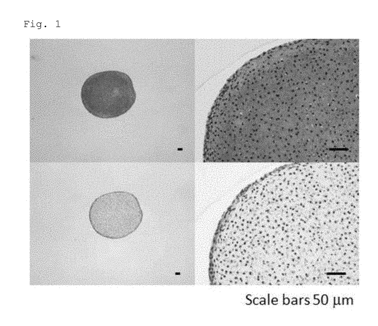
[0075]The human iPS cells were incubated with 0.5×TrypLE Select, and then detached using a cell scraper. The number of the cells was counted, and 1.0×10⁶ to 2.0×10⁶ cells were transferred to a 30-mL bioreactor (BWV-S03A, ABLE Corporation). To the bioreactor, 30 mL of StemFit AK03 (Ajinomoto) supplemented with 10 nM Y-27632 (Wako) was added, and the cells were cultured under the conditions of 37° C. and 5% CO2 for 5 days with stirring using a 6-cm magnetic stirrer (BWS-S03NOS-6, ABLE Corporation) at a rotation speed of 55 rpm. As a result, iPS cell clusters of 50 to 300 μm in diameter were obtained.

Chondrogenic Induction

[0076]The iPS cell clusters obtained by the above method were collected, and the whole or half of them were seeded o...

example 2

Human iPS Cells

[0080]The 1231A3 cell line, which was established by the method described in Nakagawa M, et al., Sci Rep. 4:3594 (2014), was provided from the Center for iPS Cell Research and Application, Kyoto University, and was used as human iPS cells.

[0081]The human iPS cells were incubated with 0.5×TrypLE Select, and then detached using a cell scraper. The number of the cells was counted, and 0.5×10⁷ to 1.0×10⁷ cells were transferred to a 100-mL bioreactor (BWV-S10A, ABLE Corporation). To the bioreactor, 100 mL of StemFit AK02N (Ajinomoto) supplemented with 10 nM Y-27632 (Wako) was added, and the cells were cultured under the conditions of 37° C. and 5% CO2 for 4 to 7 days with stirring using a magnetic stirrer (BWS-S03NOS-6, ABLE Corporation) at a rotation speed of 60 rpm. As a result, iPS cell clusters of 50 to 300 μm in diameter were obtained.

Chondrogenic Induction

[0082]The iPS cell clusters obtained by the above method were collected and seeded on four to twelve 10-cm suspen...

example 3

Human iPS Cells

[0085]The Ff-I01 cell line, which was established by the method described in Nakagawa M, et al., Sci Rep. 4:3594 (2014), was provided from the Center for iPS Cell Research and Application, Kyoto University, and was used as human iPS cells.

[0086]The human iPS cells were incubated with 0.5×TrypLE Select, and then detached using a cell scraper. The number of the cells was counted, and 0.5×10⁷ to 1.0×10⁷ cells were transferred to a 100-mL bioreactor (BWV-S10A, ABLE Corporation). To the bioreactor, 100 mL of StemFit AK03N (Ajinomoto) supplemented with 10 nM Y-27632 (Wako) was added, and the cells were cultured under the conditions of 37° C. and 5% CO2 for 4 to 7 days with stirring using a magnetic stirrer (BWS-S03NOS-6, ABLE Corporation) at a rotation speed of 60 rpm. As a result, iPS cell clusters of 50 to 300 μm in diameter were obtained.

Chondrogenic Induction

[0087]The iPS cell clusters obtained by the above method were collected and seeded on four to twelve 10-cm suspen...


PUM

PropertyMeasurementUnit
timeaaaaaaaaaa
timeaaaaaaaaaa
diameteraaaaaaaaaa

Abstract

Provided is a method for producing chondrocytes from pluripotent stem cells, the method comprising the steps of:(i) culturing pluripotent stem cells under adherent conditions in a medium containing an HMG-CoA reductase inhibitor and one or more substances selected from the group consisting of BMP2, TGFβ and GDF5, and(ii) culturing the cells obtained in step (i) under suspension conditions in a medium containing an HMG-CoA reductase inhibitor and one or more substances selected from the group consisting of BMP2, TGFβ and GDF5.Also provided is a pharmaceutical product comprising chondrocytes obtained by the method.

Description

TECHNICAL FIELD[0001]The present invention relates to a method for inducing pluripotent stem cells to differentiate into chondrocytes. The present invention also relates to a therapeutic agent comprising chondrocytes produced by the method.BACKGROUND ART[0002]Cartilage tissue is a component of the nose, ears and joints. Cartilage tissue is composed of chondrocytes and a specific extracellular matrix that contains type II, IX, and XI collagens and proteoglycans, but not type I collagen. Cartilage tissue damaged by joint injury etc. does not spontaneously heal itself, and the damage will deteriorate without repair treatment such as transplantation. For transplantation, however, cartilage tissue to be transplanted into the damaged site needs to be obtained. Transplantation of the patient's own cartilage from other body parts inevitably results in lack of cartilage tissue in these parts, and therefore, the size of the damage site that can be treated by transplantation is limited. Anothe...

Claims


Application Information

Patent Timeline
Patent Type & Authority Applications(United States)
IPC IPC(8): C12N5/077A61K35/35A61P19/04A61L27/38A61L27/54
CPCC12N5/0655A61K35/35A61P19/04A61L27/3817A61L27/54C12N2501/155C12N2501/15C12N2501/19C12N2501/71C12N2506/45A61L2430/06A61L2300/64A61L2300/412A61L27/00A61K35/32C12N2500/25C12N2500/38
Inventor TSUMAKI, NORIYUKI
Owner KYOTO UNIV
Why Eureka
Try Eureka