
Chimeric antigen receptor to which Anti-cotinine antibody is linked, and use thereof

a technology of chimeric antigen receptor and anticotinine antibody, which is applied in the direction of immunoglobulins, peptides, drugs/humans, etc., can solve the problems of short in vivo half-live, limitation of optimization conditions, and molecules with the disadvantage of no or weak cytotoxicity, etc., to suppress adverse immune side effects and induce cells

Active Publication Date: 2018-09-13
SEOUL NAT UNIV R&DB FOUND
View PDF2 Cites 3 Cited by
  • Summary
  • Abstract
  • Description
  • Claims
  • Application Information

AI Technical Summary

Benefits of technology

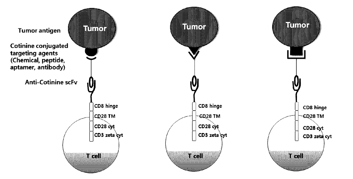
The patent is for a new type of T cell that can target and kill specific cells in the body. These cells are designed to recognize a molecule called cotinine, which is found in tobacco. The T cells are linked with an anti-cotinine antibody and can be used to treat diseases where certain cells are over-prolified. The invention also includes a way to remove the T cells if needed to prevent any negative side effects.

Problems solved by technology

While substances such as peptides, aptamers, antibodies, etc. that can be developed as biological therapeutics have excellent effects, such substances can be easily broken down in the body and rapidly released through the kidneys, and thus have short in vivo half-lives.
However, in the process of conjugating PEG, various molecular complexes are formed, and there is a limitation that optimization conditions must be established depending on binding molecules which are conjugated to PEG.
In addition, such molecules have the disadvantage of having no or weak cytotoxicity when the target object thereof is a cell (e.g., a cancer cell, etc.).
However, since conventional CAR cells have a limitation of recognizing only a single target molecule, CAR cells against haptens have been studied.

Method used


Image

  • Chimeric antigen receptor to which Anti-cotinine antibody is linked, and use thereof
  • Chimeric antigen receptor to which Anti-cotinine antibody is linked, and use thereof
  • Chimeric antigen receptor to which Anti-cotinine antibody is linked, and use thereof

Examples


example 1

on of Retrovirus Comprising Nucleic Acid Encoding Chimeric Antigen Receptor Linked with Anti-Cotinine Antibody Fragment

[0085]1-1. Preparation of Construct

[0086]First, a plasmid comprising nucleic acids each encoding an scFv fragment of an anti-cotinine antibody, a hinge domain, a transmembrane domain, and a signal transduction domain was prepared by the following method.

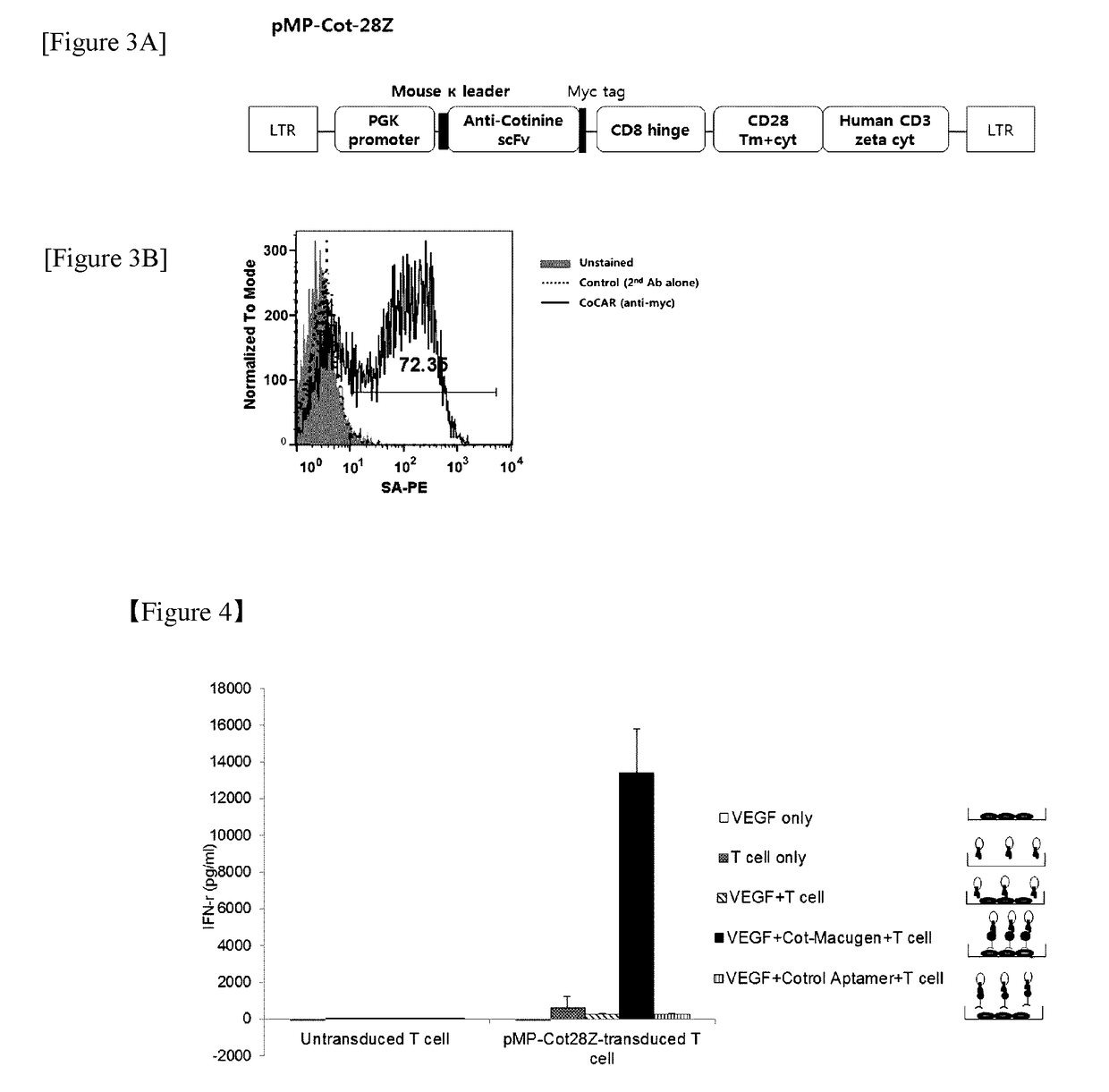
[0087]Specifically, a scFv fragment of an anti-cotinine antibody was amplified by PCR in a conventional manner from a template plasmid using a primer including a mouse Ig kappa leader sequence. The template was a plasmid generated by ligation of an anti-cotinine scFv fragment cleaved with the SfiI restriction enzyme to a pCEP4 vector (Invitrogen, US) (Clin. Chim. Acta., 2010, 411-1238). Meanwhile, the skeletal part of the chimeric antigen receptor including c-myc tag, human CD8 hinge region, transmembrane region and cytoplasmic region of mouse CD28, and a cytoplasmic region of human CD3 zeta, was amplified by PCR fro...

example 2

on of Cytotoxic T Cells with a Chimeric Antigen Receptor Linked with Anti-Cotinine Antibody Fragment Presented on Surface Thereof

[0091]Using the virus prepared in Example 1-2, cytotoxic T cells were prepared with a chimeric antigen receptor linked with anti-cotinine antibody fragment presented on the surface thereof.

[0092]First, splenocytes were isolated from B6 mice and dispensed in a cell culture container coated with 10 μg / mL of the anti-CD3 antibody (145-2C11, BD Bioscience, US) so that there were 2.5×10⁶ cells per well. The cells were supplemented with a total of 1 mL of the RPMI-1640 medium including 2 μg / mL of the anti-CD28 antibody (37.51, BD Bioscience, US), and cultured under conditions of 5% CO2 and 37° C. After 24 hours, 1 mL of the concentrated retrovirus including 2 μL of polybrene (Sigma-Aldrich, US) at 6 mg / mL was added to the cells and transduced by centrifugation for 90 minutes at 2,500 rpm using a centrifuge (Centrifuge 5810R, Eppendorf, US). Polybrene was used to...

example 3

on of Complex of Cotinine and Binding Molecule

[0094]3-1. Preparation of Complex of Cotinine and Antibody

[0095]As a binding molecule, cotinine-conjugates were prepared using rituximab (Genentech, Biogen, US), which is an anti-CD20 antibody, trastuzumab (Genentech, US), which is an anti-HER2 antibody, CT302 (Celltrion, Korea), which is an anti-influenza virus hemagglutinin antibody, and W6 / 32 (eBioscience, US), which is an anti-HLA antibody. Cotinine conjugation was performed by an EDC (1-ethyl-3-[3-dimethylaminopropyl]carbodiimide) coupling method.

[0096]First, the antibodies above were dissolved in PBS to a concentration of 25 μM. Meanwhile, trans-4-cotinine carboxylic acid (Sigma-Aldrich) was dissolved in 1 mL of the MES buffer [0.1 M MES (2-[morpholino]ethanesulfonic acid) and 0.5 M sodium chloride, pH 6.0] to a concentration of 5 mM. EDC at a concentration of 50 mM and N-hydroxysulfosuccinimide (Sulfo-NHS, Thermo Scientific, US) at a concentration of 125 mM were added to the solut...


PUM

PropertyMeasurementUnit
Cell deathaaaaaaaaaa
Cytotoxicityaaaaaaaaaa

Abstract

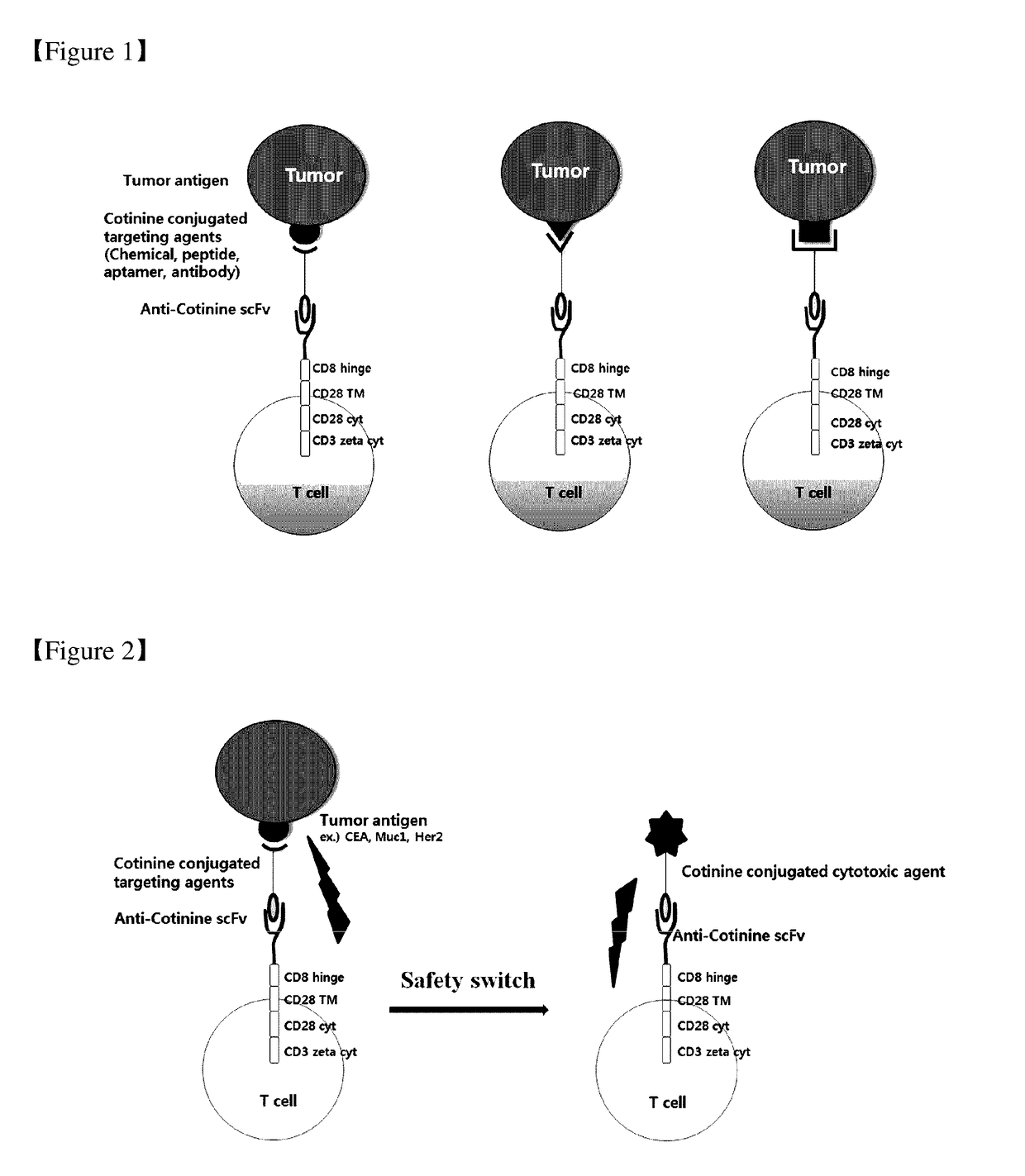
The present invention relates to chimeric antibody receptors with anti-cotinine antibodies linked, and use thereof. A T cell presenting the chimeric antibody receptor on the surface secretes interferon gamma specifically for a target molecule of a cotinine-conjugated binding molecule that is added together therewith and induces cell death of the cell expressing the target molecule by the T cell. On the contrary, by administering a cytotoxic agent conjugated with cotinine, cell death of the chimeric antigen receptor T cell is induced. Therefore, if necessary, a cytotoxic agent conjugated with cotinine can be administered to remove the chimeric antigen receptor T cells that have been already administered, thereby suppressing immune side effects due to hyperactivity of T cells. Thus, the chimeric antigen receptor to which the anti-cotinine antibody is linked can be effectively and safely used for the treatment of cancer.

Description

TECHNICAL FIELD[0001]The present invention relates to a chimeric antibody receptor linked with an anti-cotinine antibody and a use thereof.BACKGROUND ART[0002]While substances such as peptides, aptamers, antibodies, etc. that can be developed as biological therapeutics have excellent effects, such substances can be easily broken down in the body and rapidly released through the kidneys, and thus have short in vivo half-lives. In order to resolve such problem, studies have been conducted to increase the in vivo half-lives of the substances by conjugating polyethylene glycol (PEG) thereto (Veronese F. M. & Pasut G., Drug Discov. Today, 10, 1451-1458, 2005). However, in the process of conjugating PEG, various molecular complexes are formed, and there is a limitation that optimization conditions must be established depending on binding molecules which are conjugated to PEG. In addition, such molecules have the disadvantage of having no or weak cytotoxicity when the target object thereof...

Claims


Application Information

Patent Timeline
IPC IPC(8): A61K47/68C07K16/16A61K35/17C12N15/85A61K39/395C07K16/28
CPCA61K47/6819C07K16/16A61K35/17C12N15/85A61K39/39558C07K16/2818C07K16/2809C07K2317/53A61P35/00C07K2319/03C07K2319/02C07K2319/10C07K2317/73C07K2317/622C12N15/86C12N5/0636C12N2510/00C07K14/7051C12N2740/13043A61K31/704A61K38/00A61K31/7034A61K31/5513C07K2317/31C07K16/2887A61K39/4631A61K39/464482A61K2239/46A61K39/4611A61K39/4646A61K39/464406A61K2300/00A61K38/1774A61K39/395C07K2319/00C07K2317/565A61K38/17
Inventor CHOI, KYUNGHOPARK, HYUNG-BAELEE, JI EUNOH, YUMIKIM, KI-HYUNKIM, SOOHYUNKIM, HYORICHUNG, JUNHO
Owner SEOUL NAT UNIV R&DB FOUND
Learn More