Quality of Medical Images Using Multi-Contrast and Deep Learning

a multi-contrast, deep learning technology, applied in the field of medical imaging, can solve the problems of not fully knowing how to make sure the model, the parameter tuning of different images is dependent, and the model is not fully trained, so as to shorten the imaging time and improve the quality of images

Active Publication Date: 2018-10-04
THE BOARD OF TRUSTEES OF THE LELAND STANFORD JUNIOR UNIV
View PDF0 Cites 29 Cited by
  • Summary
  • Abstract
  • Description
  • Claims
  • Application Information

AI Technical Summary

Benefits of technology

[0007]To address the needs in the art, a method of shortening imaging time for diagnostic and functional imaging is provided that includes obtaining at least two input images of a subject, using a medical imager, where each input image includes a different contrast, generating a plurality of copies of the input images of the subject using non-local mean (NLM) filtering, using an appropriately programmed computer, where each input image copy of the subject includes different spatial characteristics, obtaining at least one reference image of the subject, using th...

Problems solved by technology

However, all these methods still share some disadvantages such as the dependency of parameter tuning for different images.
However, it is not clear that the model can be better trained for medical imaging, since there are much fewer data sets available for training, and deep networks typically need thousands or millions of samples due to the number of parameters in the model.
Further, it is not clear what network structure is the best for medical images due to the intrinsic properties of medical images in that they are not the same as recognizing common objects within photos.
Finally, it is not fully known how to make sure the model does not introduce artifacts that are n...

Method used


Image

  • Quality of Medical Images Using Multi-Contrast and Deep Learning
  • Quality of Medical Images Using Multi-Contrast and Deep Learning
  • Quality of Medical Images Using Multi-Contrast and Deep Learning

Examples


Embodiment Construction

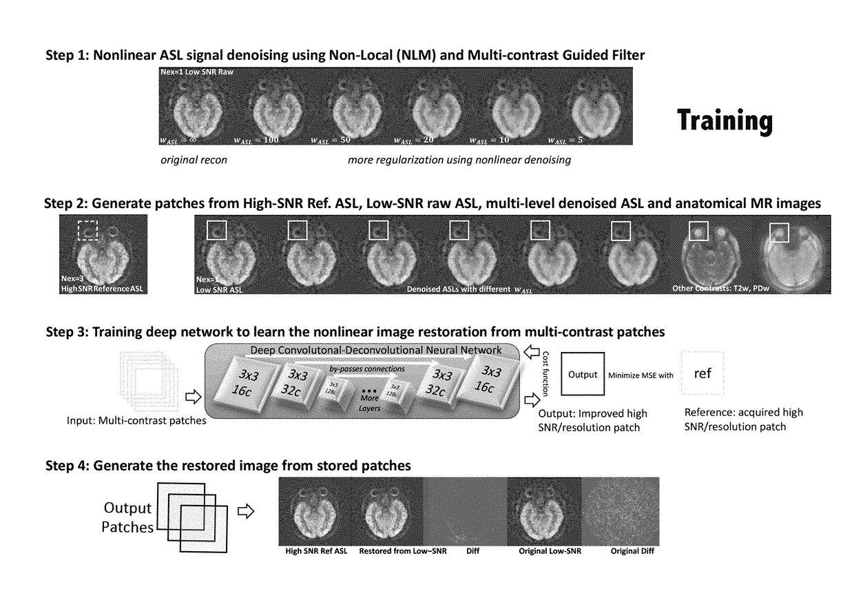
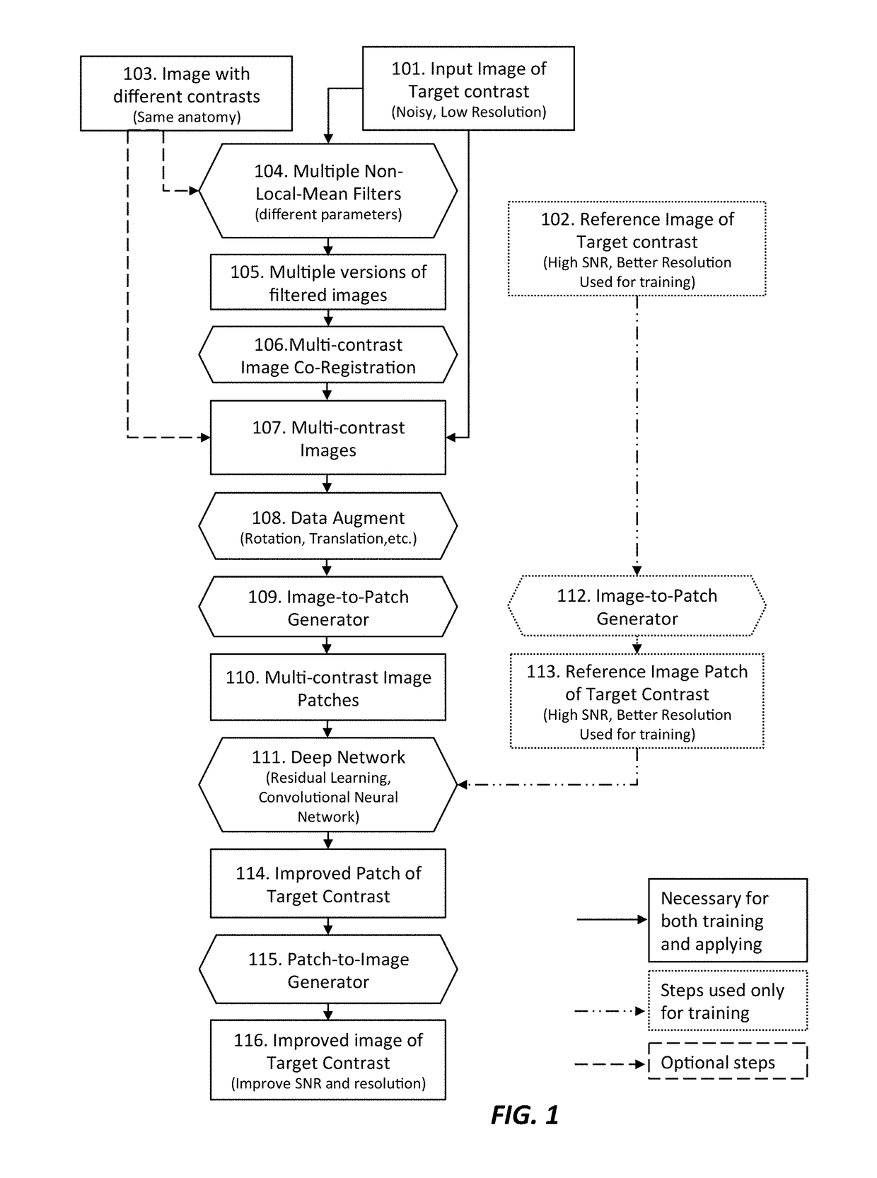
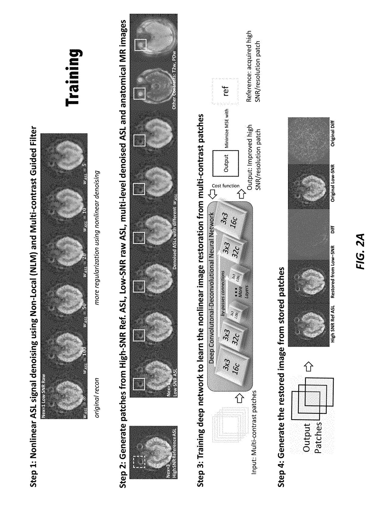
[0020]The current invention provides a method to improve the image quality of medical images. The invention provides a new end-to-end deep learning framework of taking raw image data and nonlinear filter results, which include denoised raw images with different denoising levels, and adding multi-contrast image data that have similar anatomy information with different contrasts, and generating improved image data with better quality in terms of resolution and SNR. The end-to-end framework of the current invention achieves better performance and faster speed.

[0021]In one embodiment, the invention improves the image quality of MRI imaging that typically has low SNR and resolution, for example arterial spin labeling (ASL) MRI images. The invention improves the image quality by using multi-contrast information from other images with the same anatomical structure but different contrasts, and using deep learning technique as an effective and efficient approach.

[0022]FIG. 1 shows a flow dia...


PUM


Abstract

A method of improving diagnostic and functional imaging is provided by obtaining at least two input images of a subject, using a medical imager, where each input image includes a different contrast, generating a plurality of copies of the input images using non-local mean (NLM) filtering, using an appropriately programmed computer, where each input image copy of the subject includes different spatial characteristics, obtaining at least one reference image of the subject, using the medical imager, where the reference image includes imaging characteristics that are different form the input images of the subject, training a deep network model, using data augmentation on the appropriately programmed computer, to adaptively tune model parameters to approximate the reference image from an initial set of the input and reference images, with the goal of outputting an improved quality image of other sets of low SNR low resolution images, for analysis by a physician.

Description

FIELD OF THE INVENTION[0001]The invention relates to medical imaging. More specifically the invention relates to improving the quality of medical images using multi-contrast imaging, multi-lateral filters, and deep learning methods.BACKGROUND OF THE INVENTION[0002]With medical image denoising, multiple methods have been proposed including, Gaussian filtering, wavelet filtering, and non-local means (NLM) algorithms, where experimentation has shown the NLM (possibly combining Wavelet) is the superior method. However, all these methods still share some disadvantages such as the dependency of parameter tuning for different images. In one instance, a proposed method used the redundancy in and relationships of multi-contrast images as a prior for image denoising. Related works have been used to combine a blurry and a noisy pair of images for CMOS sensors and cameras. A further implementation used Group-Sparsity representation for image denoising, which also used the multi-contrast informa...

Claims


Application Information

Patent Timeline
IPC IPC(8): G06T7/00G06T3/60
CPCG06T7/0012G06T2207/10088G06T2207/10081G06T3/60G06T5/002G06T5/20G06T5/50G06T2207/20182G06T2207/20084
Inventor ZAHARCHUK, GREGGONG, ENHAOPAULY, JOHN M.
Owner THE BOARD OF TRUSTEES OF THE LELAND STANFORD JUNIOR UNIV
Why Eureka
Try Eureka