

Method of modulating müller glia cells
a glia cell and muller technology, applied in the field of cell-based or regenerative therapy for ophthalmic diseases and disorders, can solve the problems of ineffective treatment for most patients, and achieve the effects of preventing or attenuating reactive gliosis, and enhancing or restoring retinal synaptic connectivity
- Summary
- Abstract
- Description
- Claims
- Application Information
AI Technical Summary
Benefits of technology
Problems solved by technology
Method used
Image

Examples
example 1
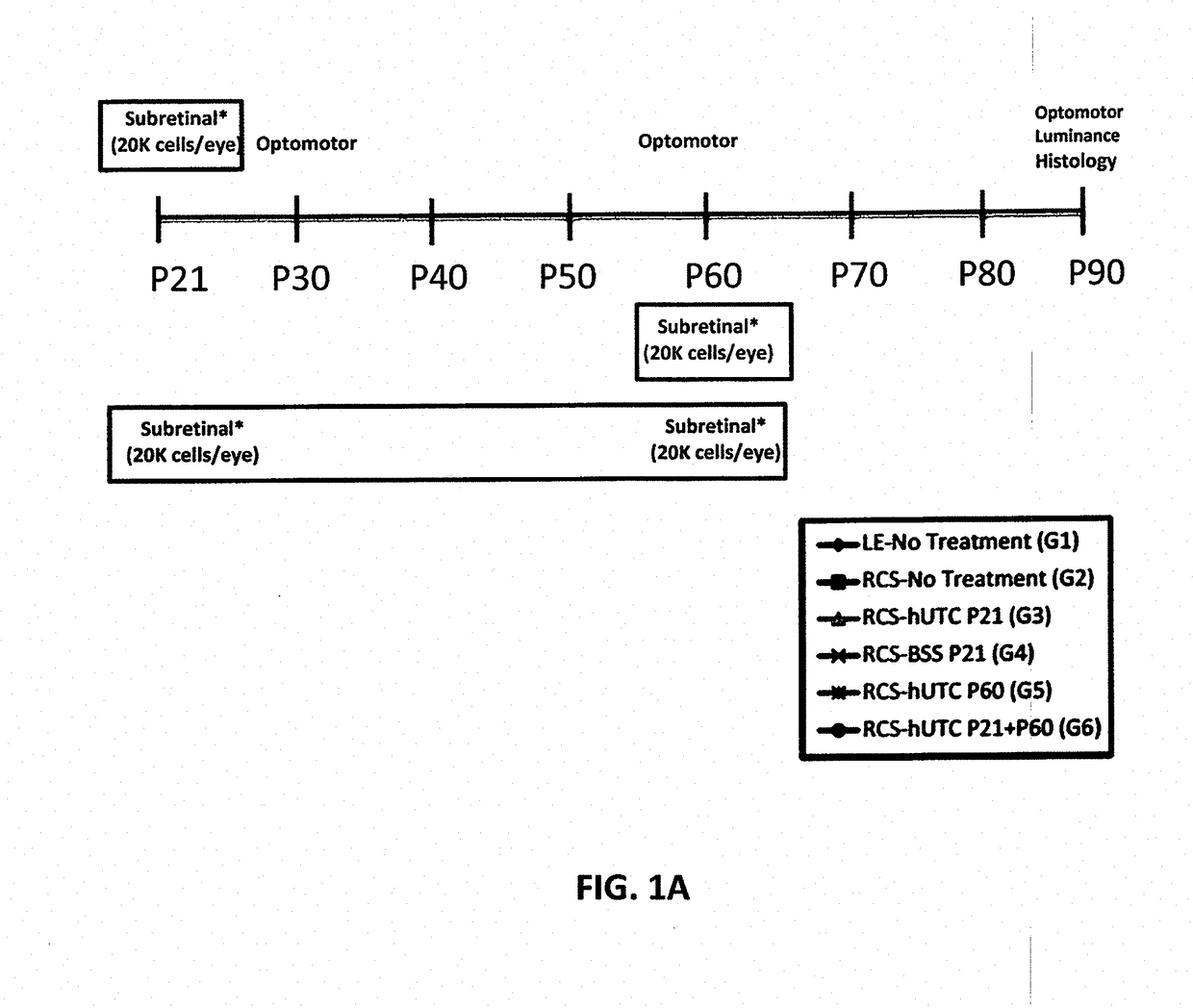
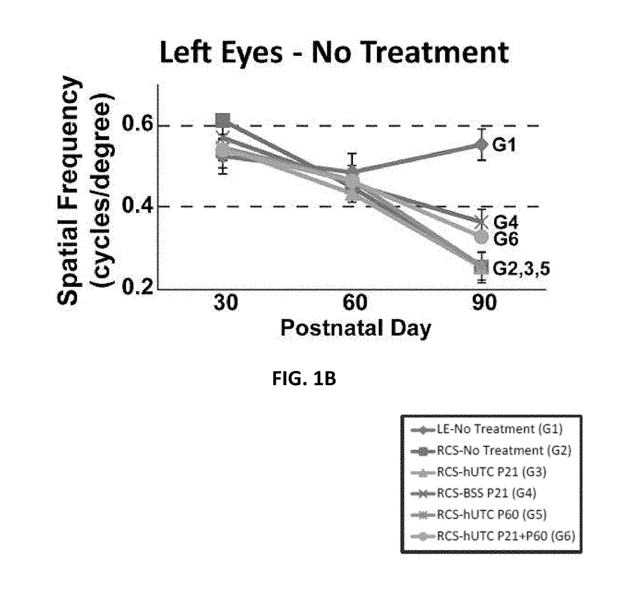
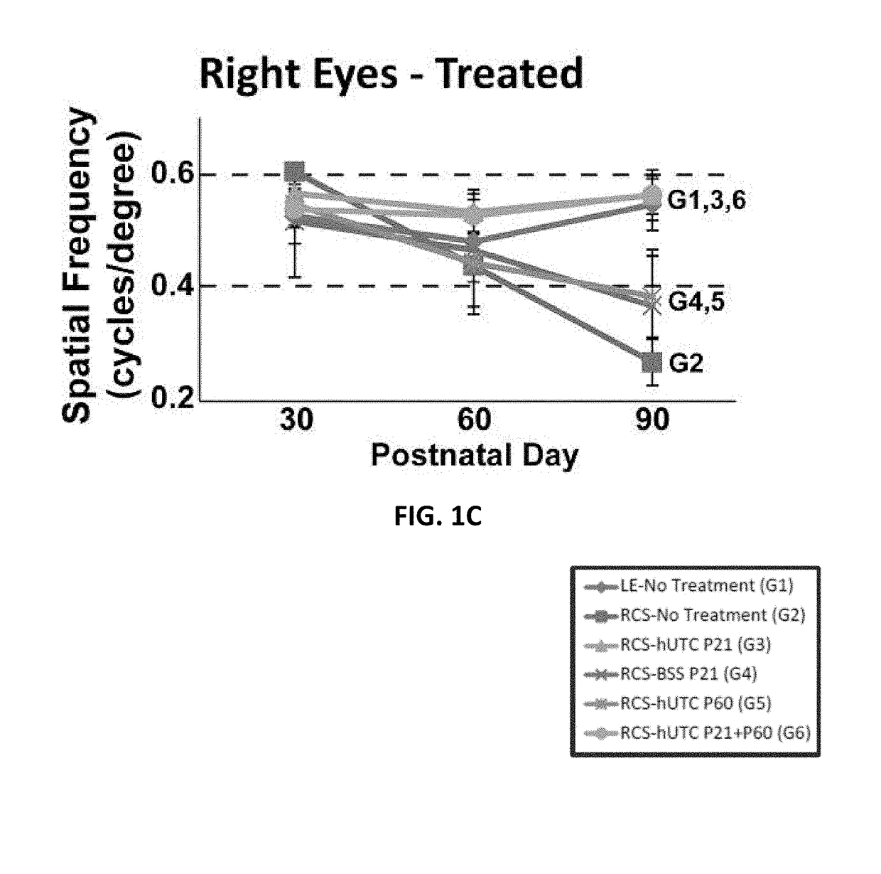
[0151]Subretinal transplantation of hUTC (administered at postnatal day 21) recovers visual function in the RCS rats (Lund et al., Stem Cells, 2007; 25; 602-611). The therapeutic effects of hUTC transplantation were gained without transdifferentiation of transplanted cells into retinal neurons. The effect of hUTC treatment during recovery of visual function was investigated.
Materials and Methods
[0152]Hutc Preparation:
[0153]hUTC were isolated and cryopreserved as described in Examples 5-11 following, and U.S. Pat. Nos. 7,524,489, 7,510,873, and 9,579,351, each incorporated by reference herein. Cryopreserved hUTC (˜31.3 population doublings; 2×10⁶ viable cells / mL) were used for the present example. On each day of injection, frozen cells (2-3 vials) were thawed at 37° C. in a water bath for ˜2 minutes. Upon thaw, cells were transferred to a single 15 mL conical tube containing 8 mL of balanced saline solution (BSS) Sterile Irrigating Solution (Alcon, Fort Wor...
example 2
Effect on Synaptic Development in RCS Rat Retina
Materials and Methods
[0173]Procedures for hUTC preparation, animals for cell transplantation, subretinal injections, visual function assessments, and retina preparation for immunohistochemistry are described in Example 1.
[0174]Identification of Synapses by Immunocytochemistry:
[0175]Retina sections were washed three times then permeabilized in PBS with 0.4% Triton-X 100 (PBST; Roche, Switzerland) at room temperature. Sections were blocked in 5% Normal Goat Serum (NGS) or Bovine Serum Albumin (BSA) in phosphate-buffered saline-triton (PBST) for 1 hr at room temperature. Primary antibodies (mouse anti-Bassoon 1:500 [RRID: AB_10618753, ADI-VAM-PS003-F, Enzo, NY], rabbit anti-mGluR6 1:150 [RRID: not applicable (n / a), RA13105, Neuromics, MN], guinea pig anti-VGlutl 1:750 [AB5905, Millipore, MA], rabbit anti-PSD95 1:500 [RRID: AB_87705, 51-6900, Invitrogen, CA], mouse anti-Gephyrin 1:250 [RRID: AB_1279448, 147-021, Synaptic Systems, Goettinge...
example 3
Effect of Synaptogenic Factors Produced by Müller Glia in the RCS Rat
[0186]This example investigates the synaptogenic signaling mediated by Müller glia in the RCS rat retina. Glia-secreted thrombospondin (TSP) family proteins play a role in excitatory synapse formation in the brain (Christopherson et al., Cell, 2005; 120: 421-433), and it has previously been reported that TSP-1 is secreted by cultured Müller glia cells in vitro.
Materials and Methods
[0187]Procedures for hUTC preparation, animals for cell transplantation, subretinal injections, visual function assessments, and retina preparation for immunohistochemistry are described in Example 1, and for identification and quantification of synapses in Example 2.
[0188]RNA Fluorescence In Situ Hybridization (FISH):
[0189]A set of FISH probes targeting either Thbs1 or Thbs2 was purchased from Stellaris (LGC Biosearch Technologies, CA). Each probe set is composed of 48 oligonucleotides (20 nucleotides each) that selectively bind to trans...
PUM
 Abstract
Description
Claims
Application Information