

System and method for medical imaging
a medical imaging and system technology, applied in the field of image processing, can solve problems such as increasing setup complexity
- Summary
- Abstract
- Description
- Claims
- Application Information
AI Technical Summary
Benefits of technology
Problems solved by technology
Method used
Image

Examples
example 1
Vision-Based Intraoperative Cone-Beam CT Stitching for Non-Overlapping Volumes
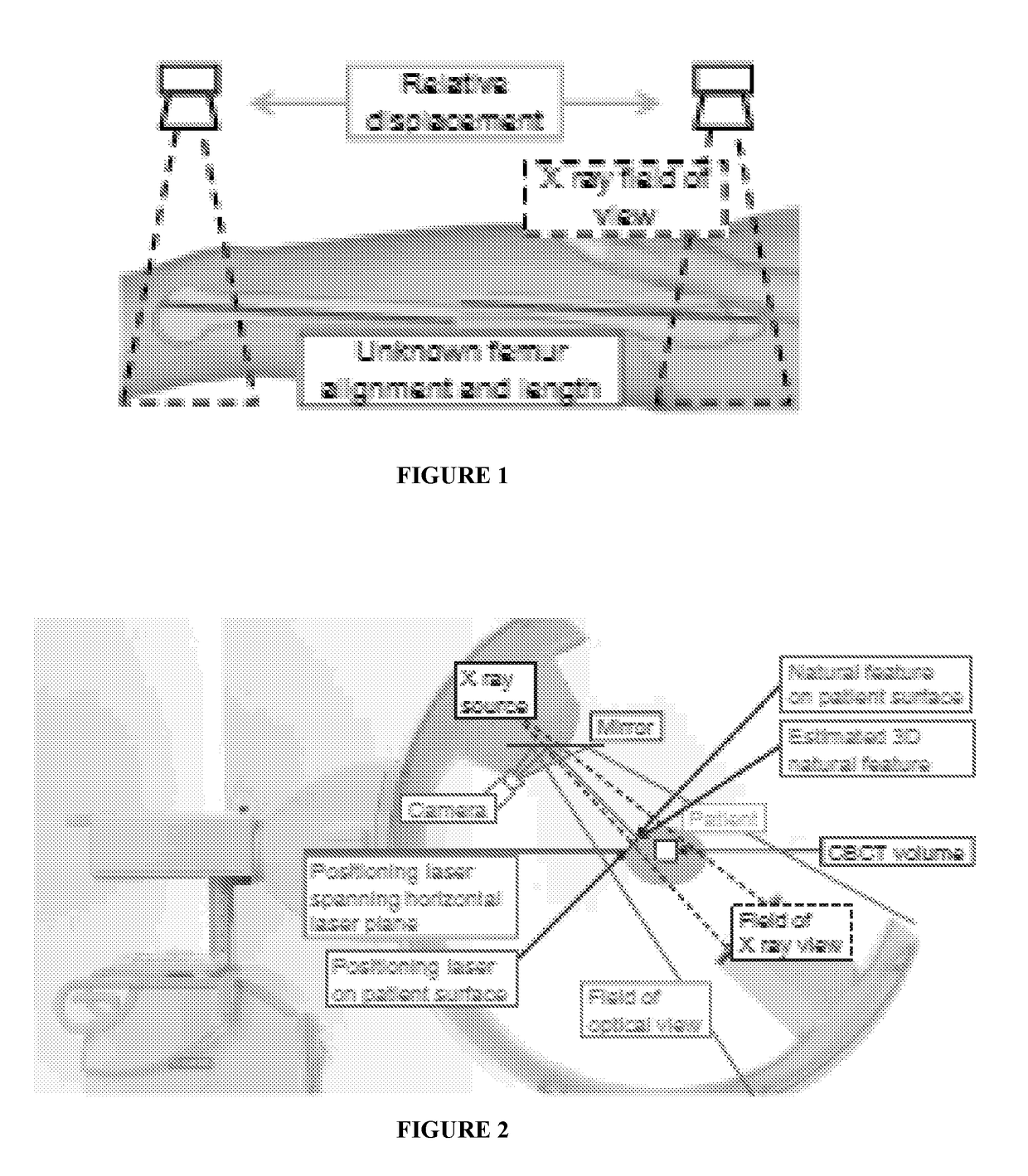
[0066]Cone-Beam Computed Tomography (CBCT) is one of the primary imaging modalities in radiation therapy, dentistry, and orthopedic interventions. While providing crucial intraoperative imaging, CBCT is bounded by its limited imaging volume, motivating the use of image stitching techniques. Current methods rely on overlapping volumes, leading to an excessive amount of radiation exposure, or on external tracking hardware, which may increase the setup complexity. We attach an optical camera to a CBCT enabled C-arm, and co-register the video and X-ray views. Our novel algorithm recovers the spatial alignment of non-overlapping CBCT volumes based on the observed optical views, as well as the laser projection provided by the X-ray system. First, we estimate the transformation between two volumes by automatic detection and matching of natural surface features during the patient motion. Then, we recover 3D inform...
example 2
Preclinical Usability Study of Multiple Augmented Reality Concepts for K-Wire Placement
[0104]Summary
[0105]Purpose
[0106]In many orthopedic surgeries, there is a demand for correctly placing medical instruments (e.g., K-wire or drill) to perform bone fracture repairs. The main challenge is the mental alignment of X-ray images acquired using a C-arm, the medical instruments, and the patient, which dramatically increases in complexity during pelvic surgeries. Current solutions include the continuous acquisition of many intra-operative X-ray images from various views, which will result in high radiation exposure, long surgical durations, and significant effort and frustration for the surgical staff. This work conducts a preclinical usability study to test and evaluate mixed reality visualization techniques using intra-operative X-ray, optical, and RGBD imaging to augment the surgeon's view to assist accurate placement of tools.
[0107]Method
[0108]We design and perform a usability study to ...
example 3
Calibration of RGBD Camera and Cone-Beam CT for 3D Intra-Operative Mixed Reality Visualization
[0171]Summary
[0172]Purpose
[0173]This work proposes a novel algorithm to register cone-beam computed tomography (CBCT) volumes and 3D optical (RGBD) camera views. The co-registered real-time RGBD camera and CBCT imaging enable a novel augmented reality solution for orthopedic surgeries, which allows arbitrary views using digitally reconstructed radiographs overlaid on the reconstructed patient's surface without the need to move the C-arm.
[0174]Methods
[0175]An RGBD camera is rigidly mounted on the C-arm near the detector. We introduce a calibration method based on the simultaneous reconstruction of the surface and the CBCT scan of an object. The transformation between the two coordinate spaces is recovered using Fast Point Feature Histogram descriptors and the Iterative Closest Point algorithm.
[0176]Results
[0177]Several experiments are performed to assess the repeatability and the accuracy of...
PUM
 Abstract
Description
Claims
Application Information