
System and Method for Measuring Intraocular Pressure and Ocular Tissue Biomechanical Properties

Active Publication Date: 2019-05-02
UNIV HOUSTON SYST
View PDF0 Cites 1 Cited by
  • Summary
  • Abstract
  • Description
  • Claims
  • Application Information

AI Technical Summary

Benefits of technology

The present invention is a system and method for measuring intraocular pressure, ocular tissue geometry, and biomechanical properties in an eye. The system includes an optical coherence tomography subsystem and an applanation tonometer subsystem in electronic communication with each other. The method involves scanning the eye with a laser source and obtaining images through optical coherence tomography. The intraocular pressure is calculated based on the obtained images. The system can also measure the elastic properties of the ocular tissue and identify any abnormalities in the eye based on the calculated intraocular pressure, ocular tissue geometry, and measured biomechanical properties. The system and method provide a non-invasive and accurate way to analyze the health of the eye.

Problems solved by technology

However, the large displacements induced by the air-puff prohibit local assessment of corneal biomechanical properties (3) and cause nonlinear biomechanical behaviors (4).
Moreover, biomechanical measurements of the cornea are confounded by other parameters, such as IOP5,6 and central corneal thickness (CCT).5,7 Thus, providing an accurate measurement of corneal mechanical parameters (e.g., Young's modulus) is not straightforward, let alone based on applanation measurements.
Nevertheless, commercially available noncontact tonometers, e.g., the ocular response analyzer and CorVis ST, can distinguish between healthy and keratoconic corneas (8), but there are conflicting results on their ability to detect corneal biomechanical changes due to therapeutic interventions such as corneal collagen crosslinking (8,9).
Therefore, it may not be entirely possible to separate the effects of corneal geometry, IOP, and corneal biomechanical properties from their respective individual measurements for corrections.
Ultrasound elastography and magnetic resonance elastography are clinically available elastographic techniques, but are not well-suited for small and thin tissues such as the cornea and the sclera, due to their relatively lower spatial resolution, poorer displacement sensitivities and need for contact-based excitation or coupling medium.
Particularly the prior art is deficient in an imaging device, system, method, and / or technique to accurately measure intraocular pressure and biomechanical properties of ocular tissues, including corneal, retinal, or scleral tissues.

Method used


Image

  • System and Method for Measuring Intraocular Pressure and Ocular Tissue Biomechanical Properties
  • System and Method for Measuring Intraocular Pressure and Ocular Tissue Biomechanical Properties
  • System and Method for Measuring Intraocular Pressure and Ocular Tissue Biomechanical Properties

Examples


example 1

Materials and Methods

Agar Phantom Samples

[0046]Tissue-mimicking agar phantoms (Difco nutrient agar, Beckton, Dickinson and Company, Sparks, Md., USA) were used. Concentrations (w / w) of agar are 1%, 1.5% and 2%, n=3 of each concentration.

Porcine Eyes

[0047]Fresh porcine eyes (Sioux-Preme Packing Company, Sioux City, Iowa; n=3) are used. Extraneous tissues such as muscles are removed from the eye-globes and all measurements were taken within 24 h of enucleation. The whole eye-globes were placed in a home-built holder for artificial IOP control (17). During testing, the eye-globes are cannulated with two 23 G needles for an artificial IOP control. One needle was connected via tubing to a pressure transducer and the other needle was connected via tubing to a microinfusion pump.

Video

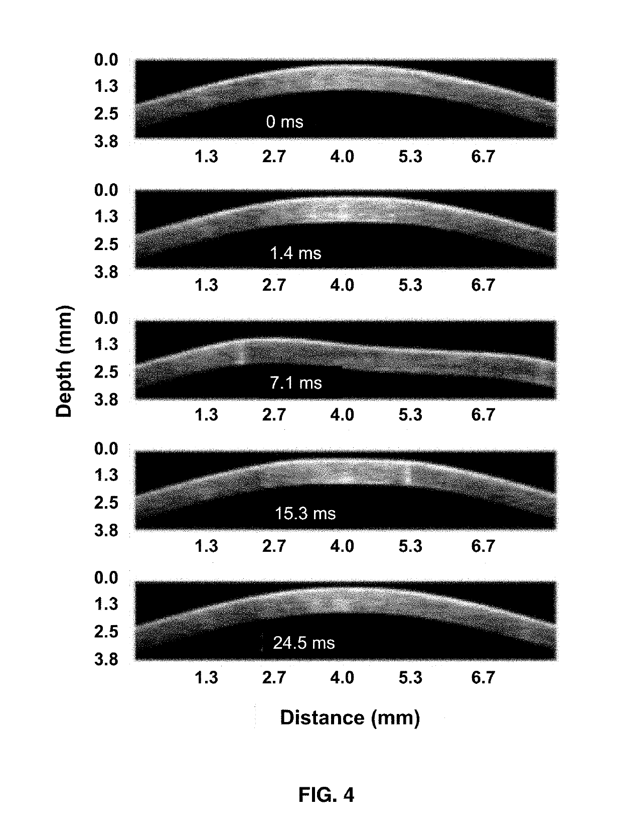
[0048]Video of noncontact applanation (Video 1) or elastic wave propagation (Video 2) was imaged at a frame rate of ˜7.3 kHz.

[0049]Video 1, MP4, 6.5 MB, dx.doi.org / 10.1117 / 1.JBO.22.2.020502.1; and

[0050]Video 2...

example 2

Optical Imaging System

Phase-Sensitive Swept-Source Optical Coherence Tomography (PhS-SSOCT) Imaging Subsystem

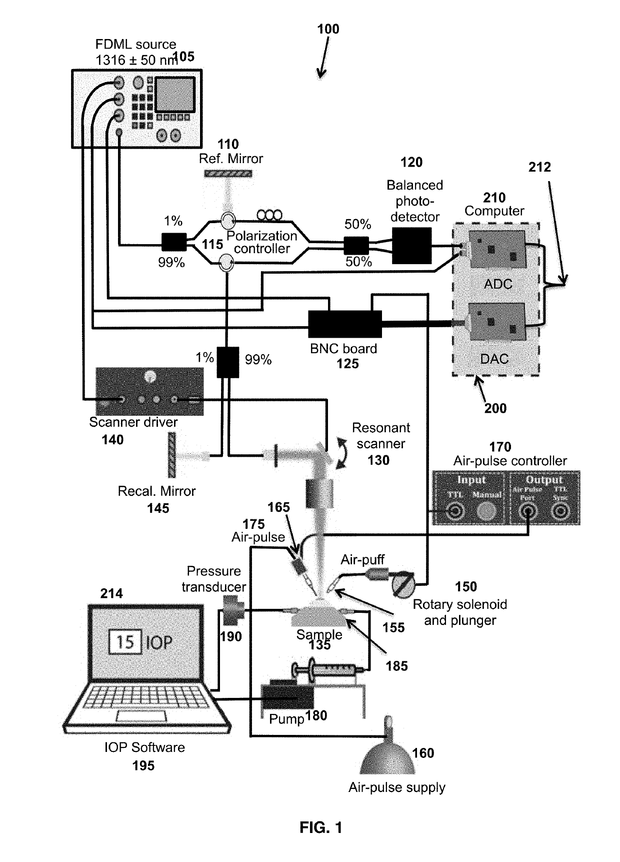
[0051]The phase-sensitive swept source Optical Coherence Tomography system (PhS-SSOCT) (see FIG. 1) imaging system utilizes a 4× buffered Fourier domain model locked (FDML) swept source laser with an A-scan or sweep rate of ˜1.5 MHz, a central wavelength of ˜1316 nm, a scan range of ˜100 nm, an axial resolution of ˜16 μm, a phase stability of ˜14 nm, and a resonant scanner at ˜7.3 kHz. The PhS-SSOCT optical imaging system is utilized to image the dynamic spatio-temporal deformation of the cornea in response to the air-puff and detect the nano to micrometer-scale displacements induced by the focused air-pulse. The PhS-SSOCT imaging system is controlled by a computer comprising at least a processor, memory, analog-to-digital (ADC) and digital-to-analog (DAC) converters, a display, and other requisite software and hardware to operate the system and at least one network connectio...

example 3

PhS-SSOCT Assessment of Elasticity of Optical Samples

Agar Phantom

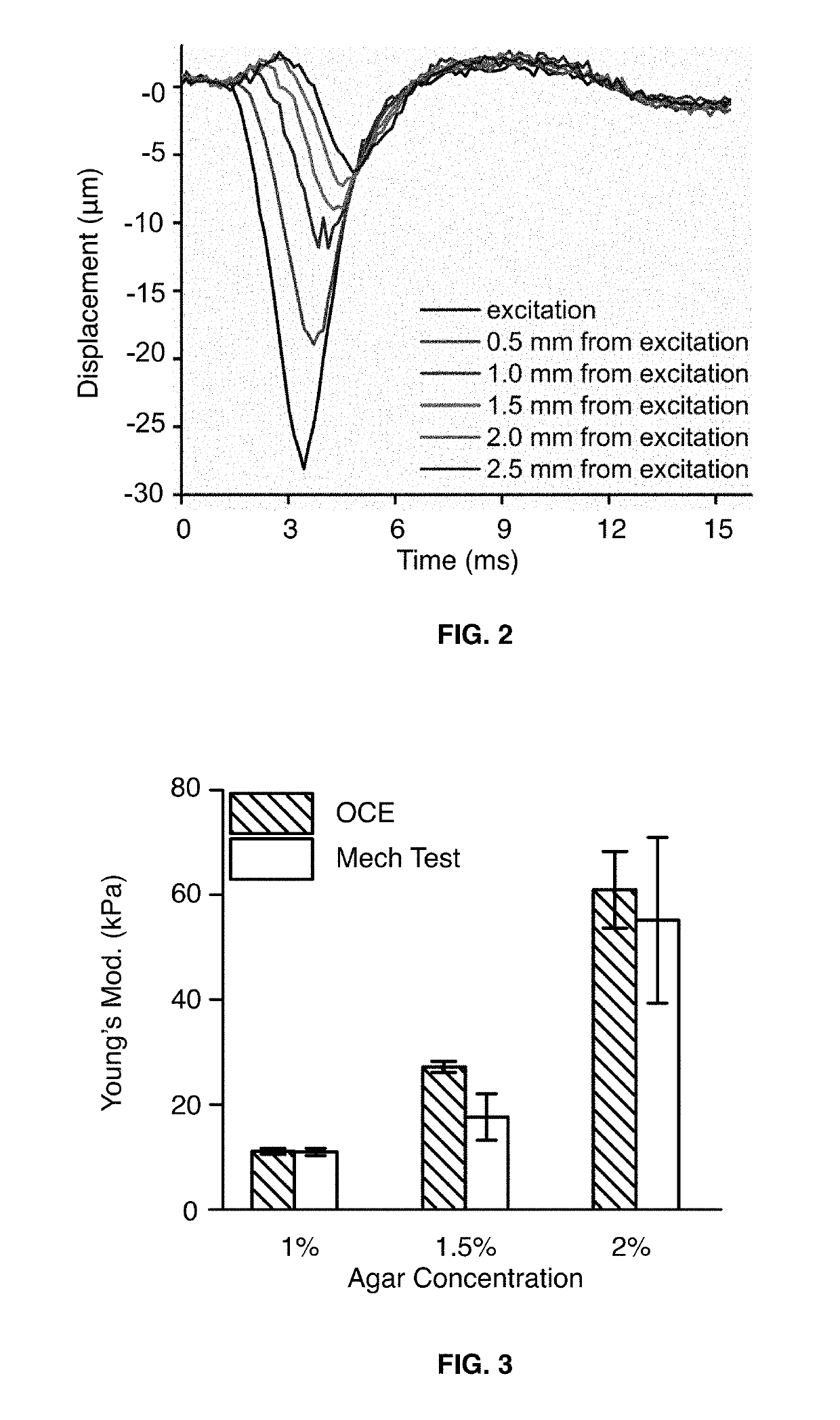
[0053]The propagation of the air-pulse induced elastic wave in a tissue-mimicking agar phantom is illustrated in FIG. 2. The temporal vertical displacement profiles at the indicated distances are plotted. The distances are at about 0.5 mm, 1.0 mm, 1.5 mm, 2.0 mm and 2.5 mm away from the air-pulse excitation. The largest displacement at all the distance appears at the time of about 3 ms, and the displacement is fully recovered after about 6 ms for all the excitation distances. The wave propagation, attenuation, and dispersion can be observed from the shape of the profiles at the various distances and can be also be used to calculate biomechanical properties.

[0054]In order to validate the optical coherence elastography technique, the stiffness of agar phantoms of various concentrations (1%, 1.5%, and 2% w / w, n=3 of each concertation) assessed by optical coherence elastography and measured by the “gold standard” uniaxial ...


PUM


Abstract

Provided herein are systems and methods to measure the intraocular pressure, ocular tissue geometry and the biomechanical properties of an ocular tissue, such as an eye-globe or cornea, in one instrument. The system is an optical coherence tomography subsystem and an applanation tonometer subsystem housed as one instrument and interfaced with a computer for at least data processing and image display. The system utilizes an air-puff and a focused micro air-pulse to induce deformation and applanation and displacement in the ocular tissue. Pressure profiles of the air puff with applanation times are utilized to measure intraocular pressure. Temporal profiles of displacement and / or spatio-temporal profiles of a displacement-generated elastic wave are analyzed to calculate biomechanical properties.

Description

CROSS-REFERENCE TO RELATED APPLICATIONS[0001]This application is a continuation under 35 U.S.C. § 120 of pending international patent application PCT / US2017 / 038799, filed Jun. 22, 2017 which claims benefit of priority under 35 U.S.C. § 119(e) of provisional application U.S. Ser. No. 62 / 353,398, filed Jun. 22, 2016, the entirety of both of which are hereby incorporated by reference.GOVERNMENT SUPPORT CLAUSE[0002]This invention was made with government support under grant numbers 1R01EY022362 and P30EY07551 awarded by the National Institutes of Health. The government has certain rights in the invention.BACKGROUND OF THE INVENTIONField of the Invention[0003]The present invention generally relates to the field of eye care systems and methods of use. More specifically, the present invention relates to a non-contact technique of measuring the intraocular pressure (IOP) and geometry of the eye-globe with optical coherence tomography (OCT) and measuring biomechanical properties of ocular ti...

Claims


Application Information

Patent Timeline
IPC IPC(8): A61B3/16A61B3/10
CPCA61B3/165A61B3/102A61B5/0066A61B3/107A61B3/18A61B5/7246A61B5/7289
Inventor LARIN, KIRILL V.SINGH, MANMOHANLI, JIASONGHAN, ZHAOLONGTWA, MICHAEL D.
Owner UNIV HOUSTON SYST
Learn More