Systems and methods for growth of intestinal cells in microfluidic devices

a microfluidic device and intestinal cell technology, applied in cell culture active agents, artificial cell constructs, instruments, etc., can solve the problems of deleterious and low-dose exposure to endocrine disrupting chemicals (edcs)

Active Publication Date: 2019-05-23
CEDARS SINAI MEDICAL CENT
View PDF0 Cites 9 Cited by
  • Summary
  • Abstract
  • Description
  • Claims
  • Application Information

AI Technical Summary

Benefits of technology

[0010]Organs-on-chips are microfluidic devices for culturing living cells in micrometer sized chambers in order to model physiological functions of tissues and organs. Continuous perfusion through the chambers allows incorporation of physical forces, including physiologically relevant levels of fluid shear stress, strain and compression, for study of organ-specific responses. Of great interest is adapting such fabrication techniques for development of a “gut-on-a-chip” capable of replicating the corresponding physiological environment, and dynamically incorporating those multiple components (microbiome, mucosa, immune components) in a manner mirroring IBD pathophysiology. Towards these aims, prior attempts have successfully relied on human intestinal epithelial cells (Caco-2) cultured in the presence of physiologically relevant luminal flow and peristalsis-like mechanical deformations. This approach allows formation of intestinal villi lined by all four epithelial cell lineages of the small intestine (absorptive, goblet, enteroendocrine, and Paneth), with enhanced barrier function, drug-metabolizing cytochrome P450 activity, and apical mucus secretion.
[0017]The use of microfluidic intestinal chips described herein improves / increases maturation of iPS derived intestinal cells. More specifically, use of such chips improves maturation efficiency, e.g. iPS cell differentiation into foregut increases numbers of cells such as synaptophysin (SYP) positive cells, and improves quality of intestinal epithelium, i.e. an epithelial layer folds into finger-like projections lined with epithelial cells of which some are separated by pit-like areas mimicking villus-like structures lined with epithelium and pit-like areas, for mimicking human intestinal microvillus when seeded with iPSC derived intestinal cells. Further, these villus structures are continuously growing as basal cells divide and move up the sides of the villi. Moreover, the folds of epithelium comprise non-epithelial intestinal cells.
[0034]As described herein, selecting proper cells before seeding on the chip provides chips mimicking intestinal epithelium (lining) having villi-like structures and a range of non-epithelial intestinal cells. During the development of the present inventions it was discovered that disassociation of enteroids into single cell suspensions then sorting cells using E-cadherin selection markers for seeding E-cadherin+ cells into the apical channel of chips, provided intestinal cell layers having finger-like projections and mimicking folding of in vivo intestinal epithelial layers with villus structures. Further, it was discovered that the use of a selection reagent for lifting cells from organoid cultures provided single cell suspensions for seeding onto chips providing equal or better quality epithelium. Thus, the use of a selection reagent can replace the cell-sorting step.

Problems solved by technology

We hypothesized that chronic low-dose exposure to endocrine disrupting chemicals (EDCs), is deleterious during early human endocrine tissue development.

Method used


Image

  • Systems and methods for growth of intestinal cells in microfluidic devices
  • Systems and methods for growth of intestinal cells in microfluidic devices
  • Systems and methods for growth of intestinal cells in microfluidic devices

Examples


example 1

Study Design

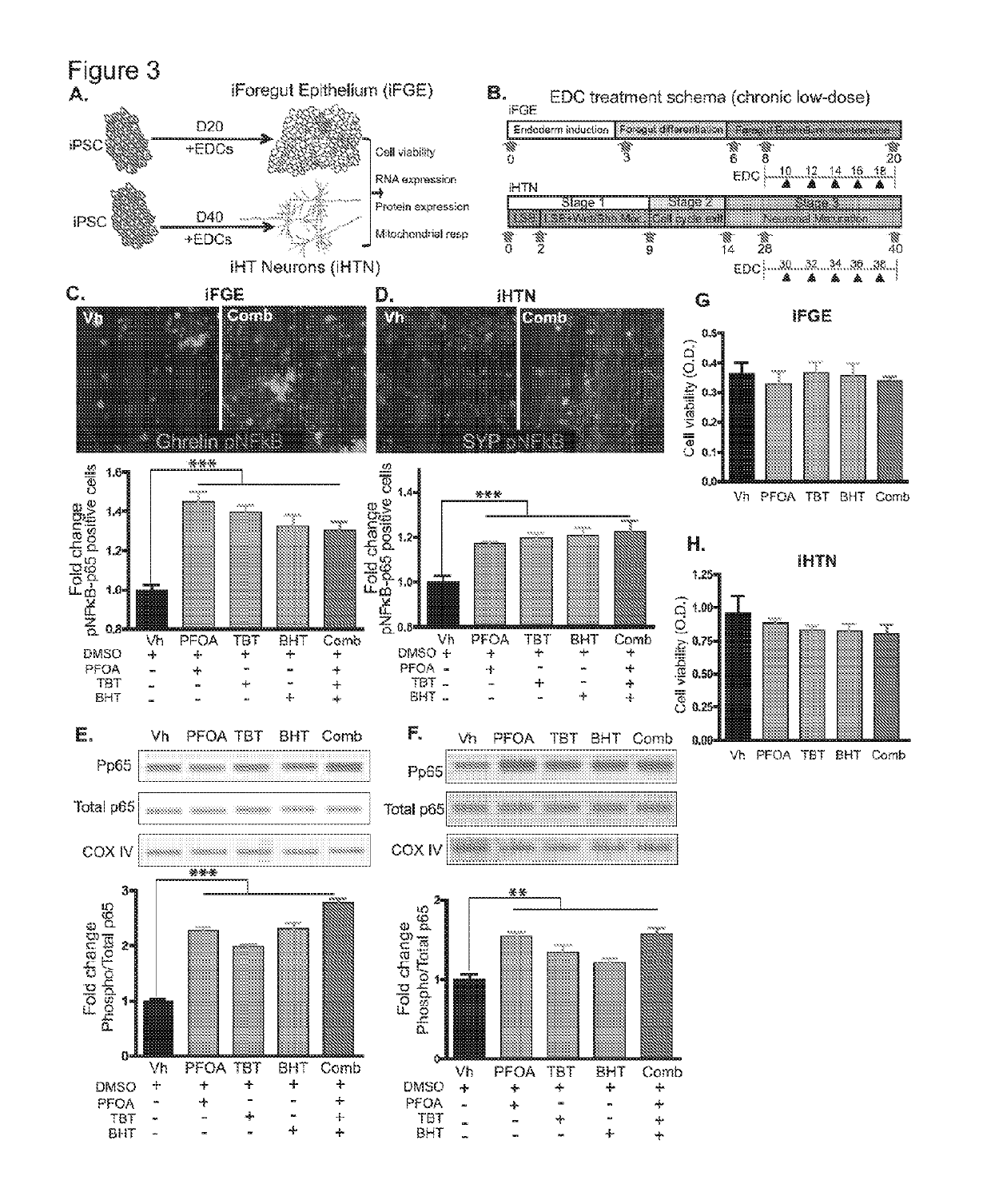
[0196]Described herein is the use of human induced pluripotent stem cells (hiPSCs) to elucidate the adverse effects and mechanisms of chronic low-dose EDC exposures on developing gut and hypothalamic neuropeptidergic neurons, and serves as a platform for mimicking the in utero exposure to EDCs. Such a screening platform can not only faithfully mimic a human model of development but also can provide invaluable insights on the developmental cues that could be disrupted by the compounds screened for.

example 2

Foregut Epithelium Differentiation (iFGE)

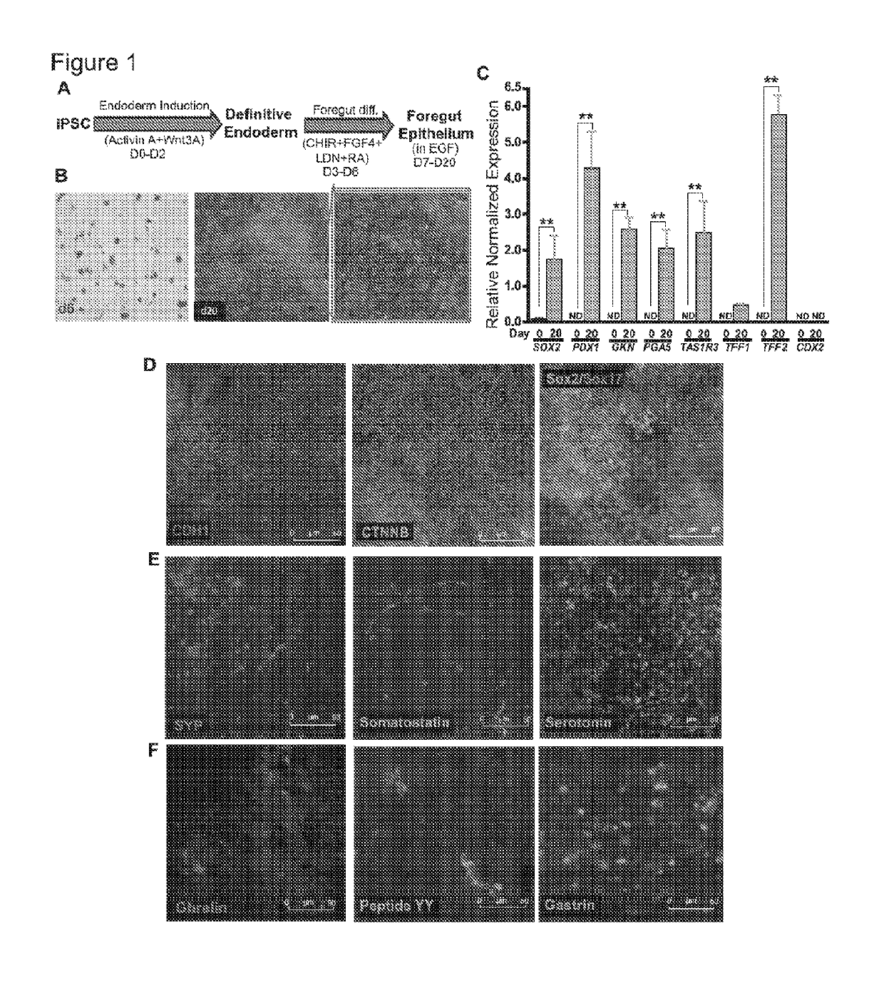
[0197]For differentiation, iPSCs were accutase-treated and plated into a 6-well Matrigel-coated dish at a density of lmillion per well in E8 medium with ROCK-inhibitor Y27632 (10 μM; Stemgent). On the next day, iPSCs were differentiated into definitive endoderm by exposing them to Activin A (100 ng / ml; R&D) and Wnt3A (25 ng / ml only on the first day; Peprotech) in RPMI 1640 (Gibco) for 3 days. During these 3 days, the cells were exposed to increasing concentrations of 0%, 0.2% and 2% defined FBS (dFBS, Hyclone). After definitive endoderm induction, the cells were directed to form foregut spheroids by culturing them for the next 3 days in Advanced DMEM / F12 medium (Gibco) containing 2% dFBS, 2 μM CHIR99021 (2 μM; Cayman), FGF4 (500 ng / ml; Peprotech), LDN (2 μM; Cayman) and retinoic Acid (2 μM; Cayman). This resulted in semi floating spheroids, which were then selectively picked and transferred on to Matrigel-coated experimental plates for furthe...

example 3

Hypothalamic Neuron Differentiation (iHTN)

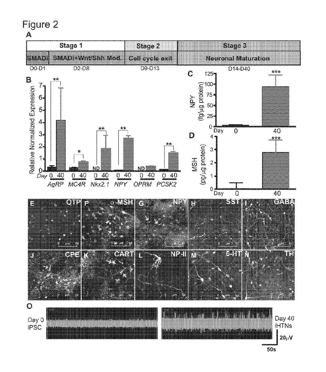
[0198]For differentiation into iHTNs, iPSCs were accutase-treated and plated as single cells in 6-well Matrigel-coated plates at a density of approx. 1 million cells / well in E8 medium with ROCK-inhibitor Y27632 (10μM; Stemgent). The next day iHTN differentiation was initiated by neuroectoderm differentiation by dual SMAD inhibition using LDN193189 (1 μM, Cayman) and SB431542 (10 μM, Cayman) and this treatment is carried on for 48 hours. This was followed by Sonic hedgehog activation by Smoothened agonist SAG (1 μM, Tocris) and purmorphamine (PMN, 1 μM, Tocris) and Wnt signaling inhibition using IWR-endo (10 μM, Cayman) from Day 3 to day 8 to direct the cells towards ventral diancephalon with regular media change every 2 days. Day 9 to Day 13 the cells are slowly made to exit cell cycle using DAPT (10μM, Cayman) in the presence of ventralizing agent retinoic acid (0.1 μM, Cayman). On Day 14, the cells were accutased and replated onto Laminin-...


PUM

PropertyMeasurementUnit
flow rateaaaaaaaaaa
flow ratesaaaaaaaaaa
flow rateaaaaaaaaaa

Abstract

Organs-on-chips are microfluidic devices for culturing living cells in micrometer sized chambers in order to model physiological functions of tissues and organs. Engineered patterning and continuous fluid flow in these devices has allowed culturing of intestinal cells bearing physiologically relevant features and sustained exposure to bacteria while maintaining cellular viability, thereby allowing study of inflammatory bowl diseases. However, existing intestinal cells do not possess all physiologically relevant subtypes, do not possess the repertoire of genetic variations, or allow for study of other important cellular actors such as immune cells. Use of iPSC-derived epithelium, including IBD patient-specific cells, allows for superior disease modeling by capturing the multi-faceted nature of the disease.

Description

FIELD OF THE INVENTION[0001]The present invention relates to a combination of cell culture systems and microfluidic fluidic systems. More specifically, in one embodiment, the invention relates microfluidic chips seeded with stem-cell-derived cells that mature and. or differentiate into intestinal cells. In one embodiment, the stems are induced pluripotent stem cells (hiPSCs) and the intestinal cells are foregut cells. In some emdodiments, such forgut chips are tested for effects of endocrine disrupting chemicals (EDCs) during critical periods in tissue development mimicking critical periods of fetal development for short and long term downstream effects. In particular, methods for use are provided for induced pluripotent stem cells (hiPSCs) to elucidate adverse effects and mechanisms of chronic low-dose EDC exposures on developing gut and hypothalamic neuropeptidergic neurons, and serves as a platform for mimicking the in utero exposure to EDCs. Morover, in yet further embodiments, ...

Claims


Application Information

Patent Timeline
Patent Type & Authority Applications(United States)
IPC IPC(8): C12N5/071C12N5/079
CPCC12N5/0679C12N5/0618C12N2501/16C12N2501/415C12N2506/45C12N2501/119C12N2501/999C12N2501/11C12N2501/13C12M23/16C12N5/0696C12N2501/24C12N2501/998C12N2535/00C12M21/08C12N2501/155C12N2501/25G01N33/5005
Inventor BARRETT, ROBERTSVENDSEN, CLIVETARGAN, STEPHAN R.WORKMAN, MICHAELSAREEN, DHRUVRAJAMANI, UTHRA
Owner CEDARS SINAI MEDICAL CENT
Why Eureka
Try Eureka