Eureka AIR delivers breakthrough ideas for toughest innovation challenges, trusted by R&D personnel around the world.

Systems and Methods for Capturing and Analyzing Pupil Images to Determine Toxicology and Neurophysiology

a system and image technology, applied in the field of system and method apparatus for capturing pupillary light reflex, can solve the problems of slow response time, commercial or consumer versions of pupilometers typically do not provide any interpretation of plr, and tests have difficulty in detecting purely synthetic toxins, so as to improve the accuracy of diagnostic outputs.

Inactive Publication Date: 2019-06-27
PUPILSCAN CORP
View PDF0 Cites 8 Cited by
  • Summary
  • Abstract
  • Description
  • Claims
  • Application Information

AI Technical Summary

Benefits of technology

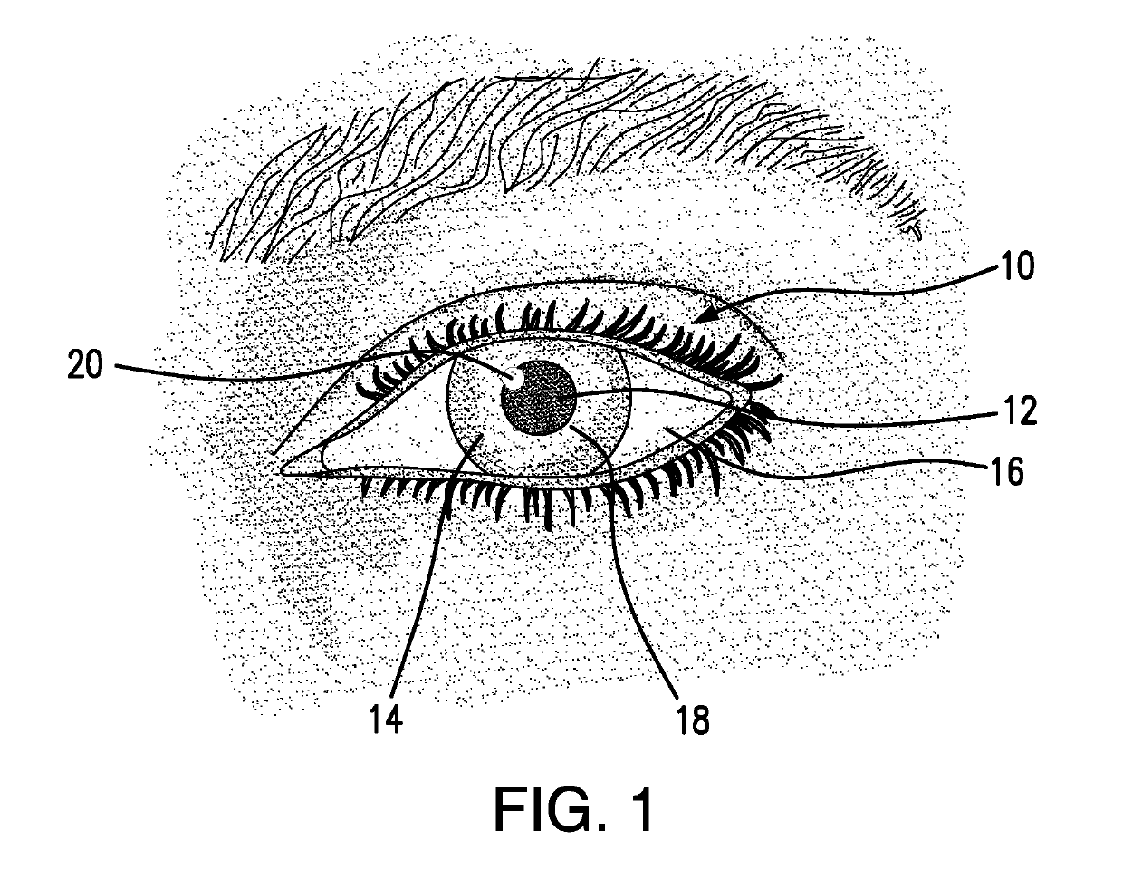
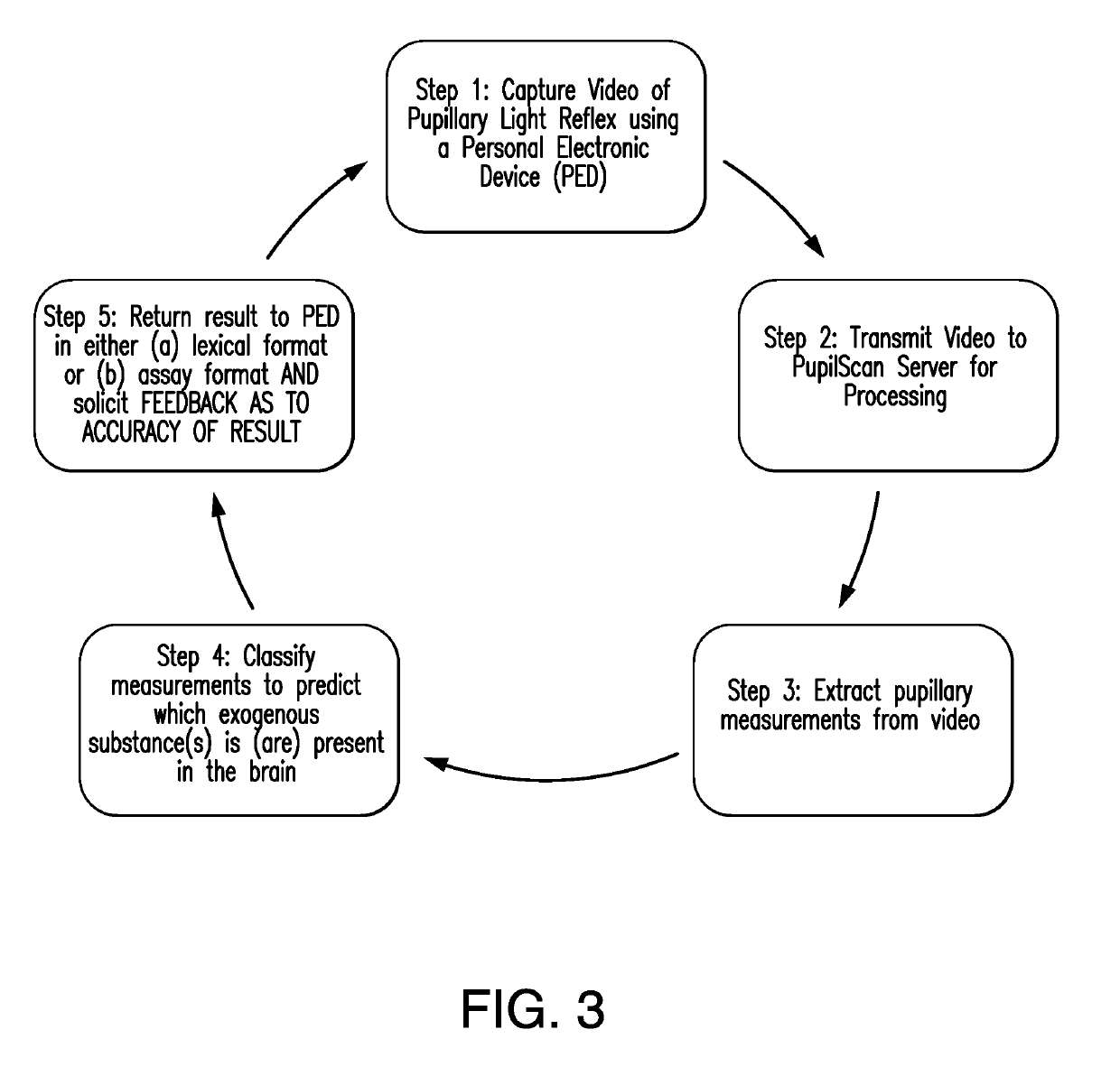
This patent describes a method and system for analyzing the pupil light reflex (PLR) in milliseconds to provide a quick result for medical professionals in clinical settings or in the field. The method uses a non-invasive method that doesn't involve physical contact with the subject's body, and it can capture and analyze the PLR using an artificial intelligence network for improved accuracy and diagnostic output. After analyzing, feedback data can be provided to the network for further accuracy improvement. The method can be implemented on a device, such as a camera and light source, and it can be a personal electronic device.

Problems solved by technology

Furthermore, commercial or consumer versions of pupilometers typically do not provide any interpretation of PLR.
1) the tests have difficulty detecting purely synthetic toxins such as fentanyl or methadone;
2) the tests reveal drug content in the blood outside of the blood-brain barrier, not neurological state or blood content inside the blood-brain barrier; and 3) the tests take time and are invasive. Fluid-based tests require fluid acquisition and then testing. Often in excess of 15 minutes is required to run the test and obtain results. By contrast, the methods that are disclosed herein below provide an immediate reading of actual neurological intoxication and provide a more relevant result, present neurological state vs. blood, saliva or urine toxin levels, in a non-invasive procedure. The methods disclosed herein below are easily used on unconscious patients in almost any environment, such as outside of a clinical setting.
In addition to a slow response time, mass spectrometry tests have a drawback akin to the assay tests.

Method used


Image

  • Systems and Methods for Capturing and Analyzing Pupil Images to Determine Toxicology and Neurophysiology
  • Systems and Methods for Capturing and Analyzing Pupil Images to Determine Toxicology and Neurophysiology
  • Systems and Methods for Capturing and Analyzing Pupil Images to Determine Toxicology and Neurophysiology

Examples


example

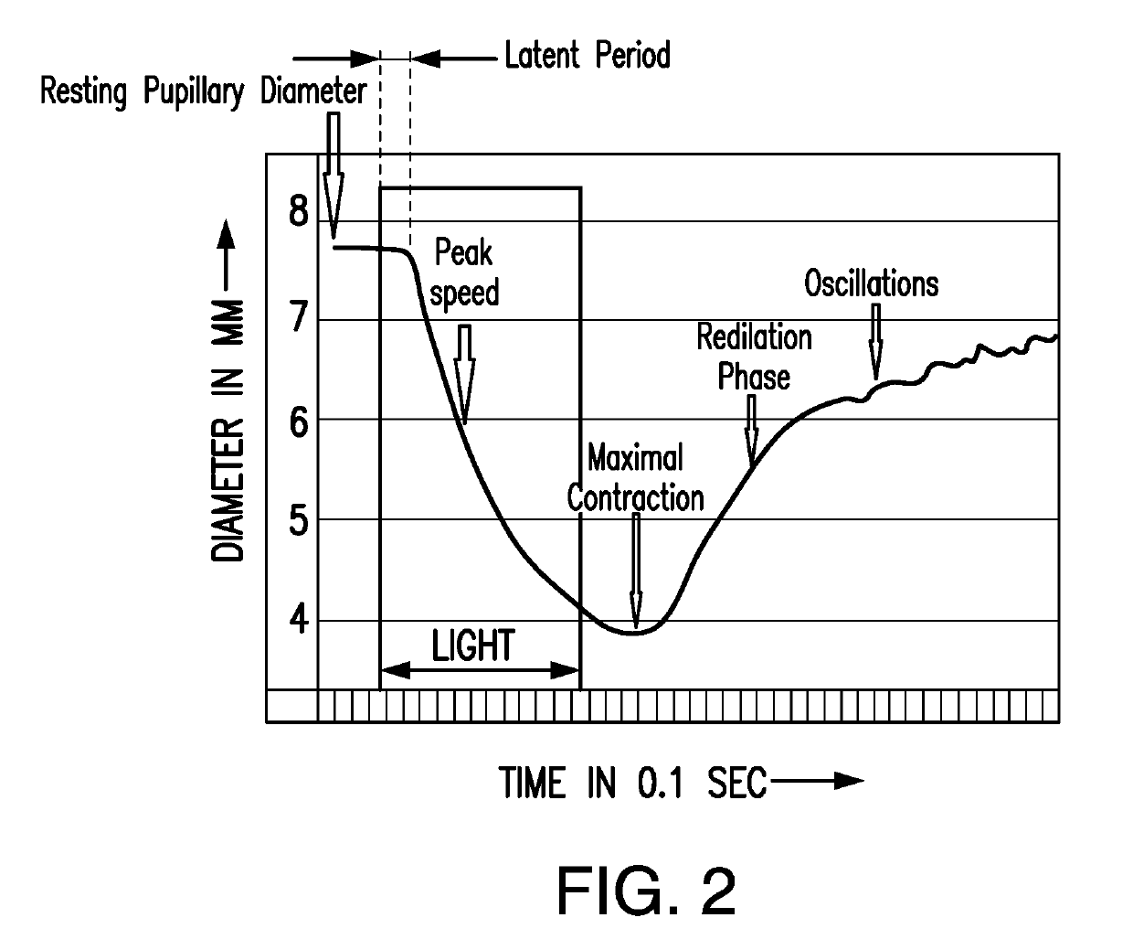
[0231]FIG. 8 is a graph depicting an actual PLR (Loewenfeld, Irene E, (1999), The Pupil, Boston, Mass., Butterworth Heineman, page 777) showing the concentration of Benzedrine (brand name under which amphetamine was marketed in the U.S. by Smith, Kline & French, now part of GlaxoSmithKline, London, UK) administered, which at 10 mg is low and has almost no sympathomimetic effect but does have a marked central effect on the pupils. The PLR after administration of the drug (broken lines) is shifted upwards and the reactions are enhanced as the increased pupil size creates a larger mechanical range for contraction. Also, the oscillations, or fatigue waves, are abolished with the amphetamine induced central stimulation. If a similar sympathomimetic drug topically were administered to the eye, with no central effect on the brain, the pupil diameter would enlarge, but there would be no effect on the fatigue waves or pupillary oscillations.


PUM


Abstract

Disclosed are systems and methods for capturing a pupillary light reflex (PLR) by capturing images of a subject's pupil, for example using a smartphone, extracting image data to determine PLR and classifying the PLR to provide an analytical output, such as a diagnosis or prognosis, of a neurological or psychiatric brain condition.

Description

CROSS-REFERENCE TO RELATED PATENT APPLICATION[0001]This patent application claims a benefit to the filing date of U.S. Provisional Patent Application Ser. No. 62 / 538,306, titled “Methods for Capturing and Analyzing Pupil Images to Determine Toxicology and Neurophysiology,” that was filed on Jul. 28, 2017. The disclosure of U.S. 62 / 538,306 is incorporated by reference herein in its entirety.FIELD OF THE DISCLOSURE[0002]Disclosed are systems, methods and apparatus for capturing pupillary light reflex (PLR) and using the PLR for analytical methods including diagnosis of a level of chemical substances in a subject and treatment of disease.BACKGROUND OF THE DISCLOSURE[0003]Observations and measurements of pupil size date to Archimedes (287-212 B.C.) and Galileo (1564-1642). The simple reflex arc of pupillary contractions to light has been studied by physicians and scientists for centuries. Retinal ganglion cells project afferent fibers via the optic nerve, optic chiasm and optic tracts t...

Claims


Application Information

Patent Timeline
Patent Type & Authority Applications(United States)
IPC IPC(8): A61B3/14A61B3/11A61B3/00A61B5/00
CPCA61B3/145A61B3/112A61B3/0025A61B5/4845A61B5/4064A61B5/7264A61B5/4836A61B3/0041A61B2560/0431A61B3/113A61B3/14A61B5/4076A61B5/1103A61B5/1128
Inventor GIOVINAZZO, VINCENT J.HOSEA, DEVIN F.VAUGHN, WILLIAM F.
Owner PUPILSCAN CORP
Why Eureka
Eureka Blog
Learn More