Gland segmentation with deeply-supervised multi-level deconvolution networks

a multi-level deconvolution network and deep-supervised technology, applied in image enhancement, medical/anatomical pattern recognition, instruments, etc., can solve the problems of difficult to be learned from data approach to generalize to all unseen cases, laborious process of manually annotating digitalized human tissue images, and simple unfavorable, so as to improve computational efficiency and weight-light learning

Inactive Publication Date: 2019-07-04
KONICA MINOLTA LAB U S A INC
View PDF0 Cites 62 Cited by
  • Summary
  • Abstract
  • Description
  • Claims
  • Application Information

AI Technical Summary

Benefits of technology

[0014]To mitigate limitations of existing technologies, embodiments of the present invention use a deep artificial neural network model that employs the DeepLab basis and the multi-layer deconvolution network basis in a unified model that allows the model to learn multi-scale and multi-level features in a deeply supervised manner. Compared with other variants, the model of the present embodiments achieves more accurate boundary location in reconstructing the fine structure of tissue boundaries. Test of the model show that it can achieve segmentation on the benchmark dataset at a level of accuracy which is significantly beyond the top ranking methods in the 2015 MICCAI Gland Segmentation Challenge. Moreover, the overall performance of this model surpasses the state-of-the-art Deep Multichannel Neural Networks published most recently, and this model is structurally much simpler, more computational efficient and weight-lighted to learn.
[0017]In another aspect, the present invention provides a method implemented on a computer for constructing and training an artificial neural network system for classification of histologic images, which includes: constructing the artificial neural network, including: constructing a primary stream network adapted for receiving and processing an input image, the primary stream network being a down-sampling network that includes a plurality of convolutional layers and a plurality of pooling layers; constructing a plurality of deeply supervised side networks, respectively connected to layers at different levels of the primary stream network to receive input, each side network being an up-sampling network that includes a plurality of deconvolutional layers; constructing a final convolutional layer connected to output layers of the plurality of side networks which have been concatenated together; and constructing a first classifier connected to the final convolutional layer and a plurality of additional classifiers each connected to a last layer of one of the side networks, wherein each of the first and the additional classifiers calculates, of each pixel of the layer to which it is connected, probabilities of the pixel belonging to each one of three classes; and training the artificial neural network using histologic training images and associated label data to obtain weights of the artificial neural network, by minimizing a loss function which is a sum of a loss function of each of the side networks calculated using output of the additional classifiers and a loss function of the final convolutional layer calculated using output of the first classifier, wherein the label data for each training image labels each pixel of the training image as one of three classes including a class for gland region, a class for boundary, and a class for background tissue.

Problems solved by technology

Manually annotating digitalized human tissue images is a laborious process, which is simply unfeasible.
Unlike natural scene images which in general have well organized and similar object boundaries, the pathological images usually have large variances due to the tissues from different body parts and the aggressiveness level of the cancer so that they are more difficult to be learned from data approach to generalize to all unseen cases.
However, the use of large receptive fields and down-sampling operator in pooling layers reduces the spatial resolution inside the deep layers and blurs the object boundaries.
FCN is well-suited for detecting the boundaries between two different classes; however, it encounters difficulties in detecting occlusion boundaries between objects from the same class, which are frequently present in pathological images.
However, Deeplab is not an end-to-end trained system, where the DCNN is trained first, and then a fully connected Conditional Random Field (CRF) is applied on top of the DCNN output as a constraint to compensate for a loss of localization accuracy due to downsampling in DCNNs.
This is mainly due to a lack of training data available in the public domain.
Some work directly uses CNN trained as pixel classifiers, which is not ideal for image segmentation tasks compared with the image-to-image prediction techniques.
Such an overly simple deconvolutional procedure is difficult to accurately reconstruct very fine and highly non-linear structure of tissue boundaries.
However, the system is overly complex.
Such an over simple deconvolutional procedure can generally lead to loss of boundary information.
This is due to the lack of good upsampling techniques in their models.
These techniques often fail to achieve satisfactory performance in challenging cases where the glandular structures are seriously deformed.
Though their performance has already improved over methods that use hand engineered features, their ability to delineate boundaries is poor and extremely inefficient in terms of computational time during inference.
Consistent good quality gland segmentation for all the grades of cancer has remained a challenge.
Thus, the approach does not fully harness the strength of DCNN of learning rich feature representations.
In addition, an observation can be made from their result that, the fuse of boundary information deteriorates the performance when applied on the challenging dataset of malignant cases.
Nevertheless, the system is overly complex.
Existing deep learning methods in this field have limited capability to accurately reconstruct highly non-linear structure of tissue boundaries.

Method used


Image

  • Gland segmentation with deeply-supervised multi-level deconvolution networks
  • Gland segmentation with deeply-supervised multi-level deconvolution networks
  • Gland segmentation with deeply-supervised multi-level deconvolution networks

Examples


Embodiment Construction

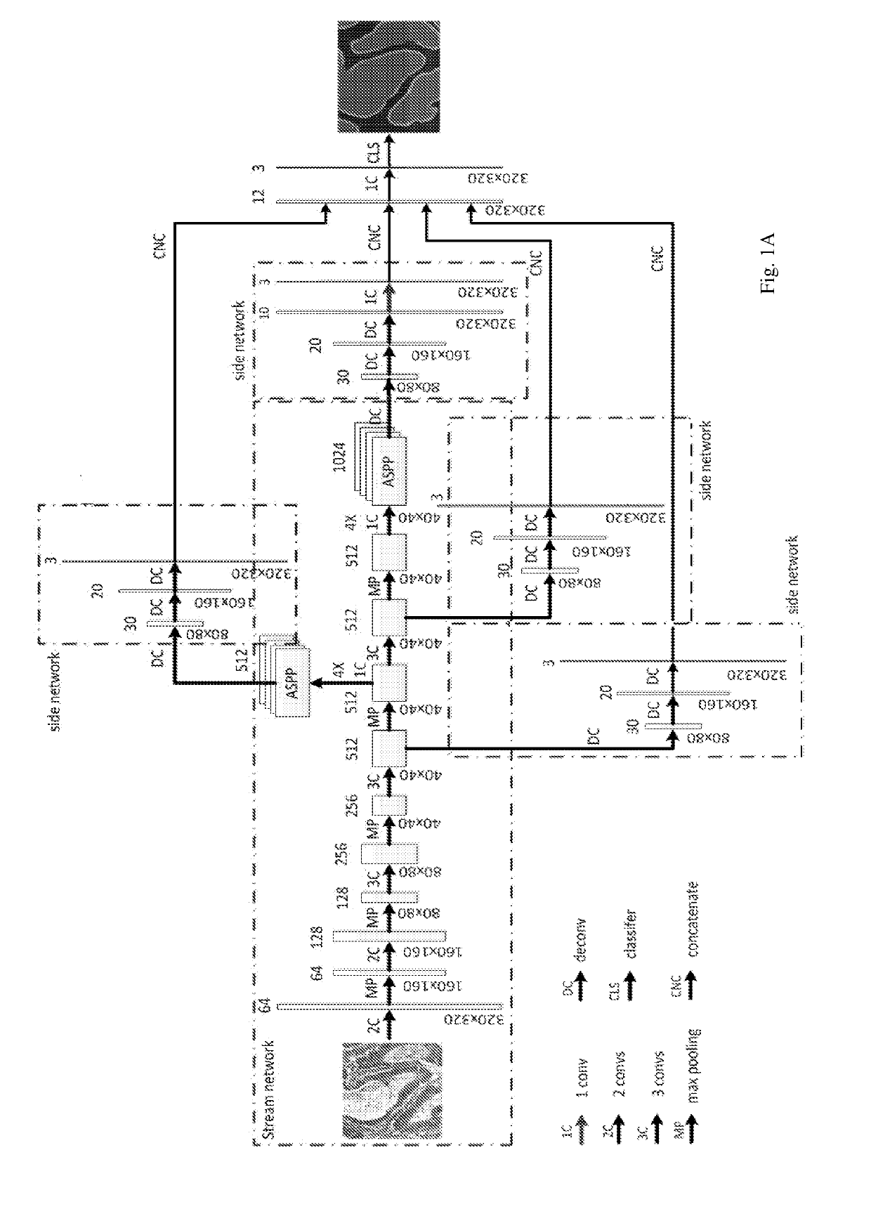
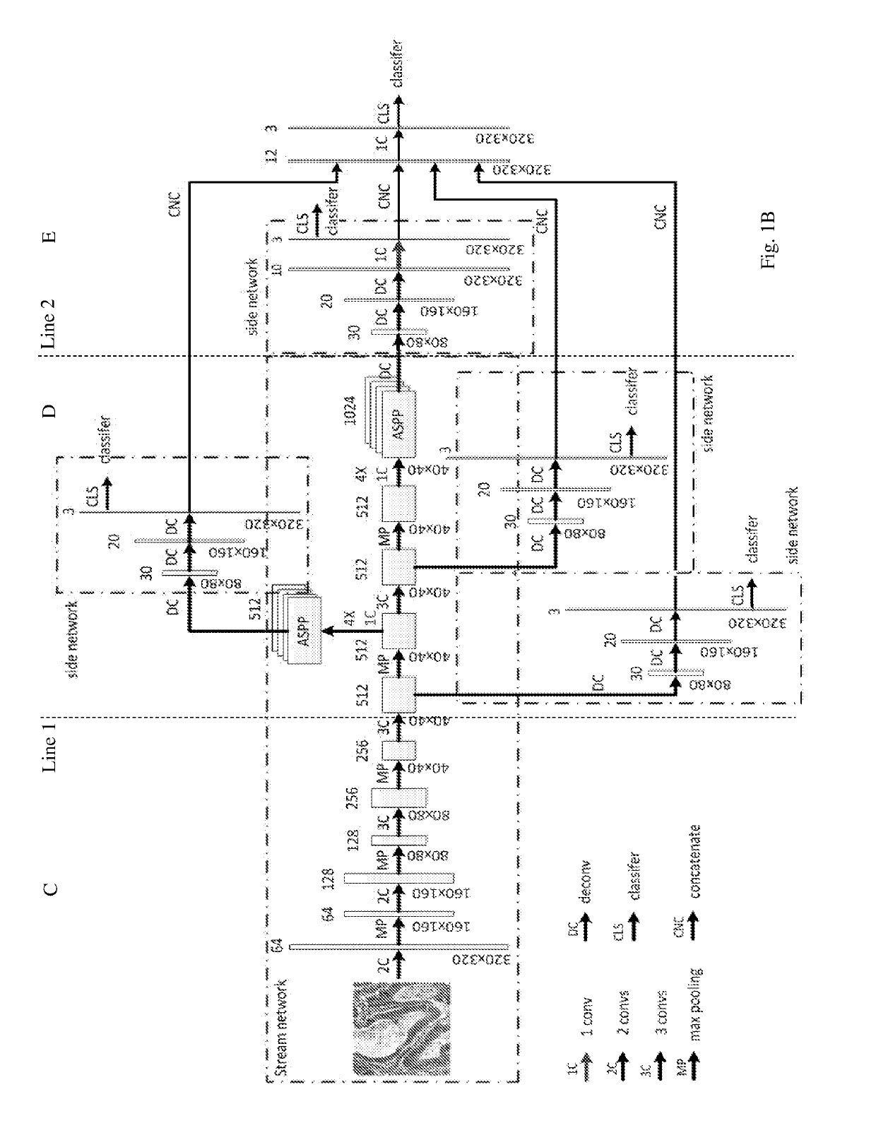
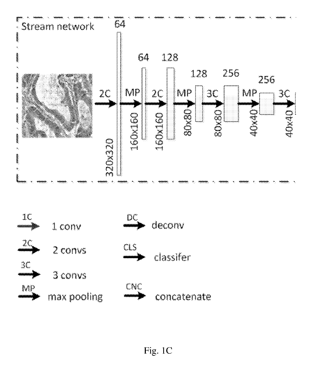
[0026]Similar to DCAN, the neural network model according to embodiments of the present invention is composed of a stream deep network and several side networks, as can be seen in FIGS. 1A-B. It, however, differs from DCAN in the following several aspects.

[0027]First, the model of the present embodiments uses DeepLab as a basis of the stream deep network, where Atrous spatial pyramid pooling with filters at multiple sampling rates allows the model to probe the original image with multiple filters that have complementary effective fields of view, thus capturing object as well as image context at multiple scales so that the detailed structures of an object can be retained.

[0028]Second, the side network of the model of the present embodiments is a multi-layer deconvolution network derived from the paper by H. Noh, S. Hong, and B. Han, Learning deconvolution network for semantic segmentation, published in arXiv:1505.04366, 2015. The different levels of side networks allow the model to p...


PUM


Abstract

Pathological analysis needs instance-level labeling on a histologic image with high accurate boundaries required. To this end, embodiments of the present invention provide a deep model that employs the DeepLab basis and the multi-layer deconvolution network basis in a unified model. The model is a deeply supervised network that allows to represent multi-scale and multi-level features. It achieved segmentation on the benchmark dataset at a level of accuracy which is significantly beyond all top ranking methods in the 2015 MICCAI Gland Segmentation Challenge. Moreover, the overall performance of the model surpasses the state-of-the-art Deep Multi-channel Neural Networks published most recently, and the model is structurally much simpler, more computational efficient and weight-lighted to learn.

Description

BACKGROUND OF THE INVENTIONField of the Invention[0001]This invention relates to artificial neural network technology, and in particular, it relates to deeply-supervised multi-level deconvolution networks useful for processing pathological images for gland segmentation.Description of Related Art[0002]Artificial neural networks are used in various fields such as machine leaning, and can perform a wide range of tasks such as computer vision, speech recognition, etc. An artificial neural network is formed of interconnected layers of nodes (neurons), where each neuron has an activation function which converts the weighted input from other neurons connected with it into its output (activation). In a learning process, training data are fed into to the artificial neural network and the adaptive weights of the interconnections are updated through the leaning process. After learning, data can be inputted to the network to generate results (referred to as prediction).[0003]A convolutional neu...

Claims


Application Information

Patent Timeline
Patent Type & Authority Applications(United States)
IPC IPC(8): G06N3/08G06N3/04G16H30/40G06K9/62G06T7/11G06T7/13
CPCG06N3/08G06N3/0454G16H30/40G06K9/628G06K9/6256G06T7/11G06T7/13G06K9/6277G06K9/6232G06K2209/05G06T2207/20081G06T2207/20084G06T7/0012G06T2207/10056G06T2207/30024G06V20/695G06V20/698G06V10/454G06V10/82G06N3/045A61B5/72G06V2201/03G06F18/213G06F18/214G06F18/2415G06F18/2431
Inventor ZHU, JINGWENZHANG, YONGMIAN
Owner KONICA MINOLTA LAB U S A INC
Why Eureka
Try Eureka