Mirnas as non-invasive biomarkers for breast cancer

a breast cancer and biomarker technology, applied in the field ofmirnas as non-invasive biomarkers for breast cancer, can solve the problems of not being routinely used for breast cancer screening and expensive imaging techniques

Pending Publication Date: 2019-07-25
HUMMINGBIRD DIAGNOSTICS GMBH
View PDF0 Cites 0 Cited by
  • Summary
  • Abstract
  • Description
  • Claims
  • Application Information

AI Technical Summary

Benefits of technology

[0034]The term “treatment”, in particular “therapeutic treatment”, as used herein, refers to any therapy which improves the health status and / or prolongs (increases) the lifespan of a patient. Said therapy may eliminate the disease in a patient, arrest or slow the development of a disease in a patient, inhibit or slow the development of a disease in a patient, decrease the frequency or severity of symptoms in a patient, and / or decrease the recurrence in a patient who currently has or who previously has had a disease. The therapeutic treatment of breast cancer is selected from the group consisting of chemotherapy, surgery, and radiotherapy, preferably chemotherapy. Preferably, breast cancer is triple-negative breast cancer (TNBC).
[0045]The present inventors analysed miRNA expression profiles of early stage breast cancer patients compared to healthy controls. They identified single miRNAs which predict breast cancer with a high specificity, sensitivity, and accuracy. The present inventors also pursued a multiple biomarker strategy by implementing sets of miRNA biomarkers for the diagnosis of breast cancer. This approach could further increase specificity, sensitivity, and accuracy and, thus, the predictive power. In addition, the present inventors identified a specific sample type, namely a blood cellular fraction comprising erythrocytes, leukocytes, and thrombocytes, as a special source of miRNAs having a high diagnostic potential.
[0063](ii) computing an algorithm or a mathematical function based on said reference levels that is suitable to distinguish between breast cancer and healthiness or to decide if breast cancer is present in the patient or not.The inventors of the present invention found that the application of a machine learning approach leads to the obtainment of an algorithm or a mathematical function that is trained by the reference levels mentioned above which allows a better discrimination between healthiness and breast cancer. In this way, the performance of patient's diagnosis can be improved.Machine learning approaches may include, but are not limited to, supervised or unsupervised analysis: classification techniques (e.g. naïve Bayes, Linear Discriminant Analysis, Quadratic Discriminant Analysis Neural Nets, Tree based approaches, Support Vector Machines, Nearest Neighbour Approaches), Regression techniques (e.g. linear Regression, Multiple Regression, logistic regression, probit regression, ordinal logistic regression ordinal probit regression, Poisson Regression, negative binomial Regression, multinomial logistic Regression, truncated regression), Clustering techniques (e.g. k-means clustering, hierarchical clustering, PCA), Adaptations, extensions, and combinations of the previously mentioned approaches.In particular, support vector machines (SVMs) are a set of related supervised learning methods which are preferably used for classification and regression. For example, given a set of training examples, each marked as belonging to one of two categories (e.g. diseased, i.e. suffering from breast cancer, or healthy, i.e. not suffering from breast cancer), an SVM algorithm builds a model that predicts whether a new example (e.g. sample to be tested) falls into one category or the other (e.g. diseased, i.e. suffering from breast cancer, or healthy, i.e. not suffering from breast cancer). A SVM model is a representation of the training examples as points in space, mapped so that the training examples of the separate categories (e.g. diseased, i.e. suffering from breast cancer, or healthy, i.e. not suffering breast cancer) are divided by a clear gap that is as wide as possible. New examples (e.g. samples to be tested) are then mapped into that same space and predicted to belong to a category based on which side of the gap they fall on (e.g. diseased, i.e. suffering from breast cancer, or healthy, i.e. not suffering from breast cancer). More formally, a support vector machine constructs a hyperplane or set of hyperplanes in a high or infinite dimensional space, which can be used for classification, regression or other tasks. A good separation is achieved by the hyperplane that has the largest distance to the nearest training data points of any class (so-called functional margin), since in general the larger the margin the lower the generalization error of the classifier.
[0094]Nucleic acid hybridization, for example, may be performed using a microarray / biochip or in situ hybridization. The microarray / biochip allows the analysis of a single miRNA as well as multiple miRNAs comprised in a blood sample from a patient. For nucleic acid hybridization, for example, the polynucleotides (probes) described herein with complementarity to the corresponding miRNAs to be detected are attached to a solid phase to generate a microarray / biochip. Said microarray / biochip is then incubated with miRNAs, isolated (e.g. extracted) from the blood sample, which may be labelled or unlabelled. Upon hybridization of the labelled miRNAs to the complementary polynucleotide sequences on the microarray / biochip, the success of hybridisation may be controlled and the intensity of hybridization may be determined via the hybridisation signal of the label in order to determine the level of each tested miRNA in said blood sample.

Problems solved by technology

Magnetic resonance imaging (MRI) of the breast could improve cancer detection in these cases, but this imaging technique is expensive and not routinely used for breast cancer screening.

Method used


Image

  • Mirnas as non-invasive biomarkers for breast cancer
  • Mirnas as non-invasive biomarkers for breast cancer
  • Mirnas as non-invasive biomarkers for breast cancer

Examples


examples

[0305]The examples given below are for illustrative purposes only and do not limit the invention described above in any way.

1. Materials and Methods

1.1 Patient Samples

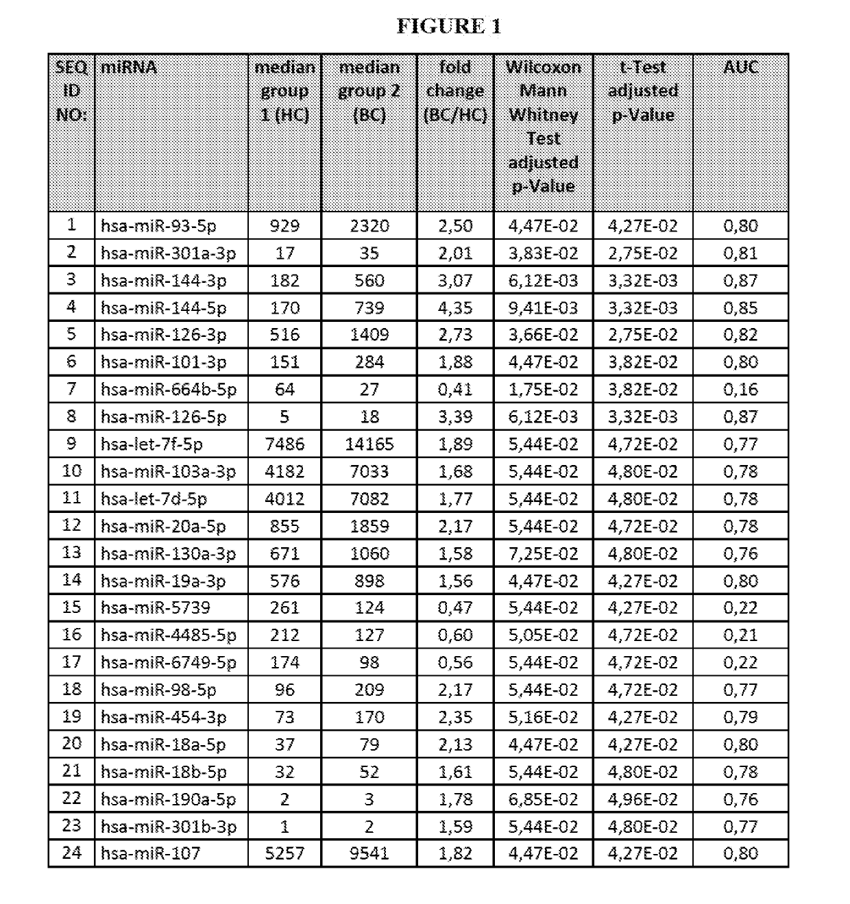
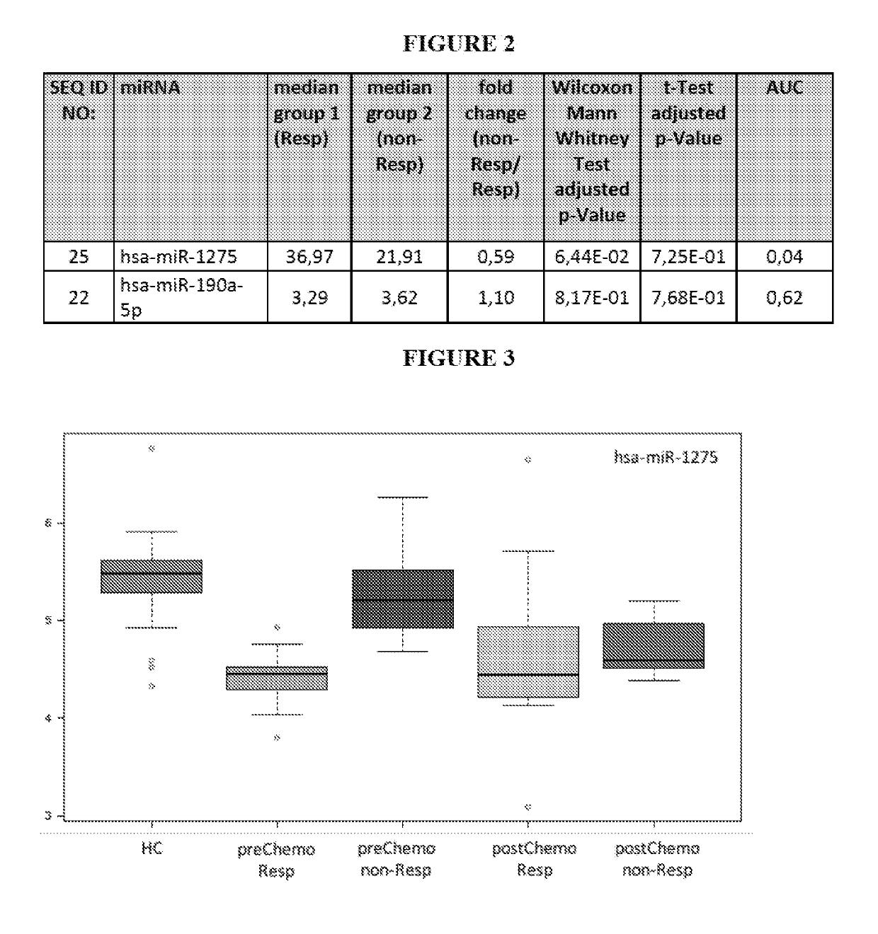
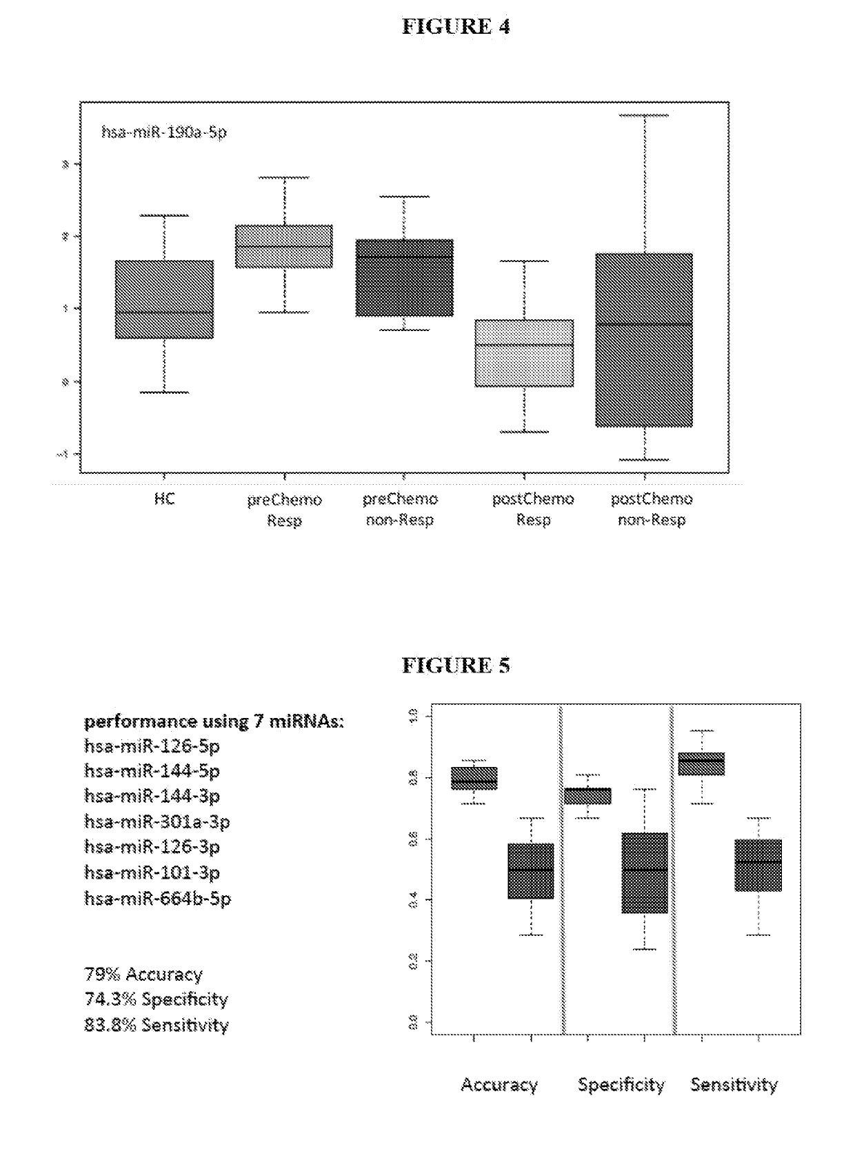
[0306]Blood was collected from subjects suffering from 21 breast cancer (in particular triple-negative breast cancer subjects, TNBC) and 21 subjects not suffering from breast cancer (healthy controls). For each of the breast cancer subject 2 samples were collected, namely one sample prior chemotherapy and one sample after being subjected to chemotherapy.

1.2 Blood Sample Collection Using PaxGene Blood RNA Tubes

[0307]For each blood donor 2.5 ml of whole blood was collected by venous puncture into a PAXgene Blood RNA Tube (PreAnalytix, Hombrechticon, Switzerland). The blood cells were derived / obtained from processing the whole blood samples by centrifugation. Herein, the blood cells from the whole blood collected in said blood collection tubes were spun down by 10 min, 5000×g centrifugation. The blood cell pellet (the cel...


PUM

PropertyMeasurementUnit
Tmaaaaaaaaaa
temperatureaaaaaaaaaa
pHaaaaaaaaaa

Abstract

The present invention relates to a method for diagnosing breast cancer in a patient or for determining whether a patient will respond to a therapeutic treatment of breast cancer. Further, the present invention relates to the use of at least one polynucleotide for detecting at least one miRNA for diagnosing breast cancer in a blood sample from a patient or for determining whether a patient will respond to a therapeutic treatment of breast cancer in a blood sample from the patient. Furthermore, the present invention relates to the use of at least one miRNA isolated from a blood sample from a patient for diagnosing breast cancer or for determining whether the patient will respond to a therapeutic treatment of breast cancer. In addition, the present invention relates to a kit for diagnosing breast cancer in a patient or for determining whether a patient will respond to a therapeutic treatment of breast cancer.

Description

[0001]The present invention relates to a method for diagnosing breast cancer in a patient or for determining whether a patient will respond to a therapeutic treatment of breast cancer. Further, the present invention relates to the use of at least one polynucleotide for detecting at least one miRNA for diagnosing breast cancer in a blood sample from a patient or for determining whether a patient will respond to a therapeutic treatment of breast cancer in a blood sample from the patient. Furthermore, the present invention relates to the use of at least one miRNA isolated from a blood sample from a patient for diagnosing breast cancer or for determining whether the patient will respond to a therapeutic treatment of breast cancer. In addition, the present invention relates to a kit for diagnosing breast cancer in a patient or for determining whether a patient will respond to a therapeutic treatment of breast cancer.BACKGROUND OF THE INVENTION[0002]Breast cancer (BC) is one of the leadin...

Claims


Application Information

Patent Timeline
Patent Type & Authority Applications(United States)
IPC IPC(8): C12Q1/6886C12Q1/6851
CPCC12Q1/6886C12Q1/6851C12Q2600/158C12Q2600/178C12Q2545/113
Inventor KAHRAMAN, MUSTAFALAUFER, THOMASKELLER, ANDREASROSKE, ANNE
Owner HUMMINGBIRD DIAGNOSTICS GMBH
Why Eureka
Try Eureka